ISSN Print: 2164-3024
DOI: 10.4236/ojrad.2018.83017 Aug. 20, 2018 150 Open Journal of Radiology
Primary Chondrosarcoma of the Chest Wall—
A Case Report
Cyriac George
1, D. Diallo
2, F. Velez-Cubian
3, J. Fontaine
4, M. Bui
4, T. Rose
41Radiology Department, University of South Florida, Tampa, USA 2General Surgical Department, University of South Florida, Tampa, USA
3Thoracic Surgical Department, Moffitt Cancer Center, Tampa, USA
4Pathology Department, Moffitt Cancer Center, Tampa, USA
Abstract
A 60-year-old Hispanic male presented to his primary care physician office with an asymptomatic, but palpable right anterior chest wall mass. Initial work up of the finding included a CT scan of the chest which revealed a non-calcified, solid right anterior chest wall mass with invasion of the ante-rior fifth rib and intercostal space. The patient was presented at multidiscip-linary conference with the patient’s primary physician, a medical oncologist, radiologist, pathologist and oncologic surgeon in attendance. The decision was to perform surgical resection of the mass to treat this primary mesen-chymal malignancy. The anterior aspect of the fifth rib and intercostal mus-cles were resected with negative margins. Pathology confirmed the mass to be a low-grade chondrosarcoma. Due to the low-grade nature, low metastatic potential and negative margins of the tumor, the decision was made not to pursue adjuvant chemotherapy or radiation therapy. The patient made full recovery.
Keywords
Chondrosarcoma, Chest Wall Tumors, Cartilaginous Tumors
1. Introduction
Chondrosarcomas are the most common primary chest wall malignancies and the second most common malignant bone tumors. The diagnosis and manage-ment warrant a multidisciplinary approach that includes the primary physician, medical oncologist, surgeon, pathologist and radiologist. Multi-modality imag-ing includimag-ing CT, MRI and PET along with histopathological tissue analysis can
How to cite this paper: George, C., Diallo, D., Velez-Cubian, F., Fontaine, J., Bui, M. and Rose, T. (2018) Primary Chondrosar-coma of the Chest Wall—A Case Report. Open Journal of Radiology, 8, 150-158. https://doi.org/10.4236/ojrad.2018.83017
Received: July 14, 2018 Accepted: August 17, 2018 Published: August 20, 2018
Copyright © 2018 by authors and Scientific Research Publishing Inc. This work is licensed under the Creative Commons Attribution International License (CC BY 4.0).
http://creativecommons.org/licenses/by/4.0/
C. George et al.
DOI: 10.4236/ojrad.2018.83017 151 Open Journal of Radiology
be used to determine a conclusive diagnosis of chondrosarcoma.
2. Case Presentation
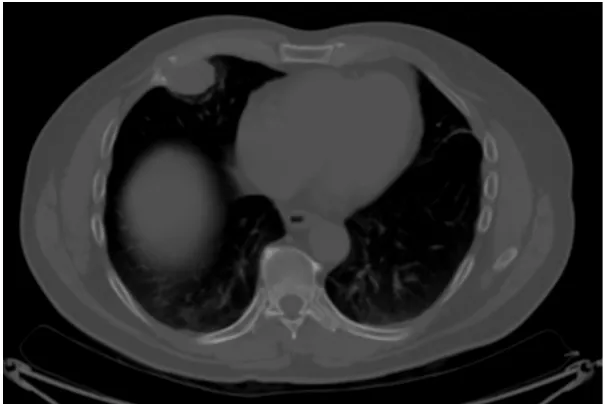
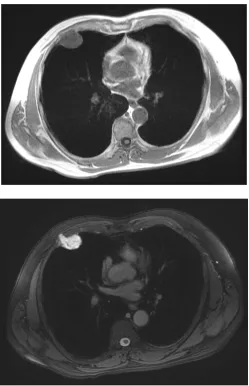
A 60-year-old Hispanic male presented to his primary care physician office with an asymptomatic, but palpable right anterior chest wall mass. The patient has no significant past medical history except for a clavicular fracture sustained during soccer. Physical examination revealed an asymptomatic 4 cm soft tissue palpable mass of the right anterior chest wall. The skin was intact, no erosive markings were present. The patient’s laboratory work up was unremarkable. Radiology work up included CT chest, MRI of the chest and PET/CT. His CT chest axial series on bone windows demonstrated a 2.8 × 3.8 × 2.9 cm pleural based soft tissue mass eroding through the right anterior fifth rib (Figure 1). There was mild compression of the nearby lung parenchyma, however no pulmonary le-sions were identified. Further characterization of the mass by MRI contrast en-hanced images of the chest revealed a well-circumscribed pleural based T1 hy-po-intense avidly enhancing soft tissue mass in the right anterior chest wall in-volving the right anterior fifth rib and into the overlying sub-pectoral soft tissues
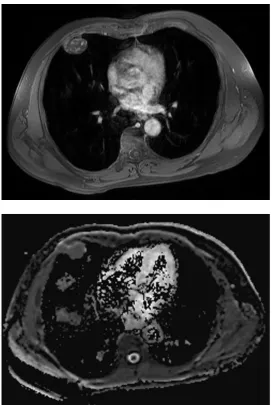
(Figure 2). The mass also demonstrated restricted diffusion which is indicative
of high cellularity (Figure 3 and Figure 4). PET/CT imaging revealed mild hypermetabolic activity of the mass with maximum SUV of 1.9 (Figure 5).
The patient underwent a right-sided video-assisted thoracoscopy (VATS) with right chest wall resection and reconstruction with an advancement muscle flap. No synthetic material or plates were used. The patient tolerated the operation without complications and had an uneventful recovery.
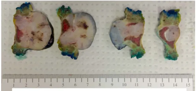
[image:2.595.222.526.491.693.2]Gross pathology of the fifth rib resection revealed a firm, oval, tan-white co-lored 3.7 LR × 3.3 CC × 3.2 AP cm solid mass (Figure 6). Cut surfaces of the mass were gray-white, focally mucinous and cartilaginous (Figure 7). A small
DOI: 10.4236/ojrad.2018.83017 152 Open Journal of Radiology Figure 2. Axial post-contrast T1 weighted MRI image is noted for avid enhancement of the destructive anterior chest wall mass.
[image:3.595.249.500.496.683.2]C. George et al.
[image:4.595.237.511.67.473.2]DOI: 10.4236/ojrad.2018.83017 153 Open Journal of Radiology
Figure 4. Apparent diffusion imaging correlate with hypointense signal of the mass.
[image:4.595.239.509.508.682.2]DOI: 10.4236/ojrad.2018.83017 154 Open Journal of Radiology Figure 6. Gross pathology of the resected right fifth rib revealed a firm, oval, tan-white colored 3.7 × 3.3 × 3.2 cm solid mass.
Figure 7. Cross sectional gross pathology shows the tumor invading the soft tissue arising from the benign cartilage.
portion of the mass was submitted for intraoperative frozen section diagnosis. The mass was serially sectioned from lateral to medial in 3 - 4 mm intervals into 9 slices. The mass grossly involved the bone and cartilage of the rib and abut the anterior and posterior surfaces. The mass measured 7 mm from the inferior soft tissue margin, 8 mm from the superior soft tissue margin, 5.6 cm from the later-al bone margin and 5.9 cm from the medilater-al bone margin.
3. Diagnosis
[image:5.595.214.532.301.449.2]C. George et al.
DOI: 10.4236/ojrad.2018.83017 155 Open Journal of Radiology
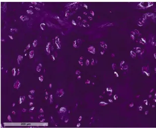
Figure 8. Micropathology slide low power magnification analysis was consistent with Grade I chondrosarcoma.
Figure 9. Micropathology slide high power magnification analysis was consistent with
Grade I chondrosarcoma.
[image:6.595.215.533.71.328.2] [image:6.595.214.530.374.634.2]DOI: 10.4236/ojrad.2018.83017 156 Open Journal of Radiology
negative for re-occurrence.
4. Discussion
4.1. Clinical Findings
Chondrosarcomas are the most common primary chest wall malignancies and the second most common malignant bone tumors. They are more commonly seen in males, usually presenting between the 4th-7th decades of life [1] [2] [3]. Patients can present with progressive pain or palpable soft tissue mass. Devel-opment of pathologic fractures is common at initial presentation. The tumors are commonly clinically aggressive. The diagnosis and management warrant a multidisciplinary approach that includes the primary physician, medical oncolo-gist, surgeon, pathologist and radiologist. Surgical resection is the most common treatment with optional neoadjuvant or adjuvant chemotherapy and/or radia-tion therapy for high grade or dedifferentiated chondrosarcoma with positive margins.
4.2. Radiologic Findings
Radiologic screening of chondrosarcomas involves multimodality characteriza-tion which is determined according to the American College of Radiology (ACR) appropriateness criteria [4]. The criteria are based upon a grading system which has the following values: rating system of 1 - 9, where 9 is the most appropriate measure. The initial imaging modality of localized bone tumors is plain radio-graphy of the region of interest, rating 9. Computed tomoradio-graphy (CT) with and without contrast, is rating 9 if the lesion cannot be adequately characterized on radiographs. Magnetic resonance imaging (MRI) with and without contrast, is rating 8 if the lesion is indeterminate or a lytic lesion. Technetium-99m (Tc-99m) bone scan, rating 5 if better localization of the lesion is needed. Posi-tron emission tomography (PET), is rating 1. PET is not part of the initial screening of lesions. It is reserved for staging and evaluating of metastatic le-sions.
Chondrosarcomas of the rib are most commonly found at the costochondral junction [1]. Radiographically they may be seen as a lytic lesion with endosteal scalloping, cortical thinning or thickening and irregular margins. Calcifications of the mass commonly have a ring and arcs configuration, however, they may be punctate as well. Soft tissue extension is relatively common as demonstrated by our patient’s case.
exten-C. George et al.
DOI: 10.4236/ojrad.2018.83017 157 Open Journal of Radiology
sion while high grade lesions generally have non-calcified areas with soft-tissue extension. CT performed with intravenous contrast may demonstrate a mild pe-ripheral rim and septal enhancement.
Magnetic resonance imaging (MRI) is excellent at characterizing bone mar-row involvement, soft tissue extension and components. Chondrosarcoma le-sions tend to demonstrate a lobular morphology. Marrow replacement may ap-pear as low to intermediate signal intensity on T1 weighted images. Non-mineralized components may have high signal intensity on T2 weighted images due to their high water content, while mineralized matrices have low signal intensity with all MR pulse sequences.
Positron emission tomography combined with computed tomography (PET/CT) provides a functional information of the tumor with analysis of active glucose consumption. The higher the metabolic activity the higher the glucose consumption and hence the PET/CT signal. In general, malignant cells have higher metabolic rate relative to the normal soft tissue background. The active lesions will display a high signal which is indicative of active disease.
4.3. Pathologic Findings
Chondrosarcoma has chondromyxoid matrix with nuclear pleomorphism, mito-sis and necromito-sis [5]. Permeation of medullary and cortical bone and soft tissue invasion are indicative of malignancy. In addition, the prognosis of chondrosar-coma depends on tumor grade. Grade I is the best prognostic factor without evidence of metastases; Grade II and III are with evidence of metastatic disease with Grade III having the worst outcomes [6]. Chondrosarcomas can originate centrally from the bone or peripherally within the cartilage cap. Central chon-drosarcomas are somewhat genetically different from peripheral chondrosarco-mas. Central lesions are usually diploid and peripheral lesions are aneuploidy. However, both lesions share the same histology. Other rare chondrosarcoma subtypes include dedifferentiated, mesenchymal and clear cell chondrosarcoma. The dedifferentiated subtype is associated with a very low survival rate due to often distant metastatic involvement at time of initial diagnosis. Mesenchymal chondrosarcomas are usually bi-morphic consisting of low grade cartilaginous cells and hyper cellular small, uniform and undifferentiated cells that resemble Ewing’s sarcoma cells. Clear cell chondrosarcomas generally have a good prog-nosis with complete surgical eradication. Some chondrosarcomas can arise from enchondroma, osteochondroma or extra skeletal myxoid subtypes [6].
5. Conclusion
evalua-DOI: 10.4236/ojrad.2018.83017 158 Open Journal of Radiology
tion of the site with chest CT every 6 months for the first 2 - 3 years followed by yearly follow-up chest CT for a total of 5 - 10 years for this patient.
Conflicts of Interest
The authors declare no conflicts of interest regarding the publication of this pa-per.
References
[1] Murphey, M.D., et al. (2003) From the Archives of the AFIP: Imaging of Primary Chondrosarcoma: Radiologic-Pathologic Correlation. Radio Graphics, 23, 1245-1278. https://doi.org/10.1148/rg.235035134
[2] Ollivier, L., Vanel, D. and Leclere, J. (2003) Imaging of Chondrosarcomas. Cancer Imaging, 4, 36-38. https://doi.org/10.1102/1470-7330.2003.0022
[3] Varma, D.G., Ayala, A.G., Carrasco, C.H., Guo, S.Q., Kumar, R. and Edeiken, J. (1992) Chondrosarcoma: MR Imaging with Pathologic Correlation. Radio Graphics, 12, 687-704.
[4] Kransdorf, M.J., et al. (2018) ACR Appropriateness Criteria Soft-Tissue Masses.
Journal of the American College of Radiology, 15, S189-S197. https://doi.org/10.1016/j.jacr.2018.03.012
[5] Brien, E.W., Mirra, J.M. and Luck Jr., J.V. (1999) Benign and Malignant Cartilage Tumors of Bone and Joint: Their Anatomic and Theoretical Basis with an Emphasis on Radiology, Pathology and Clinical Biology. II. Juxtacortical Cartilage Tumors.
Skeletal Radiology, 28, 1-20. https://doi.org/10.1007/s002560050466