ORIGINAL RESEARCH
Diffusion-Weighted MR Imaging Lesions after
Filter-Protected Stenting of High-Grade
Symptomatic Carotid Artery Stenoses
R. du Mesnil de Rochemont S. Schneider B. Yan A. Lehr M. Sitzer J. Berkefeld
BACKGROUND AND PURPOSE: The clinical efficacy of filter devices in internal carotid artery (ICA) stent placement has been a matter of controversy. The aim of this retrospective study was to assess the number and extent of cerebral emboli, as represented by new lesions on diffusion-weighted MR imaging (DWI), in patients treated with filter-protected carotid stent placement.
METHODS: Standard DWI (B0⫽1000) was performed within 48 hours before and 48 hours after filter-protected carotid stent placement in 50 patients with symptomatic, high grade (⬎70%), athero-sclerotic ICA stenosis. Number, extent, and vascular territory of new DWI lesions after stent place-ment were assessed by consensus of 2 experienced neuroradiologists. Multifactorial statistical anal-ysis was performed to determine risk factors associated with DWI lesions.
RESULTS: New punctate DWI lesions with a median diameter of 2 mm were detected in 14 of 50 cases in the territory of the stented ICA and in 7 of 50 cases in other vascular territories. Median lesion load was 1 lesion (range, 1–15) per positive case in the stented ICA and 1 lesion (range, 1–7) in other vascular territories. All DWI lesions were clinically asymptomatic. Because of 1 hyperperfusion syndrome with temporary brain swelling, the 30-day stroke and death rate was 2%. Ageⱖ70 years was the only significant predictor for new DWI lesions, whereas sex, degree and site of stenosis, vascular risk factors, and stent and filter type showed no significant correlation.
CONCLUSIONS:New DWI lesions after filter-protected carotid stent placement are substantially more frequent in the ipsilateral ICA territory compared with other vascular territories. Therefore, intraluminal filters cannot completely protect the brain from procedure-related embolization. However, individual lesion load and the risk of clinically relevant ischemia is low.
N
europrotective filters for internal carotid artery (ICA) stent placement have been developed to a high technical standard and are increasingly used for prevention against ce-rebral embolic complications. The current generation of filters from different manufacturers (Table 1) have pore sizes be-tween 100 and 150m, which are small enough to capture embolic particles detached during the stent placement proce-dure and large enough for the continuation of blood flow. This is especially relevant for patients with poor cerebrovascular collateral supply, because filters are better tolerated than pro-tection devices that use temporary balloon occlusion. The ef-fectiveness of filter devices has been examined in experimental studies that have shown elimination of up to 96% of embolic particles with sizes greater than or equal to pore size generated in vitro or during stent placement of endarterectomy specimens.1,2However, the recent studies of carotid stent placement without filter protection reported low rates of microemboli detected by transcranial Doppler3 and low incidence of stroke.4As a result, the clinical effectiveness of filter devices has been questioned. In addition, the usage of filters may in-crease the incidence of arterial thrombosis, vasospasm, or dissection.5,6
On the other hand, the incidence of stroke after carotid stent placement is low at 3% to 5%; stroke as a sole marker for
cerebral emboli is insensitive, and therefore only a large ran-domized study could prove the clinical benefits of filter-pro-tected against unprofilter-pro-tected carotid stenting, if stroke is used as the only endpoint. Therefore, alternative surrogate markers for embolic events, especially new lesions on diffusion-weighted imaging (DWI), has assumed increasing importance in the detection of subclinical microembolic events.7
To assess the extent of microembolic events and cerebral ischemia during filter-protected carotid stent placement, we have compared the DWI obtained before and after carotid stent placement to assess the number, size, and location of new lesions. Furthermore, statistical analysis was used to correlate the occurrence of DWI lesions with age, sex, degree, and site of the stenosis, technical features of the stent procedure as well as with factors indicating the status of atherosclerotic disease of the patient.
Materials and Methods
Between year 2001 and 2005, 76 patients with symptomatic, high grade, atherosclerotic carotid artery stenoses were treated by carotid stent placement with the use of a filter as an embolic protection de-vice. Fifty consecutive patients who had DWI within 48 hours before and after stent placement were included in the study. Patients with strokes within the first 24 hours after onset of symptoms and those with contraindication to MR imaging were excluded from the study. Patients with incorrect timing of MR imaging or with incomplete MR imaging examinations were also excluded from evaluation.
MR imaging examinations were performed, with the use of stan-dard head coils, on a Siemens Magnetom Vision 1.5T (Siemens, Er-langen, Germany). DWI with echo-planar imaging (EPI) sequence (B0⫽1000) was obtained in axial and coronal sections. The number Received July 15, 2005; accepted after revision October 12.
From the Institute of Neuroradiology (R.M.R., S.S., B.Y., J.B.) and Department of Neurology (A.L., M.S.), University of Frankfurt/Main, Germany.
Address correspondence to Dr. Richard du Mesnil de Rochemont, Institut fu¨r Neuroradi-ologie, Klinikum der Johann Wolfgang Goethe-Universita¨t, Schleusenweg 2-16, 60 528 Frankfurt am Main, Germany.
INTERVENTIONAL
ORIGINAL
and vascular territory of new poststent lesions were determined by consensus of 2 experienced neuroradiologists. The maximal diame-ters of DWI lesions were measured. Statistical analysis by using2test, and multivariate regression with backward elimination correlated the occurrence of de novo DWI lesions with age, sex, and site and degree of stenoses. The risk factors arterial hypertension, diabetes, actual smoking, and hypercholesterolemia and the presence of coronary heart disease (CHD) and peripheral arterial occlusive disease (pAOD) as markers for generalized atherosclerosis were also included in the statistical analysis. Time of the procedure since the introduction of filter protection in our institute in 2001, as well as stent and filter type as potential influence factors for the occurrence of DWI lesions, were also analyzed.
Carotid stent placement was performed according to a study pro-tocol approved by the local review board and ethics committee. Pa-tients gave their written informed consent to participate. PaPa-tients withⱖ70% symptomatic ICA stenosis (North American Symptom-atic Carotid Endarterectomy Trial [NASCET] criteria) and older than 50 years were selected for carotid stent placement by an interdiscipli-nary consensus among neurologists, vascular surgeons, and neurora-diologists. Concomitant medical treatment included a combination of 100 mg of aspirin and 75 mg of clopidogrel started at least 3 days before carotid stent placement and continued for 3 months afterward. All stent procedures were done under local anesthesia with mon-itoring by anesthesiologists. After stent placement, the patients were monitored in a neurologic intermediate care unit for the first 24 hours.
Diagnosis of a carotid stenosis was established by Doppler and duplex sonography. Transfemoral arterial approaches were per-formed in all cases. A diagnostic angiogram of the common carotid artery in question was done to confirm the presence of high-grade stenosis and to make the final decision for stent placement.
A heparin bolus of 100 to 150 U/kg was given immediately before the interventional part of the procedure to increase the activated clot-ting time (ACT) to a minimum of 300 seconds. An 8F guiding cath-eter was introduced into the common carotid artery below the ste-notic plaque. The stenosis was passed with the filter system, and the filter was deployed in the high cervical ICA below the skull base. The filter type was chosen according to availability and technical features. In subtotal stenoses (95% by NASCET criteria), we preferred the Spi-der monorail filter (ev3, Plymouth, Minn) because of the low crossing profile of the delivery catheter, which is able to pass tight residual lumen. If vascular curves within the filter deployment zone in the high cervical ICA were present, we used concentric filters such as Accunet (Guidant, Santa Clara, Calif) or EmboShield (MedNova, Abbott, Gal-way, Ireland) instead of eccentric filters such as EPI FilterWire EX, FilterWire EZ (Boston Scientific, Galway, Ireland) or Spider filters (ev3). The patency of the filter was checked by repeated contrast in-jections. Filter complications such as technical failure, vasospasm, dissection, filter obstruction, or thrombosis were registered.
In general, predilation, stent deployment, and postdilation should be performed under filter protection. It is noteworthy that the study protocol allowed predilation with a 3-mm balloon without protection if the passage of the filter system was deemed impossible. All stenoses with a diameter equal or lower than the diameter of the stent delivery system were predilated with a 3- to 20-mm percutaneous translumi-nal coronary angioplasty (PTCA) balloon (Crossail; Guidant, Santa Clara, Calif). Self-expanding carotid stents, either carotid Wallstents (Schneider Boston Scientific, Galway, Ireland) or segmented nitinol stents (Acculink; Guidant) were deployed. Wallstents were preferred in patients with straight course of the carotid artery, whereas the more flexible segmented nitinol stent was used in angulated or curved bi-furcations or in cases with significant differences between the diame-ters of internal and common carotid artery.
All stents were postdilated maximally to the measured diameter of the normal ICA. In most of the cases, a 5- to 20-mm percutaneous transluminal coronary angioplasty balloon (Viatrac; Guidant) was used, and postdilation was regarded as successful if a residual narrow-ing below 30% was proved on control angiograms.
The filter was retrieved and checked for macroscopic particles, which were registered if visible. Finally, angiograms of the carotid bifurcation and the intracranial circulation were performed to dem-onstrate the reconstruction of the carotid lumen and to exclude mac-roembolic complications.
Clinical and sonographic evaluation of the patient was done by independent neurologists before and immediately after the proce-dure, at discharge, and at 3 months after the intervention. Clinical complications were classified according to the Modified Rankin Scale (mRS). Table 1 shows the filter types used for carotid stent placement in this series.
Statistical significance of nonparametric data were determined by using the Wilcoxon-Whitney-Mann U test and the2test. The cor-relation between the occurrence of new DWI lesions and procedural and patient-based risk factors was evaluated with multivariate logistic regression analysis with backward elimination.
Results
Patient and Procedure Characteristics
Forty-eight patients, 35 men and 13 women with a median age of 70 years (range, 50 – 87 years) had 50 symptomatic carotid stenoses with median degree of stenosis of 85% (range, 70%– 95%, NASCET criteria). All were treated by filter-protected carotid stent placement. There were 2 patients with bilateral stenoses who had neurologic symptoms and intervention at different times.
All 50 carotid stent procedures fulfilled the inclusion crite-ria for this study and the patients had DWI examinations within an interval of 48 hours before and 48 hours after tech-nically successful filter-protected carotid stent placement. All
Filter types used in this study
Material Design
Nitinol Filaments⫹Synthetic Membrane
Nitinol Basket
Concentric Eccentric
Type Accunet AngioGuard EmboShield EPI FilterWire EX FilterWire EZ Spider
Manufacturer Guidant Cordis Abbott Boston Scientific Boston Scientific ev3
Porus size 115m 100m 150m 80m 110m 110m
Crossing profile 3.2F 3.2F 3.7F 3.9F 3.2F 3.2F
patients had filter protection during all steps of the angioplasty procedure with the exception of 1 patient in whom predilation without protection was required to allow the passage of the filter system through a 95% stenosis. Thirty-eight Acculink stents and 12 carotid Wallstents were implanted. No severe technical complications were observed. Five patients showed slow flow after filter deployment as a result of vasospasm or guidewire-induced vessel kinking. Occlusion of the filter by debris or clot without flow in the carotid artery after postdila-tion was observed in 3 cases, 2 with EPI filters and 1 with FilterWire EZ. Three of 8 patients with filter-induced restric-tion of flow showed temporary neurologic symptoms that completely disappeared after retrieval of the filter. Eleven of 50 filters (22%) contained macroscopic particles with sizes up to 1.2 mm. Table 1 shows the technical details of the carotid stent procedures.
DWI Evaluation
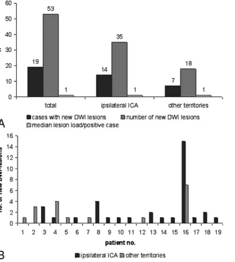
Fourteen of 50 cases (28.4%) with technically successful ca-rotid stent placement showed punctate new DWI lesions in the territory of the ipsilateral ICA. With a single exception (a pa-tient with small cerebellar infarcts with a maximal diameter of 12 mm), none of the other lesions exceeded a maximal diam-eter of 4 mm (median, 2 mm; range, 1– 4 mm). Median lesion load on the ipsilateral side was 1 lesion (range, 1–15) per pos-itive case. Seven of 40 patients showed a median of 1 lesion (range, 1–7) in vascular territories independent of the stented ICA. Two patients showed simultaneous lesions in the ipsilat-eral carotid and other vascular territories. Only 1 patient with severe aortic arch atherosclerosis showed multiple emboli in several vascular territories. Fig 1 illustrates the occurrence and distribution of DWI lesions. Fig 2 shows MR images of 2 cases with typical and maximal extension of embolic events.
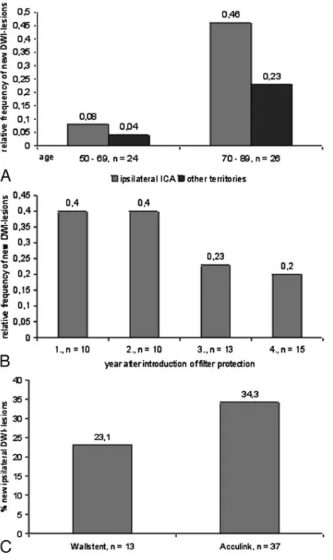
There were significantly more new DWI lesions in elderly patients equal or above the median age of 70 years (70 – 89 years) compared with a younger group aged between 50 and 69 years (P⫽.01; Fig 3). Multivariate regression did not dem-onstrate any statistically significant correlation between sex, site, and percentage of stenosis or vascular risk factors and the presence of CHD or pAOD and the occurrence of new DWI lesions.
In the first 2 years after introduction of filter protection, we observed a statistically nonsignificant trend toward more DWI lesions than in the most recent 2 years. There was also a trend toward a higher incidence of lesions after implantation of niti-nol stents compared with the Wallstent (Fig 3). This was sta-tistically not significant, and no clear correlation between filter type and filter complications was found. In addition, the pa-tient who was treated with predilation without filter protec-tion showed no new DWI lesions.
During early follow-up, 1 patient developed new neuro-logic deficits and seizures 2 days after carotid stent placement. On MR imaging, there was evidence of vasogenic reperfusion edema. It is noteworthy that DWI showed no lesions and helped to distinguish reperfusion phenomenon from cerebral infarct. The patient’s symptoms, which included worsening of pre-existing right-sided hemiparesis and aphasia, regressed within 2 weeks. At the 3-month follow-up, there remained only minor residual deficit with no relevant impairment of daily activities (mRS grade 2).
There were no periprocedural deaths, and the 30-day stroke and death rate was 1 of 50 (2%). One patient developed an asymptomatic 70% in-stent-restenosis during the first 3 months; all other stents remained patent.
Discussion
In agreement with previous studies,8we have observed that new DWI lesions occur frequently after carotid stent place-ment even with the use of protective devices.9,10Some of these lesions have been detected in vascular territories independent of the stented carotid artery and are probably related to cath-eterization procedure and the placement of a large-caliber guiding catheter. The frequency and extent of these clinically silent DWI lesions in regions other than the stented vascular territories are within the range described for diagnostic cere-bral angiography.11,12Air embolism, thrombus formation, or detachment of particles from atherosclerotic vessel walls dur-ing catheterization are the most probable causes of these mi-croembolic lesions, which cannot be influenced by the use of a protective device.13
With MRI, new DWI lesions in the territory of the stented ICA are detected more frequently than emboli on diagnostic intra-arterial angiograms. Our results in a group of patients with filter protection show that the frequency of occurrence of new DWI lesions is within the range described by other au-thors.8-10We cannot exclude the possibility that a proportion of DWI lesions are due to the catheterization procedure before the deployment of the filter device. Other potential sources of microemboli are the passage of the filter device through the stenosis, incomplete apposition of the filter, passage of
parti-Fig 1.A,Number of new diffusion-weighted imaging (DWI) lesions after filter-protected carotid stent placement and median lesion load of DWI positive cases in different vascular territories.
[image:3.585.300.538.43.308.2]cles through the filter pores, and loss of material during cap-ture and retrieval of the filter.
Embolic signals have been observed by transcranial Dopp-ler during all phases of the carotid stent procedure. A recently published study detected even more signals with the use of filter devices. However, the rate of macroembolic complica-tions in filter-protected stent placement was lower than in the unprotected group.3With a median of 1 punctate ischemia per DWI positive case, the lesion load in our study is low, and a shower of multiple emboli occurred in only 1 case. Additional involvement of DWI lesions in vascular territories, which are not treated with a stent, led to the assumption that catheter-ization of the atherosclerotic aortic arch was the most proba-ble cause of this subclinical complication. Low number and extension of lesions in the ipsilateral carotid territory do not support hypotheses that the filter itself is a source of additional embolism by thrombogenicity, material loss through the filter membrane, or movement of the device.3,14,15 Macroscopic particles captured in the filter membrane add weight to the claim that filter devices are effective. The rate of filter compli-cations and DWI-positive cases has decreased over time with increasing experience of the operators and technical improve-ments of filter systems. Filter obstruction was mainly observed with the first generation of the EPI filter, which had a pore size of only 80m.16Filter movement during the intervention as potential source of microemboli or vasospasm occurred
mainly during the initial learning curve and can be widely avoided with improved technique and the use of monorail devices. Increased age was the only significant risk factor for the occurrence of DWI lesions, probably because of a higher proportion of patients with widespread atherosclerosis. A clear influence by other vascular risk factors or presence of CHD and pAOD could not be detected. This may occur be-cause the subgroups of patients presenting with different en-tities become relatively small.
The trend toward more lesions with the use of segmented nitinol stents may be explained by the fact that these stents were favorably used in technically more difficult cases with tortuosity of the carotid artery. A clear difference between dif-ferent filter devices was not detectable. The low DWI lesion load corresponds to the absence of clinically detectable cere-bral ischemia and larger territorial infarcts that might have been prevented by filters containing macroscopic particles.
The limitation of this study was the lack of a control group, which was not included in the protocol for ethical reasons. Comparison of the results with centers working without filter protection may be helpful in this regard. At the moment, there is no level 1 evidence to support the mandatory use of filter protective devices in carotid stent placement. We hope that subgroup analyses of larger randomized carotid endarterec-tomy versus stent studies will contribute to this topic. A fur-ther possible limitation of this study was that we might have
Fig 2.Range of expression of diffusion-weighted imaging (DWI) lesions after filter-protected internal carotid artery (ICA) stent placement.
AandB,DWI the day before and the second day after stent placement of a right ICA stenosis. Upper limit of a small cortical infarct in the right middle cerebral artery (MCA) territory is visible on both images (arrow). Typical appearace of 2 new punctate lesions in the right parietal lobe (B).
[image:4.585.53.529.41.420.2]selected only cases suitable for easy filter placement. But in our patient group, the median carotid stenosis grade of 85%, me-dian age of 70 years, and comorbidity profile similar to that of the SAPPHIRE study17 were arguments against this assumption.
Conclusions
Low lesion load and lesion extent and absence of embolic strokes in our cases is encouraging in that filters are effective in the prevention of macroembolic ischemia during carotid stent procedures. However, the higher incidence of new DWI-le-sions in the stented ICA territory indicates that distal filters are not perfect in the prevention of subclinical procedure-related ischemia. Elderly patients in particular seem to be at a higher risk. From the current data, it remains unclear whether cath-eter access to the angioplasty site or complications of filter devices or carotid stents are mainly responsible for the occur-rence of new DWI lesions.
References
1. Muller-Hulsbeck S, Jahnke T, Liess C, et al.Comparison of various cerebral protection devices used for carotid artery stent placement: an in vitro exper-iment.J Vasc Interv Radiol2003;14:613–20
2. Ohki T, Roubin GS, Veith FJ, et al.Efficacy of a filter device in the prevention of embolic events during carotid angioplasty and stenting: an ex vivo analysis.
J Vasc Surg1999;30:1034 – 44
3. Vos JA, van den Berg JC, Ernst SM, et al.Carotid angioplasty and stent placement: comparison of transcranial Doppler US-data and clinical out-come with and without filtering cerebral protection in 509 patients.Radiology
2005;234:493–99
4. Sztriha LK, Voros E, Sas K, et al.Favorable early outcome of carotid artery stenting without protection devices.Stroke2004;35:2862– 66
5. Cremonesi A, Manetti R, Setacci F, et al.Protected carotid stenting: clinical advantages and complications of embolic protection devices in 442 consecu-tive patients.Stroke2003;34:1936 – 41
6. Ohki T, Veith FJ.Critical analysis of distal protection devices.Semin Vasc Surg
2003;16:317–25
7. Lovblad KO, Pluschke W, Remonda L, et al.Diffusion-weighted MRI for mon-itoring neurovascular interventions.Neuroradiology2000;42:134 –38 8. Roh HG, Byun HS, Ryoo JW, et al.Prospective analysis of cerebral infarction
after carotid endarterectomy and carotid artery stent placement by using dif-fusion-weighted imaging.AJNR Am J Neuroradiol2005;26:376 – 84 9. Jaeger H, Mathias K, Drescher R, et al.Clinical results of cerebral protection
with a filter device during stent implantation of the carotid artery.Cardiovasc Intervent Radiol2001;24:249 –56
10. Flach HZ, Ouhlous M, Hendriks JM, et al.Cerebral ischemia after carotid intervention.J Endovasc Ther2004;11:251–57
11. Bendszus M, Koltzenburg M, Burger R, et al.Silent embolism in diagnostic cerebral angiography and neurointerventional procedures: a prospective study.Lancet1999;354:1594 –97
12. Hahnel S, Bender J, Jansen O, et al. [Clinically silent cerebral embolisms after cerebral catheter angiography].Rofo2001;173:300 – 05
13. Bendszus M, Koltzenburg M, Bartsch AJ, et al.Heparin and air filters reduce embolic events caused by intra-arterial cerebral angiography: a prospective, randomized trial.Circulation2004 12;110:2210 –15
14. Castellan L, Causin F, Danieli D, et al.Carotid stenting with filter protection. Correlation of ACT values with angiographic and histopathologic findings.
J Neuroradiol2003;30:103– 08
15. Muller-Hulsbeck S, Stolzmann P, Liess C, et al.Vessel wall damage caused by cerebral protection devices: ex vivo evaluation in porcine carotid arteries. Ra-diology2005;235:454 – 60
16. Berkefeld J, du Mesnil de Rochemont R, Sitzer M, et al. [Distal protection devices in carotid stent].Radiologe2004;44:991–97
17. Yadav JS, Wholey MH, Kuntz RE, et al. Protected carotid-artery stenting ver-sus endarterectomy in high-risk patients.N Engl J Med2004;351:1493–501
Fig 3.Diagrams showing potential influence factors for the occurrence of diffusion-weighted imaging (DWI) lesions after carotid stent placement.
A,Dependence between different age groups and DWI lesions was statistically significant (P⫽.01).
B,Trend toward lower rates of lesions with increasing experience of operators (n.s.).
[image:5.585.53.288.42.437.2]