REVIEW ARTICLE
Resectability Issues with Head and Neck Cancer
D.M. Yousem K. Gad R.P. Tufano
SUMMARY: Head and neck surgeons often rely on imaging to determine if a neoplasm is resectable. Many of the critical issues are outlined in theAmerican Joint Committee on Cancer Staging Manual, wherein T4a and T4b head and neck cancers are defined as resectable and unresectable, respectively. Even within the T4a advanced resectable classification, there are critical determinants that define whether the surgical option is such that major morbidity and mortality could be expected. This review article examines the imaging literature to determine the accuracy and diagnostic criteria of different modalities for evaluating these critical T4a and T4b factors, which include the following: 1) arterial encasement, 2) prevertebral fascia involvement, 3) mediastinal infiltration, 4) tracheal and esophageal extension, 5) laryngeal cartilage penetration, 6) pre-epiglottic fat involvement, 7) dural spread, 8) bone (mandible/maxilla and skull base) infiltration, 9) perineural spread, 10) orbital involvement, and 11) brachial plexus invasion. For the most part, the studies find MR imaging with higher sensitivity but lower specificity than CT. An ever-increasing role for PET/CT is suggested. Imaging is of great value in the determination of resectability issues listed previously for head and neck cancers, with the possible exception of prevertebral fascia involvement.
I
n 2002, the American Joint Commission on Cancer (AJCC) revised the T-staging classifications of head and neck can-cers.1The most advanced staging, the T4 classification, was separated in large part by subsite of the head and neck mucosal system into T4a and T4b designations. The purpose of this separation into T4a and T4b was to emphasize the poor prog-nosis and high rate of unresectability in T4b classification tu-mors. Most patients with T4b tumors, therefore, were gener-ally categorized as unresectable, and the suggestion was that they should preferentially be offered medical therapy, be it with chemotherapy, radiation, or a combination thereof. On the other hand, the T4a tumors were also lesions that would require extensive surgery but were still classified as resectable tumors. (Table 1).In the evaluation of a patient with head and neck cancer, a conflict exists between what it would take to completely resect an advanced cancer versus the impact such a resection would have on the patient’s quality of life and self-image. Head and neck cancers are analogous to brain tumors; although it may be possible to obtain a gross total resection, the morbidity may create a compromised quality of life of questionable additional value. Many patients have to think long and hard about the prospects of the surgical deformities of the face with respect to head and neck cancer before deciding whether to go with sur-gical treatment versus combination chemotherapy—radia-tion therapy (hereafter called “medical therapy”). Would you take the surgical option that requires an orbital exenteration if it only leads to a 10% improvement in 5-year prognosis over another treatment that spares the orbit? Would you be willing to have your entire larynx removed and have to speak via ar-tificial means if hypothetically, the improvement in prognosis over medical therapy is to increase 5-year survival from 20% to 35%? Obviously, these decisions are individual and based on self-image, career issues, age, and future expectations.
For these reasons, the critical issues regarding head and
neck cancer resectability, both from the standpoint of long-term prognosis as well as the morbidity of the patient, must be considered closely. The purpose of this review article is to dis-cuss the imaging findings regarding those critical issues that are the basis for the patient designation as 1) unresectable, 2) resectable with limited “collateral damage” and morbidity, or 3) resectable but requiring extensive complicated surgery with these quality-of-life issues.
It is important to understand that unresectability does not necessarily imply incurability. The classic example of this is nasopharyngeal carcinoma, with which one can have extensive disease into the intracranial compartment, yet chemotherapy and radiation therapy may be curative. Additionally, the his-tology of the tumor may be critical, with some histologies (like adenoid cystic carcinoma) providing long-term survival even in “unresectable” diseases. Whereas the subsequent discussion of the AJCC criteria refers to squamous cell carcinoma (SQCC), it is clear that many patients who have lesions such as lymphoma can have no evidence of residua after therapy, even with initial “unresectable disease” (vascular encasement, pre-vertebral musculature infiltration, or mediastinal infiltration) before therapy.
T4b Issues
The AJCC classification includes 3 repetitive criteria for the T4b cancers across most aerodigestive system sites (Table 1). These are 1) vascular encasement and invasion, 2) prevertebral space invasion, and 3) invasion of mediastinal structures.
Vascular Encasement
The process of vascular invasion was extensively studied by Niimi et al2in 26 surgical and autopsy specimens of SQCC of the oral and maxillofacial regions. They observed mechanical disruption of the vascular walls by carcinoma cell nests, which subsequently invaded the lumen as clusters of cells. At the vascular invasion sites, inflammatory reactions were seen around carcinoma cell nests, inside as well as outside the lu-mina, with microthrombotic reactions due to endothelial injury.
The diagnostic value of CT in detecting vascular invasion by head and neck malignancies has been assessed by different groups. Yu et al3described 6 types of vascular involvement of From The Russell H. Morgan Department of Radiology and Radiological Sciences, Division
of Neuroradiology (D.M.Y., K.G.), and the Department of Otolaryngology (R.P.T.), Johns Hopkins Medical Institution, Baltimore, MD.
Dr. Khaled Gad’s work on this topic was supported in part by the Egyptian government.
the carotid artery and jugular vein on CT in 43 patients with head and neck malignant tumors. The highest accuracy (84.1%) was recorded in 2 types: compression and deforma-tion of the common carotid artery (CCA) or internal carotid artery (ICA) and partial fat or fascia deletion between tumor and the CCA or ICA. Circumferential vessel-wall involvement of greater than 180° on CT had a sensitivity as low as 18.5%. The authors concluded that the accurate diagnosis of carotid artery involvement by CT was difficult.
Yoo et al4evaluated 34 patients with head and neck cancer who underwent carotid artery resection based on the clinical impression of tumor invasion. They used a single CT criterion of 180° circumferential tumor attachment. However, they found that clinical assessment was as predictive as CT for tu-mor invasion. Moreover, Nix and Coatesworth5compared the CT scans and the operative findings of 196 patients with an upper aerodigestive tract SQCC and indicated that CT signif-icantly overestimated carotid artery invasion.
Ultrasonography is routinely used in the evaluation of the carotid arteries and other superficial structures of the neck
because of its noninvasiveness, reliability, and cost-effective-ness. Mann et al6developed a helpful ultrasonographic staging system (Table 2) for carotid artery involvement based on find-ings from 41 patients with extensive metastatic neck disease. Five patients with stage III and IV underwent transcranial Doppler (TCD) sonography to determine crossflow in the middle cerebral artery (MCA) with compression testing of the CCA. They concluded that a crossflow circulation of the MCA by using TCD demonstrating greater than 90% of flow velocity obtained under normal conditions within 20 seconds after ex-ternal compression of the carotid artery allows safe resectabil-ity of this vessel.
Vascular encasement was evaluated by Yousem et al7with regard to MR imaging findings suggestive of unresectable ar-terial encasement. This study used MR imaging and looked at the extent of circumferential involvement of the vascular wall, intraluminal tumor, and focal signal-intensity abnormalities in the vascular wall compared with the gold standard of surgi-cal evaluation. The study population included 29 patients with SQCC in which the tumor was believed to be fixed clinically to the CCA or ICA or in close proximity to the carotid or verte-bral artery.
The authors found that the single criterion of involvement of 270° or more of the circumference of the carotid artery was accurate in predicting the surgeon’s inability to peel the tumor off the carotid artery in 100% of the cases.7This included 10 positive cases and 19 negative cases for vascular invasion. For those cases in which the artery could not be salvaged, tempo-rary balloon occlusion was performed with resection of the tumor, and histopathologic confirmation was performed for vascular invasion. In the entire dataset of 49 patients and 53 arteries, there were 6 false-positive cases that were not SQCC, including 4 chordomas. However, for the SQCC, the 270° rule was 100% accurate. Intraluminal tumor, though specific for vascular infiltration and unresectability, was infrequently seen and not very sensitive.
Subsequently numerous articles have been published dem-onstrating similar findings that the circumferential involve-ment of the vessel (Figs 1 and 2) is most useful in determining that a tumor is unresectable on the basis of vascular infiltra-tion. Most of these studies include cases of both primary tu-mor involvement of the encased vessel as well as infiltration from adjacent adenopathy.
Prevertebral Fascia Involvement
[image:2.585.301.533.59.185.2]Another critical criterion for the T4b classification is the pres-ence of prevertebral fascia involvement, defined clinically by Table 1: T4a versus T4b tumor classifications for various sites of
head and neck cancers1
Site T4a T4b
Oral cavity Tumor invades adjacent
structures, bone, muscle of tongue, maxillary sinus, facial skin
Tumor invades masticator space, pterygoid plates, or skull base and/or encases internal carotid artery
Oropharynx Tumor invades the
larynx, deep muscle of tongue, medial pterygoid, hard palate, or mandible
Tumor invades lateral pterygoid muscle, pterygoid plates, lateral nasopharynx, or skull base or encases carotid artery
Hypopharynx Tumor invades thyroid/
cricoid cartilage, hyoid bone, thyroid gland, esophagus, or central compartment
Tumor invades prevertebral fascia, encases carotid artery, or involves mediastinal structures
Supraglottis, glottis, subglottis
Tumor invades through the thyroid cartilage and/or invades tissues beyond the larynx (e.g. trachea or esophagus)
Tumor invades prevertebral fascia, encases carotid artery, or invades mediastinal structures
Maxillary sinus, nasal cavity, ethmoid sinus
Tumor invades anterior orbital contents, skin of cheek, pterygoid plates, infratemporal fossa, cribriform plate, sphenoid or frontal sinus
Tumor invades orbital apex, dura, brain, middle cranial fossa, cranial nerves other than V2, nasopharynx, or clivus
Salivary glands Tumor invades skin,
mandible, ear canal, and/or facial nerve
Tumor invades skull base and/or pterygoid plates and/or encases carotid artery
Thyroid Tumor grows outside
[image:2.585.52.286.66.483.2]the thyroid capsule into subcutaneous tissue, larynx, trachea, esophagus, recurrent laryngeal nerve Tumor invades prevertebral fascia or encases carotid arteries or mediastinal vessels
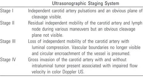
Table 2: Sonographic classification of vascular resectability6
Ultrasonographic Staging System
Stage I Independent carotid artery pulsations and an obvious plane of
cleavage visible.
Stage II Residual independent mobility of the carotid artery and lymph
node during various maneuvers but an obvious cleavage plane not visible.
Stage III Loss of independent mobility of the carotid artery with
luminal compression. Vascular boundaries no longer visible and circular encroachment of the vessel is presumed.
Stage IV Gross invasion of the carotid artery with and without
intraluminal tumor present associated with impaired flow velocity in color Doppler US.
REVIEW
fixation of the tumor to the prevertebral musculature. Trying to strip tumor from the longus colli/capitis muscle complex has been shown to be fraught with difficultly and does not improve a patient’s long-term prognosis because there is often residual disease. In addition, the presence of prevertebral in-filtration is associated with a high rate of retropharyngeal and other site adenopathy, again leading to a worse prognosis. Fix-ation to the prevertebral musculature is difficult to evaluate clinically because it requires a manual assessment of the mo-bility of the tumor with the muscular column and vertebral column. The tumors that usually infiltrate the prevertebral space are ones that are arising from the posterior mucosal wall, be it from nasopharynx, oropharynx, hypopharynx or, rarely, extensive laryngeal, thyroid, or esophageal cancers.
Hsu et al8demonstrated that the preservation of a retro-pharyngeal high-signal-intensity fat stripe on sagittal or axial T1-weighted scans served as an excellent indicator that tumors had not become fixed to the prevertebral fascia or muscula-ture. This finding makes sense in that mucosal lesions largely must cross the retropharyngeal space, usually containing fat, lymph nodes, and connective tissue, to broach the longus colli/ capitis muscle complex (Fig 3). The width of this fat stripe is variable from patient to patient and from superior to inferior, being more abundant in the superior region. Hsu et al found that 39 of 40 patients with preservation of the retropharyngeal fat stripe on T1-weighted scans showed absence of infiltration of the prevertebral musculature. Thus, this is a relatively spe-cific finding for excluding prevertebral musculature invasion.
Loevner et al9evaluated the findings that would suggest the presence of prevertebral musculature invasion. These in-cluded abnormal longus muscle concavity, abnormal T2 hy-perintensity in the prevertebral musculature, abnormal con-trast enhancement in the prevertebral musculature, and an irregular border to the tumor’s interface with the prevertebral musculature. The authors found that none of the 4 findings evaluated showed an accuracy rate greater than 60%. Al-though a criterion might show sensitivity as high as 88% (as in bright T2-weighted signal intensity), that same criterion had specificity as low as 14%. The striking finding of the study, however, occurred in 7 patients who had all 4 abnormalities— loss of the fat stripe, nodularity within the prevertebral mus-culature, abnormal signal intensity in the prevertebral muscu-lature, and gadolinium enhancement of the prevertebral musculature. Four of the 7 patients did not show prevertebral musculature infiltration when evaluated at surgery.
It is useful to understand the surgeon’s evaluation of the infiltration of the prevertebral fascia in the operating room. In most cases, the surgeon who suspects prevertebral muscula-ture infiltration attempts to assess the fat plane on the side opposite the suspected region of prevertebral invasion. He or she therefore, first creates a plane with dissector and/or finger on the normal contralateral side, hoping to extend that plane of dissection across to the opposite side. If that plane of dis-section cannot be created and there is tumor preventing the dissection, then the surgeon will close the wound and refer the patient for medical therapy having deemed the patient unre-sectable. Loevner at al9concluded that this surgical or endo-scopic assessment (in which the tumor can be palpated and manipulated to assess fixation without surgical dissection) should continue to be the standard of care at this time, with limited assistance by imaging unless there is an intact high-signal-intensity fat stripe on the T1-weighted scans as per Hsu et al.8
Subsequently, other authors have evaluated patients with prevertebral muscular invasion and nasopharyngeal cancer. They have found that in these patients with fascial invasion, in whom surgical treatment is not offered because standard ra-diation and chemotherapy are so effective and surgery incurs unacceptable morbidity, the prognosis is much worse. The chance of local recurrence and lymphadenopathy is much higher, and the 5-year prognosis is much worse. Even hema-togenous metastases are increased in patients with nasopha-ryngeal carcinoma and prevertebral infiltration.10
Fig 1.Carotid encasement; adenoid cystic carcinoma.A, Note the circumferential
involve-ment of the left cavernous artery by a soft-tissue mass with narrowing of the carotid artery in this patient who had adenoid cystic carcinoma from the sinonasal cavity.B, This patient also demonstrates erosion of the clivus with bony infiltration of the skull base.
Fig 2.Carotid fixation. This patient who had recurrent thyroid cancer in the left side of the
neck demonstrates effacement of the fat planes around the left CCA laterally (black arrow). Nonetheless, the tumor does not extend greater than 270° around the circumference of the carotid artery, and the tumor was able to be stripped off the carotid artery at surgery. Note also the tumor abutting the anterior scalene muscle on the left side (white arrow). In many instances, the head and neck surgeons believe that this is a contraindication for further surgery due to the potential for brachial plexus involvement.
Fig 3.Prevertebral muscular infiltration.
[image:3.585.53.288.40.291.2]Mediastinal Invasion
The third criterion for unresectability in the T4b category is mediastinal invasion. This is obviously much less common in patients with suprahyoid aerodigestive system masses but can be seen on rare occasions in patients with subglottic laryngeal, hypopharyngeal, thyroid, and esophageal cancers. The criteria for mediastinal invasion have not been well studied however, empirically; infiltration of mediastinal fat and vascular inva-sion of their supra-aortic vessels and/or the aortic arch in a circumferential manner would be indicative of mediastinal invasion. In many instances, head and neck surgeons may de-fine an inferior border below which they feel they can no longer resect tumor without the participation of a thoracic surgeon. This is usually stated in terms of the sternal notch in that tumors below the sternal notch are more difficult to resect via a cervical approach than those above the sternal notch. Mediastinal invasion may include tracheal and esophageal infiltration.
Wang et al11evaluated the accuracy of 3 MR imaging cri-teria in the prediction of tracheal invasion in 67 patients with thyroid carcinoma.12Thirty healthy subjects and 1 cadaver underwent MR imaging of the trachea as a reference standard. Criteria of invasion were 1) soft-tissue signal intensity in the cartilage, 2) intraluminal mass, or 3) circumferential tracheal abutment of 180° or greater. There were 7 false-positive find-ings using these criteria. The authors indicated that a soft-tissue signal intensity was normally seen in healthy tracheal cartilage and sometimes represented tumor extension be-tween the cartilaginous rings without invasion. The highest accuracy (90%) was achieved when using a combination of any of the 3 findings yielding sensitivity of 100% (23/23) and specificity of 84% (37/44). These findings were associated with a much worse overall prognosis from thyroid cancer.11
Karwowski et al13demonstrated the use of intraoperative ultrasonography (IOU) in the identification of recurrent thy-roid cancer in 13 patients. They inserted the sterile-covered transducer directly into the skin incision. Areas of recurrent tumor were seen as hypoechoic masses with increased color Doppler flow, indicating their vascular nature. IOU was par-ticularly useful in detecting paratracheal tumor or thyroid car-tilage invasion by nodules 20 mm or less.
Intraluminal infiltration of the trachea or the esophagus is a late sign of their involvement by neck tumors. King et al14 evaluated MR imaging in the staging of papillary carcinoma of the thyroid with operative findings as a gold standard.15The small number of cases (14 patients) and the use of the single criterion of intraluminal invasion for tracheal and esophageal involvement led to the detection of tracheal involvement in 50% (1 of 2) and esophageal involvement in 0% (0 of 1) of cases by MR imaging.
In a study of 22 patients with periesophageal neck masses, Roychowdhury et al16indicated that a circumferential mass of ⬎270° or focal T2 signal intensity abnormality of the esopha-geal wall on MR imaging suggests the presence of esophaesopha-geal invasion with a specificity of 100% and 86%, respectively. On the other hand, an intact fat plane, absence of wall thickening, and no T2 wall-signal-intensity abnormality imply that the esophagus is not invaded, with a sensitivity of 100% for all 3 criteria.
In a case control study by Chen et al,17CT and MR imaging
of 78 patients with head and neck cancer was retrospectively analyzed. According to pathology and follow-up findings, pa-tients were divided into a case group (N⫽32) with esophageal inlet invasion and a control group (N⫽46) without invasion. The distance between the posterior aspect of the cricoid carti-lage and the anterior aspect of the vertebra (d-CV) was mea-sured and compared between the 2 groups. The authors indi-cated that a d-CV of greater than 1.0 cm is an optimal criterion for esophageal inlet invasion by advanced head and neck car-cinomas, with a statistical significance between the 2 groups. Accuracy was 79% and 80% on CT and MR imaging, respectively.
van den Hoed et al18found a positive correlation between CT prediction of unresectability based on mediastinal involve-ment and patient outcome in 85 patients with esophageal car-cinoma. Criteria used for the diagnosis of aortic invasion were 1) obliteration of peri-aortic fat and 2) an angle of contact ⬎45° with the aorta. Median survival was 21 and 8 months, respectively, for tumors considered CT resectable or unresectable.
Koda et al19compared the accuracy of intra-aortic endo-vascular sonography (IES) with that of CT in the diagnosis of aortic invasion in 28 patients with esophageal carcinoma who subsequently underwent surgical exploration. A criterion of obliteration of the outer hyperechoic layer of the aorta was used to diagnose invasion on sonography, whereas a contact angle of at least 90° with loss of periaortic fat was considered positive for invasion on CT. Accuracy was 100% for the pres-ence of invasion by using IES and 89% for CT.
Endoscopic ultrasonography (EUS) is an imaging tech-nique that is gaining popularity in esophageal cancer staging. Its great value relies on the ability to visualize in detail the 4 layers of the esophagus as well as the outer periesophageal fat. In a study by Wildi et al,2017 patients presented with lower neck masses and suspected esophageal invasion on CT and were evaluated by EUS, which demonstrated esophageal inva-sion in 4 patients and pleural invainva-sion in 1 patient. The re-maining 12 patients (71%) showed no visible invasion on EUS, with a resultant change in the plan of management.
Criteria with Respect to Surgical Planning
Laryngeal Cartilage Invasion
have investigated extensively the criteria for the presence or absence of cartilage invasion by using CT and MR imaging criteria.
Agada et al31examined data from 38 patients with SQCC of the larynx. When comparing CT staging with histopathology, they found that 45% of the patients were erroneously over-staged to T4 on the basis of laryngeal cartilage invasion as judged by the radiologic sign of cartilage sclerosis.
Becker et al26looked at several CT laryngeal imaging find-ings that could suggest cartilage infiltration including 1) ex-tralaryngeal tumor, 2) sclerosis, 3) tumor adjacent to nonos-sified cartilage, 3) serpiginous contour, 4) erosion or lysis, 5) obliteration of marrow space, 6) cartilaginous blowout, and 7) bowing. Although the authors were able to identify several criteria that had either a high sensitivity or a high specificity, no 1 criterion had both sensitivity and specificity over 70% with regard to the thyroid cartilage. The findings were a little more optimistic with regard to the presence of invasion of cricoid or arytenoid cartilage, in which sclerosis and through and through involvement were the most accurate criteria (Figs 4 and 6).
The reason that the thyroid cartilage is the most difficult one of the larynx to assess is because it often has areas of chon-drification and ossification in contiguity with each other. Al-though ossified cartilage has a different attenuation than tu-mor and therefore is tu-more readily assessed, chondrified cartilage may be isoattenuated to the adjacent neoplasm, mak-ing the assessment that much more difficult. Although the resolution of the multidetector CT (MDCT) is far superior to that of MR imaging currently, the soft-tissue contrast to dis-criminate between the 2 types of tissue is the weakness of MDCT.
The MR assessment of cartilaginous invasion requires eval-uation of thin-section T2-weighted fat-suppressed scans and thin-section T1-weighted post– gadolinium-enhanced scans. MR imaging studies have suggested that high signal intensity within the boundaries of the cartilage on the T2-weighted fat-suppressed scans and contrast enhancement into the cartilage, demonstrated as high signal intensity against a dark back-ground of suppressed ossified fat, low-signal-intensity bone of the ossified cartilage, and low-signal-intensity chondrified
cartilage, are indicative of infiltration. Using these criteria, Zbaren et al, Castelijns et al, and Becker et al21-25 demon-strated a high sensitivity though low specificity for cartilage infiltration with MR imaging.
CT criteria have high specificity but low sensitivity and a mean accuracy of approximately 75%– 80% versus MR imag-ing, in which there is a high sensitivity but low specificity and accuracy rates of, on average, 80%– 85%. The implications are quite important; a false-positive prediction of cancer infiltrat-ing cartilage on MR imaginfiltrat-ing may result in inadvertent re-moval of the larynx when the cancer might have been curable with larynx-sparing medical therapy or limited surgery. The cosmetic and functional deformities of total laryngectomy have long-ranging implications as described previously. On the other hand, using a study that has low sensitivity (CT) could lead to deferral of surgery, incomplete surgery, or inap-propriate radiation therapy with either tumor remaining or the inability to cure the patient.
Nishiyama et al32studied the value of single-photon emis-sion tomography (SPECT) compared with that of radiologic examination (CT and/or MR imaging) in the detection of car-tilage invasion in patients with laryngohypopharyngeal can-cer. They suggested that superimposed early bone and tumor dual-isotope SPECT images may be sufficient in the diagnostic evaluation of cartilage invasion by superimposing tumor loca-tion (from tumor SPECT) onto the osseous structures (shown on bone SPECT) with an accuracy of almost 90% (17 of 19 cases).
Pre-epiglottic Fat Invasion
One of the factors determining the potential for the perfor-mance of conservation laryngeal surgery for cancer is the ex-tent of invasion of the pre-epiglottic fat.33,34When there is extensive pre-epiglottic fat infiltration, the hyoid bone is often at risk for infiltration. The hyoid bone is critical for the su-pracricoid surgical laryngeal conservation procedure, which requires a pexy between the hyoid bone and the cricoid carti-lage to allow reconstruction. This pexy procedure is required to pull the incised portions of the larynx superiorly so that the arytenoid cartilage can appose the tongue base with or without the epiglottis to create laryngeal airway closure during
swal-Fig 4.Gross infiltration of the laryngeal cartilage. Erosion of the right side of the cricoid cartilage (long arrow) is well demonstrated in this patient with laryngeal carcinoma with subglottic
extension. Additionally, the anterior margin of the right thyroid cartilage (short arrow) appears to be invaded.
Fig 5.Laryngeal cartilage sclerosis. Axial postcontrast CT scan at the level of the true vocal cord shows sclerosis of the right arytenoid cartilage (arrow), associated with thickening of
the true vocal cord. This finding is suggestive of cartilaginous invasion or perichondrial reaction.
Fig 6.Arytenoid sclerosis in laryngeal carcinoma. Axial CT scan with bone windows shows a sclerotic right arytenoid cartilage (arrow) in a patient with true vocal cord cancer. Despite
[image:5.585.56.533.45.179.2]lowing. When the epiglottis is preserved, the procedure is called a cricohyoidoepiglottopexy, whereas reconstruction without the epiglottis is called cricohyoidopexy. This is a pro-cedure that can be performed for tumors with limited thyroid cartilage invasion be it from the supraglottic or glottic primary sites.
The presence of pre-epiglottic fat infiltration was evalu-ated with MR imaging by Loevner et al.35The assessment was best made on sagittal T1-weighted or axial T1-weighted scans, in which the high-signal-intensity fat is an excellent background with which to evaluate neoplastic infiltration. Using the substitution of soft-tissue signal intensity for the pre-epiglottic fat, the authors studied 40 patients with su-praglottic laryngeal or pharyngeal carcinomas in which pre-epiglottic fat invasion was suspected. The accuracy rate of the MR evaluation was 90% with 4 false-positive cases, which were related to either partial volume averaging, peri-tumoral edema extending into the pre-epiglottic fat, para-glottic fat infiltration that was mistakenly called pre-epi-glottic fat, and minor salivary gland ectopic tissue mistaken for neoplasm.
The extent of pre-epiglottic fat infiltration is not just im-portant from the standpoint of surgical options.33In addition, it has been shown that pre-epiglottic fat infiltration by neo-plasm increases the risk of cervical adenopathy of the primary tumor. When the pre-epiglottic fat is infiltrated from a supra-glottic laryngeal carcinoma, there is also the possibility that a cuff of tissue from the tongue base may also need to be re-sected. This resection has the attendant morbidity of swallow-ing dysfunction and poor airway preservation.
Dural Infiltration
Another issue with regard to sinonasal cavity cancer or other cancers that may infiltrate the skull base is the presence of dural infiltration (Fig 7). Dural infiltration can be considered a preliminary step before potential parenchymal invasion. Therefore, its detection is critical, particularly when determin-ing if craniofacial surgery is required. Craniofacial surgery re-quires the combined efforts of a neurosurgeon/skull base sur-geon as well as a head and neck surgical oncologist.
Dural infiltration versus reactive change, even for tumors such as meningiomas, can be problematic to differentiate on the basis of imaging findings. Although some studies have sug-gested that the dural tail of a meningioma is largely due to neoplasm, others have suggested that the dural tail may, in part, be due to reactive vascular tissue in the dura. This can be assessed at frozen section; however, most surgeons have adopted the philosophy of removing the entire dural tail of a meningioma rather than potentially leaving tumor cells behind.
With respect to dural infiltration by a malignant neoplasm, it is critically important to be able to give a patient a reasonable sense of the likelihood of a complete resection once a com-bined craniofacial approach is considered. To go through the surgery unnecessarily or to opt to not go through the surgery and potentially leave residual surgically treatable tumor be-hind is a difficult decision to make.
Eisen et al36evaluated 23 patients in whom dural infiltra-tion by facial head and neck neoplasm was suspected. The authors measured the thickness of the dura, classified the dura in terms showing of nodular versus linear enhancement, and
Fig 7.Dural and intracranial extension of mass.A, Axial T2-weighted scan demonstrates soft-tissue mass infiltrating the
[image:6.585.54.536.61.361.2]also assessed the presence of pial enhancement. Although there were a limited number of positive cases in this study, the authors found that when the linear dural enhancement was greater than 5 mm in thickness, the likelihood of neoplastic infiltration of the dura was very high. If there was nodularity to the enhancing dura or if there was pial enhancement, cancer-ous infiltration was present histopathologically. However, lin-ear dural enhancement less than or equal to 5 mm in thickness correlated with a benign histology in the study. The finding of this study that thin linear enhancement need not suggest neo-plastic infiltration has been reproduced in numerous settings. Please note that there are numerous reasons for linear dural enhancement of the meninges beyond neoplasm, including infectious, vascular, and inflammatory etiologies.
Bone Infiltration
The presence of infiltration of the skull base and/or the facial bones is also critical to the assessment of the extent of surgery required for resection of tumors. One site that has extensive importance with respect to the surgical approach to tumor resection is infiltration of the mandible or maxilla from oral cavity or oropharyngeal carcinoma (Figs 8 –10). The recon-struction of the mandible, if it has to be completely or partially resected, often requires bone grafting from another site. This is accomplished with simultaneous surgical teams, one harvest-ing and preparharvest-ing the donor site while the other is involved in the surgical resection of the cancer. An unexpected discovery of bony disease while in the operating room renders the recon-struction procedure problematic. Planning for the possibility of a complicated reconstruction is critical to achieve the best functional outcome for the patient.
Mandibular Invasion
Brown37-41described the mechanisms of cancer invasion of the mandible in a detailed report based on an extensive review of 57 related articles. He indicated that the route of tumor entry to the mandible is at the point of abutment, which is often the junction of the attached and reflected mucosa below the crest of the alveolar ridge, with a positive relationship be-tween the infiltrative pattern of the disease and the size of the primary tumor. He noted that preferential tumor spread along the inferior alveolar nerve or the bone marrow is unusual, and this characteristic argues against the inclusion of the neuro-vascular bundle in marginal resections of the mandible.
Chung et al42investigated the ability of MR imaging to predict neoplastic infiltration of the mandible in patients with oral cavity and oropharyngeal carcinomas. The authors found that MR imaging had high sensitivity but relative low specific-ity in this arena. The authors used findings of replacement of the normal high-intensity bone marrow at T1-weighted scan-ning, the presence of cortical erosions and contrast enhance-ment in the cortex or marrow, or high signal intensity on T2-weighted scanning in the bone marrow to identify neoplastic infiltration. False-positive findings were identified in those in-dividuals who had undergone recent dental extractions or ra-diation therapy or who had infectious inflammatory odonto-genic disease. In these cases, the marrow signal intensity often was abnormal, and there was abnormal contrast enhancement present. In those individuals who had undergone recent ex-tractions, the presence of cortical erosion from neoplasm could not be distinguished from tooth-extraction defects. Os-teoradionecrosis also accounted for a false-positive study. However the MR imaging demonstrated that no false-negative
Fig 8.Post– gadolinium-enhanced scan through the floor of the mouth demonstrates a contrast-enhancing mass (arrow) infiltrating the right side of the mandible and extending into the
subcutaneous tissue. There is an associated mass at the right lateral base of the tongue. Note infiltration of the mandible extends across the midline.
Fig 9.Infiltration of the mandible with retropharyngeal spread of tumor. Axial T1-weighted scan demonstrates a large mass emanating from the right palatine tonsil extending along the
floor of the mouth posteriorly to infiltrate the right side of the mandible. Note that the signal intensity of the right side of the bone marrow is dark, replacing the bone marrow fat. There is effacement of the retropharyngeal fat on the right side, with tumor abutting the longus colli/capitis muscle complex.
Fig 10.Sarcoma growing into the left maxilla. Postcontrast fat-suppressed images demonstrate soft-tissue enhancement emanating from the retromolar trigone region (black arrow), with
[image:7.585.50.534.50.254.2]findings existed (100% sensitivity). This included a total of 22 patients who had mandibular resections.
Using the same criteria of MR imaging in a larger group of patients43with oral or oropharyngeal SQCC, Bolzoni et al44 identified mandibular invasion in 16 patients, with the tumor confined only to the cortex in 6. They reported a sensitivity, specificity, and accuracy of 93%, 93%, and 93% respectively.
With the advent of 0.5-mm MDCT scanning of the head and neck, subtle erosions of the cortex of the mandible can readily be ascertained by axial scanning with multiplanar re-constructions. New literature has suggested that the sensitivity and specificity of CT for mandibular invasion by neoplasm has improved with the higher resolution afforded by MDCT. Nonetheless, it is expected that the sources for false-positive studies (ie, dental extraction defects, radiation fibrosis, and osteoradionecrosis) in the MR imaging arena might still pro-voke an inaccurate CT scan. Positron-emission tomography (PET) scanning may be of some utility in these settings; how-ever, it has not been extensively investigated to date.
Brockenbrough et al45evaluated the diagnostic accuracy of DentaScan (dental CT software) in predicting mandibular in-vasion in 36 patients with SQCC of the oral cavity by using histopathology as a gold standard. With DentaScan, a thin axial dataset of images can be reformatted into multiple illus-trative panoramic and cross-sectional views that allow better ultrastructural visualization of the mandible without intrave-nous contrast administration. Using a section thickness of 1–1.25 mm, the authors could identify bone invasion in 21 of 22 patients (sensitivity, 95%). However, they had 3 false-pos-itive results (specificity, 79%). The relatively low specificity was partly related to the small number of patients without bone invasion (14 of 36).
Compared with CT, MR imaging appears less specific in assessing the presence and extent of mandibular invasion. In a recent work by Imaizumi et al,4351 patients with oral SQCC were evaluated by using MR imaging and axial CT for the presence and extent of mandibular invasion. In 35 patients in whom axial (5-mm-thick) CT was difficult to evaluate, a ded-icated dental CT (DentaScan) examination was performed by using 1-mm-thick images. The specificity of CT was higher than that of MR imaging in all aspects of evaluation, including mandibular cortical invasion (88%, 54%), bone marrow in-volvement (88%, 81%), and inferior alveolar canal invasion (96%, 70%) for both CT and MR imaging, respectively.43The 12 false-positive diagnoses of mandibular cortical invasion by MR imaging were due to a chemical shift artifact that obscured the cortical line. MR imaging also had 14 false-positive diag-noses of inferior alveolar canal invasion, which was explained by the presence of inflammation surrounding the tumor, which shows similar signal intensity.
Jungehulsing et al46 compared different imaging tech-niques, including SPECT, with histopathology for the predic-tion of mandibular invasion in 35 patients with oral SQCC. With a semiquantitative assessment of technetium Ic99m methylene diphosphonates uptake by the affected part of the mandible compared with an unaffected part, they found that SPECT could correctly predict 11 of 12 cases of mandibular invasion with no false-positive results.
PET/CT fusion is a relatively new technology that com-bines the precise structural information of CT with the
sensi-tive metabolic information of PET into 1 diagnostic image. Babin et al47evaluated mandibular invasion in 8 patients with oral/oropharyngeal carcinoma by using PET/CT fusion. They proved the technique to be 100% sensitive and 83% specific compared with histopathology. Findings were interesting, but further larger studies may be needed to confirm those prelim-inary results.
Skull Base Invasion
In a study from Korea, Roh et al48retrospectively analyzed the data of 119 patients with nasopharyngeal carcinoma (NPC). Skull base invasion was reported in 46 (38.6%) patients. In terms of prognostic values, the authors recommended further subdivision of NPC with skull base invasion into 4 groups in ascending order of prognostic importance: 1) simple skull base erosion, 2) minimal involvement of either anterior or posterior cranial nerves, 3) multiple involvement of both groups of cranial nerves, and 4) intracranial extension.
Yu et al49demonstrated different CT features in 58 patients with maxillofacial tumors invading the skull base. The main manifestations observed on CT were foraminal enlargement, bone thinning, erosion, and displacement. The following structures were involved in descending order of frequency: the roof of the pterygoid process of the sphenoid bone, the greater wing of sphenoid, the sphenoid body and sinus, the petrous apex, the clivus, and the articular surface of the temporal squamosa.
Xie et al50,51compared MR imaging with CT in 63 patients with NPC. They found that MR imaging was able to detect skull base erosion more easily than CT (23 and 15 cases for MR imaging and CT, respectively) with a noticeable impact of MR imaging on the accuracy of the staging of NPC.
The same parameters for infiltration of the mandible can be applied to other locations at the skull base. Once again, replacement of bone marrow high signal intensity on T1-weighted scans, erosion of the bone as demonstrated by con-trast-enhancing tissue against a fat-suppressed background, and high-signal-intensity marrow edema have been used in this regard. Unfortunately, there are numerous pathologic sources of low signal intensity in the bone marrow including chronic anemias, HIV infection, hematologic disorders, fibro-sis dysplasia, Paget disease, osteomalacia, vitamin deficiency sources, as well as postherapeutic effects.52Recent traumatic injuries to the bone may also demonstrate enhancement, high signal intensity on T2-weighted scans, and replacement of high-signal-intensity bone marrow fat on T1-weighted scans. This has been equally problematic when evaluating the spinal column.
Stambuk et al53demonstrated a model of skull base inva-sion with NPC. They evaluated 11 children (12–17 years of age) who presented with NPC. Because pediatric NPC is gen-erally not suspected clinically until late in the disease process, they found that all patients except 1 (91%) had skull base invasion by the time of radiographic evaluation (CT and/or MR imaging). Widening of the petroclival fissure was present in 8 (73%) patients. The tumor had extended to involve the pterygopalatine fossa in 2 (18%), and the sphenoid sinus in 3 (27%) cases. Clival invasion was seen as abnormal high T2 signal intensity on fat-saturated MR imaging.
diffusion-weighted scanning and calculating apparent diffusion coeffi-cient (ADC) values to distinguish neoplastic infiltration (with its lower ADC) versus non-neoplastic infiltration (with higher ADC values).54,55This has been applied with both diffusion-weighted imaging, diffusion line scanning, and calculations of ADC values for spinal compression fractures and has resulted in modest levels of accuracy. These same perimeters may be applied to the skull base.
Perineural Spread or Tumor
Another phenomenon that may affect the therapeutic decision making in dealing with head and a neck cancer is the presence of perineural spread of tumor. In some cases perineural spread of tumor refers to tumoral infiltration of the nerve itself (Fig 10) as opposed to spread on the nerve by using the nerve as a scaffold for neoplastic spread. Perineural spread in the context of this article will incorporate both explanations as well as tumoral spread through the neural foramina of the skull base. Many theories have been proposed to understand the exact mechanism of perineural spread of tumors. Tumor cells are known to spread along the perineural connective tissue or the endoneural plane. Neural cell adhesion molecule (N-CAM) expression has been shown to be positively correlated with perineural spread in a variety of tumors. Gandour-Edwards et al56demonstrated the expression of N-CAM in 93% (14 of 15 patients) of adenoid cystic carcinomas that showed perineural spread. What is most interesting, Vural et al57found the same percentage (93%, 38 of 41 patients) of expression of N-CAM 3 years later in SQCC with perineural spread.
Perineural spread on CT is demonstrated by the enlarge-ment of the neural foramina and/or soft-tissue infiltration of the fat adjacent to or within the neural foramina.58-66 Soft-tissue infiltration of the foramen rotundum, for example, can be assumed to be neoplastic infiltration in most CT settings, given a tumor with a propensity to access the second division of cranial nerve V. In a similar vein, foramen ovale infiltration (Fig 11) implies a third division of cranial nerve V infiltration. Contrast-enhanced CT rarely shows actual enhancement of tumor along the nerve; however, this can be readily identified on fat-suppressed T1-weighted postcontrast MR imaging. The
CT evidence is by inference, whereas there can be direct visu-alization of enhancement of a nerve and tumor around the nerve with MR imaging. MR imaging may also demonstrate infiltration of the fat of the foramina at the skull base or the trigeminal ganglion on the basis of T1-weighted scanning demonstrating the soft-tissue low-intensity signal that re-places the white-signal-intensity fat.
Although adenoid cystic carcinoma is the histologic neo-plasm most commonly associated with perineural spread, at a rate of approximately 60%, SQCC, basal cell carcinoma, mu-coepidermoid carcinoma, lymphoma, neurotropic mela-noma, rhabdomyosarcoma, and metastasis may certainly demonstrate this phenomenon.
Adenoid cystic carcinoma populates salivary gland neo-plasms; therefore, infiltration of cranial nerves V and VII is the most commonly seen. Spread along cranial nerve XII, cranial nerve VII, and cranial nerve V by oral cavity or retromolar trigone cancers is another commonly identified process with SQCC. In fact, the T4b classification for oral cavity and oro-pharyngeal carcinoma specifically addresses invasion of the lateral pterygoid muscle, the pterygoid plates (bone and nerves of the pterygopalatine fossa), the skull base, and the carotid artery.
Chang et al67identified 8 cases of malignant melanoma at 2 institutions that had MR imaging evidence of perineural spread with confirmatory tissue sampling. Five cases (63%) were desmoplastic melanomas. Enhancement of at least 1 branch of the trigeminal nerve was demonstrated in all cases and at least 1 other cranial nerve in 5 cases. Other findings included abnormal enhancement and soft-tissue thickening of the cavernous sinus, Meckel cave, and/or the cisternal portion of the trigeminal nerve.
There are few case reports in the literature describing lym-phomatous infiltration of the mandibular canal (along the in-ferior alveolar nerve). Yamada et al68reported a case of non-Hodgkin lymphoma, which had slowly grown into the mandibular canal with continuous dilation to an average of 15-mm width with peripheral bone sclerosis and no destruction.
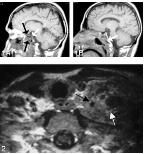
Moorjani and Stockton69reported a very rare case of
des-Fig 11.Nasopharyngeal carcinoma with infiltration of the prevertebral musculature, skull base, and cavernous sinus.A, Postcontrast fat-suppressed T1-weighted scan shows a large
[image:9.585.53.534.42.214.2]moplastic fibroma of the mandible with perineural spread along the third division of trigeminal nerve, which appeared enlarged and showed abnormal enhancement along its course in the masticator space, through the wide foramen ovale, and in Meckel cave.
The cranial nerves to the orbits (cranial nerves III, IV, and VI to the extraocular muscles and V for sensation) are uncom-monly infiltrated with neoplasm; however, infiltration may be seen as enhancing nerves in patients with plexiform schwnomas, lymphoma, and sarcoidosis. Skin cancers over the an-terior face may infiltrate all 3 divisions of cranial nerve V and can represent a significant therapeutic predicament for some-one with what was initially expected to be a relatively small skin lesion.
Imaging studies of perineural spread include works by the MD Anderson Cancer Center group (Ginsberg, Ginsberg and Eicher, Ginsburg and DeMonte, and Ginsburg et al58,59,62-65), which have shown detection rates of approximately 70%. Cer-tainly microscopic disease that occurs along the cranial nerves may escape imaging surveillance. The propensity for “skip re-gions” in what appears to be noncontiguous tumor cells pre-sents another daunting task for the radiologic evaluation. Rarely, intracranial spread with enhancement of the cisternal portion of the cranial nerves may be visualized. When one is suspecting microscopic perineural spread, it is difficult for the radiation therapist to assign portals for the extent of the tu-mor. By incorporating a greater amount of the intracranial contents and brain stem, the radiation oncologist risks the toxicity to those cranial nerves and/or to the visual apparatus with respect to the optic nerves and optic chiasm. Again, quite often the issue is “At what cost, cure?” This problem is espe-cially apparent with adenoid cystic carcinoma, which is a rel-atively indolent malignancy in which, even without therapy, long-term survival measured in terms of decades may occur. The added benefit of therapy, though not inconsequential, is also not overwhelming with respect to improved 20-year survivals.
Gandhi et al70described, in a review article, the different patterns of perineural spread on MR imaging. Direct signs included cranial nerve enlargement, irregularity, excessive en-hancement, and foraminal enlargement with destruction in advanced disease. The authors also addressed the importance of careful observation of the normal intraforaminal fat pads on T1-weighted scans, which can be obliterated with tumor infiltration. Muscle denervation is an indirect sign of cranial nerve involvement. Swelling, abnormal T2 signal intensity, and enhancement can be seen with acute-subacute denerva-tion, whereas chronic muscle denervation results in fatty re-placement and muscle atrophy.
Orbital Invasion
Sinonasal carcinomas are dangerous from the standpoint of their proximity to the orbit as well as the skull base (Fig 12). Orbital infiltration is generally limited by the tough periorbita, which functions as an excellent barrier to the spread of neo-plasm, similar to that of the periosteum. Unfortunately, the periorbita is not readily identifiable even in high-resolution MR imaging or thin-section CT. Peritumoral edema can lead to abnormal signal intensity within muscles and abnormal contrast enhancement of muscles, which simulates neoplasm on MR imaging. This potential for false-positive results is less of an issue with CT.
Invasion of the orbits by head and neck tumors can occur either by using the route of skull base foramina and sinuses or by direct extension through the orbital bone (Fig 13). In a large study of 562 patients with nasopharyngeal carcinoma, Luo et al71identified orbital involvement by using CT and/or MR imaging in 18 patients. The route from the pterygopalatine fossa and the inferior orbital fissure into the orbital cavity was the most common route of invasion (n ⫽13), followed by ethmoid and/or sphenoid sinuses into the orbit (n⫽4). In 1 patient, the exact route could not be identified.
Eisen et al72examined the accuracy of CT and MR imaging in predicting tumor invasion of the orbit. Nineteen
preopera-Fig 12.AandB, Orbital infiltration. Postcontrast
fat-sup-pressed T1-weighted scans show soft-tissue infiltration in the superior medial aspect of the left orbit, with irregular margins in the extraconal fat. Although there are portions of the tumor in which there still appears to be periosteum containing the tumor, the irregular margins seen more an-teriorly (arrow) suggest infiltration requiring orbital surgery.
Fig 13.Orbital infiltration.A, Soft tissue is seen
[image:10.585.53.529.44.290.2]tive CT scans and 17 preoperative MR images from patients with cancer at risk of orbital invasion were retrospectively re-viewed in correlation with pathologic and intraoperative as-sessment. Eleven criteria involving the tumor, orbit, and na-solacrimal fossa were used in each study. Tumor adjacent to the periorbita was the most sensitive predictor of orbital inva-sion (90%) for both CT and MR imaging. The highest positive predictive values were achieved with extraocular muscle in-volvement manifested as high signal intensity on T2-weighted scans or enhancement on MR imaging (100%) and orbital fat infiltration (80% on MR imaging, 86% on CT). No one crite-rion was more than 79% accurate in predicting orbital inva-sion, whereas nasolacrimal invasion was predicted with an ac-curacy of 89%. CT was more accurate than MR imaging in 7 of 9 criteria.
Brachial Plexus Infiltration
Related to perineural spread of tumor is direct invasion of the brachial plexus. Because head and neck surgical oncologists are less comfortable with the implications of resecting tumor from the nerves that feed the upper extremities, these discus-sions are often made in consultation or in collaboration with neurosurgeons. In most instances, the head and neck surgeon will deem the patient inoperative if the patient has any symp-toms whatsoever referable to the brachial plexus or if the an-terior scalene muscle is infiltrated by the neoplasm. Anan-terior scalene muscle infiltration is based on loss of the normal at-tenuation appearance and fatty striations of the muscle on CT, loss of the fat plane between the middle-posterior scalene complex and the anterior scalene muscle through which the brachial plexus passes, or direct displacement of the visualized portion of the brachial plexus (Fig 14). This is better identified with MR imaging, in which high signal intensity on T2-weighted imaging in the muscle, gadolinium enhancement of muscle, or enhancement or abnormal T2W signal intensity along the brachial plexus roots, trunks, divisions, cords, and branches is seen.
In a retrospective review of the clinicopathologic data of 71 patients with cancer and brachial plexopathy, Thyagarajan et al73indicated that the presence of a mass adjacent to the bra-chial plexus was highly predictive of tumor infiltration and was the most useful feature in distinguishing infiltration from radiation plexopathy; increased T2 signal intensity was com-monly seen in both groups.
Ohta74reported a rare case of brachial plexus infiltration by non-Hodgkin lymphoma. Findings included soft-tissue masses with abnormal T2 hyperintensity signal intensity along the brachial plexus. Scintigraphy was superior to MR imaging
in detecting the abnormality, showing increased uptake of gal-lium-67 along the course of the brachial plexus.
Recently, Graif et al75used ultrasonography to evaluate 28 patients with different varieties of brachial plexus pathology. Most of the patients underwent surgical confirmation. On scanning along the nerve course from its exit at the vertebral foramina down to the axilla and in the presence of some tech-nical limitations, the authors could depict different patterns of sonographic abnormalities. Among 10 patients with second-ary tumors, 6 patients with primsecond-ary tumors, and 6 patients with associated radiation fibrosis, findings were categorized as either focal hypoechoic masses adjacent to the nerves with fusiform nerve thickening or diffuse nerve thickening. Despite a false-negative rate of 29% of cases, some of these cases showed tumors that were small (⬍12 mm) posttraumatic le-sions. Ultrasonography may be useful as part of the preoper-ative evaluation of brachial plexus infiltration.
Resectability Issues in Patients Who Have Undergone Previous Treatment
Combined chemoradiation regimens for advanced-stage head and neck cancer have gained popularity during the last decade. The advent of new treatment protocols and novel chemother-apeutic agents, along with refinements in the field of radiation oncology, have made it possible to treat advanced-stage head and neck cancer for cure while avoiding the morbidity of sur-gical intervention. Tumors deemed unresectable are now be-ing treated in this fashion as well, with curative intent. In the last few years, postoperative adjuvant chemoradiation has been recommended for patients who have undergone primary surgical intervention with histopathologic criteria that are as-sociated with a high risk of locoregional recurrence.76
The evaluation of a patient who has been previously treated for advanced head and neck cancer and now has documented cancer recurrence or persistence poses many diagnostic and therapeutic challenges. These patients typically can no longer receive any further radiation and chemotherapy at curative doses. Surgical salvage rates are poor. Early detection of tumor recurrence or persistence is a key factor in improving disease control rates and survival rates for patients undergoing surgi-cal salvage treatment. The same issues that have been eluci-dated earlier with regard to resectability still apply. The prob-lem with this patient subset is in distinguishing treatment effect from tumor recurrence or persistence.
PET/CT fusion technology has been especially helpful in this patient subset. Surgery and chemoradiation therapy both result in anatomic changes that are difficult to characterize purely by conventional imaging (CT, MR) alone.
Conven-Fig 14.A, Nodal metastases from breast cancer (short
white arrow) are seen to infiltrate the anterior scalene muscle (a) and the hypodense brachial plexus (long white arrow) on this enhanced axial CT image. A tongue of nodal disease indents the plexus beyond the anterior scalene muscle.
[image:11.585.54.372.43.166.2]tional imaging is typically obtained within a 2- to 3-month period after completion of treatment to establish a baseline for which all future studies will be compared. If this baseline scan or any future scan demonstrates any change or irregularity that raises the suspicion for cancer recurrence or persistence, then PET/CT may be ordered to help characterize those changes on the basis of the degree of metabolic activity.77If tumor recurrence or persistence is still suspected, then a de-tailed evaluation with the patient under anesthesia with pos-sible biopsy is warranted.
Defining the extent of tumor recurrence or persistence in this patient population is key to the surgeon and patient in determining resectability and the likelihood of surgical salvage with any meaningful long-term survival.
Summary
Imaging can be very helpful in the determination of resectabil-ity for head and neck cancers. The decision to attempt exten-sive and significantly morbid resections for tumor control must rest with the surgeons and patients and should be, in part, based on the most accurate imaging techniques available. MR imaging benefits from a high sensitivity but is prone to false-positive findings, a failure that may be addressed through the judicious use of PET/CT. High-resolution MDCT with multiplanar reconstructions is another alternative in address-ing the critical issues of vascular encasement, skull base or perineural infiltration, cartilaginous invasion, orbital involve-ment, mediastinal (including trachea and esophagus) spread, and dural disease. Imaging is less successful in issues of pre-vertebral fixation and early cartilaginous invasion.
Acknowledgment
Laurie Loevner is credited for numerous contributions to the concepts in this manuscript as well as the head and neck liter-ature on cancer resectability.
References
1. Greene FL, Page DL, Fleming ID, eds.AJCC Cancer Staging Manual. 6th ed. Philadelphia: Lippincott Raven; 2002
2. Niimi K, Yoshizawa M, Nakajima T, et al.Vascular invasion in squamous cell carcinomas of human oral mucosa.Oral Oncol2001;37:357– 64
3. Yu Q, Wang P, Shi H, et al.Carotid artery and jugular vein invasion of oral-maxillofacial and neck malignant tumors: diagnostic value of computed to-mography.Oral Surg Oral Med Oral Pathol Oral Radiol Endod2003;96:368 –72 4. Yoo GH, Hocwald E, Korkmaz H, et al.Assessment of carotid artery invasion
in patients with head and neck cancer.Laryngoscope2000;110:386 –90 5. Nix PA, Coatesworth AP.Carotid artery invasion by squamous cell carcinoma
of the upper aerodigestive tract: the predictive value of CT imaging.Int J Clin Pract2003;57:628 –30
6. Mann WJ, Beck A, Schreiber J, et al.Ultrasonography for evaluation of the carotid artery in head and neck cancer.Laryngoscope1994;104:885– 88 7. Yousem DM, Hatabu H, Hurst RW, et al.Carotid artery invasion by head and
neck masses: prediction with MR imaging.Radiology1995;195:715–20 8. Hsu WC, Loevner LA, Karpati R, et al.Accuracy of magnetic resonance
imag-ing in predictimag-ing absence of fixation of head and neck cancer to the preverte-bral space.Head Neck2005;27:95–100
9. Loevner LA, Ott IL, Yousem DM, et al.Neoplastic fixation to the prevertebral compartment by squamous cell carcinoma of the head and neck.AJR Am J Roentgenol1998;170:1389 –94
10. Feng AC, Wu MC, Tsai SY, et al.Prevertebral muscle involvement in nasopha-ryngeal carcinoma.Int J Radiat Oncol Biol Phys2006;65:1026 –35.
11. Wang JC, Takashima S, Takayama F, et al.Tracheal invasion by thyroid carcinoma: prediction using MR imaging.AJR Am J Roentgenol2001;177: 929 –36
12. Takashima S, Matsushita T, Takayama F, et al.Prognostic significance of mag-netic resonance findings in advanced papillary thyroid cancer.Thyroid2001; 11:1153–59
13. Karwowski JK, Jeffrey RB, McDougall IR, et al.Intraoperative ultrasonography improves identification of recurrent thyroid cancer.Surgery2002;132:924 –29 14. King AD, Ahuja AT, To EW, et al.Staging papillary carcinoma of the thyroid: magnetic resonance imaging vs ultrasound of the neck.Clin Radiol2000;55: 222–26
15. Bayles SW, Kingdom TT, Carlson GW.Management of thyroid carcinoma invading the aerodigestive tract.Laryngoscope1998;108:1402– 07
16. Roychowdhury S, Loevner LA, Yousem DM, et al.MR imaging for predicting neoplastic invasion of the cervical esophagus.AJNR Am J Neuroradiol2000;21: 1681– 87
17. Chen B, Yin SK, Zhuang QX, et al.CT and MR imaging for detecting neoplastic invasion of esophageal inlet.World J Gastroenterol2005;11:377– 81 18. van den Hoed RD, Feldberg MA, van Leeuwen MS, et al.CT prediction of
irresectability in esophageal carcinoma: value of additional patient positions and relation to patient outcome.Abdom Imaging1997;22:132–37
19. Koda Y, Nakamura K, Kaminou T, et al.Assessment of aortic invasion by esophageal carcinoma using intraaortic endovascular sonography.AJR Am J Roentgenol1998;170:133–35
20. Wildi SM, Fickling WE, Day TA, et al.Endoscopic ultrasonography in the diagnosis and staging of neoplasms of the head and neck.Endoscopy2004;36: 624 –30
21. Zbaren P, Becker M, Lang H.Staging of laryngeal cancer: endoscopy, com-puted tomography and magnetic resonance versus histopathology.Eur Arch Otorhinolaryngol1997;254(suppl 1):S117–122
22. Zbaren P, Becker M, Lang H.Pretherapeutic staging of hypopharyngeal carcinoma: clinical findings, computed tomography, and magnetic resonance imaging compared with histopathologic evaluation.Arch Otolaryngol Head Neck Surg1997;123:908 –13
23. Zbaren P, Becker M, Lang H.Pretherapeutic staging of laryngeal carcinoma: clinical findings, computed tomography, and magnetic resonance imaging compared with histopathology.Cancer1996;77:1263–73
24. Castelijns JA, Becker M, Hermans R.Impact of cartilage invasion on treatment and prognosis of laryngeal cancer.Eur Radiol1996;6:156 – 69
25. Becker M, Zbaren P, Laeng H, et al.Neoplastic invasion of the laryngeal cartilage: comparison of MR imaging and CT with histopathologic correla-tion.Radiology1995;194:661– 69
26. Becker M, Zbaren P, Delavelle J, et al.Neoplastic invasion of the laryngeal cartilage: reassessment of criteria for diagnosis at CT.Radiology1997;203: 521–32
27. Becker M, Moulin G, Kurt AM, et al.Atypical squamous cell carcinoma of the larynx and hypopharynx: radiologic features and pathologic correlation.Eur Radiol1998;8:1541–51
28. Becker M, Moulin G, Kurt AM, et al.Non-squamous cell neoplasms of the larynx: radiologic-pathologic correlation.RadioGraphics1998;18:1189 –209 29. Becker M.Neoplastic invasion of laryngeal cartilage: radiologic diagnosis and
therapeutic implications.Eur J Radiol2000;33:216 –29
30. Becker M.Larynx and hypopharynx.Radiol Clin North Am1998;36:891–920, vi 31. Agada FO, Nix PA, Salvage D, et al.Computerised tomography vs. pathological staging of laryngeal cancer: a 6-year completed audit cycle.Int J Clin Pract
2004;58:714 –16
32. Nishiyama Y, Yamamoto Y, Yokoe K, et al.Superimposed dual-isotope SPECT using 99mTc-hydroxymethylene diphosphonate and 201Tl-chloride to assess cartilage invasion in laryngohypopharyngeal cancer.Ann Nucl Med2004;18: 527–32
33. Yousem DM, Tufano RP.Laryngeal imaging.Neuroimaging Clin N Am2004; 14:611–24
34. Yousem DM, Tufano RP.Laryngeal imaging.Magn Reson Imaging Clin N Am
2002;10:451– 65
35. Loevner LA, Yousem DM, Montone KT, et al.Can radiologists accurately pre-dict preepiglottic space invasion with MR imaging?AJR Am J Roentgenol1997; 169:1681– 87
36. Eisen MD, Yousem DM, Montone KT, et al.Use of preoperative MR to predict dural, perineural, and venous sinus invasion of skull base tumors.AJNR Am J Neuroradiol1996;17:1937– 45
37. Brown JS, Lowe D, Kalavrezos N, et al.Patterns of invasion and routes of tumor entry into the mandible by oral squamous cell carcinoma.Head Neck2002;24: 370 – 83
38. Brown JS, Lewis-Jones H.Evidence for imaging the mandible in the manage-ment of oral squamous cell carcinoma: a review.Br J Oral Maxillofac Surg
2001;39:411–18
39. Brown JS, Grew NR.Predicting mandibular invasion in mouth cancer.Clin Otolaryngol21:265–268.Clin Otolaryngol Allied Sci1997;22:558
40. Brown JS, Browne RM.Factors influencing the patterns of invasion of the mandible by oral squamous cell carcinoma.Int J Oral Maxillofac Surg1995;24: 417–26
41. Brown J.Mechanisms of cancer invasion of the mandible.Curr Opin Otolar-yngol Head Neck Surg2003;11:96 –102
42. Chung TS, Yousem DM, Seigerman HM, et al.MR of mandibular invasion in patients with oral and oropharyngeal malignant neoplasms.AJNR Am J Neu-roradiol1994;15:1949 –55
assessing mandibular invasion of squamous cell carcinoma in the oral cavity.
AJNR Am J Neuroradiol2006;27:114 –22
44. Bolzoni A, Cappiello J, Piazza C, et al.Diagnostic accuracy of magnetic reso-nance imaging in the assessment of mandibular involvement in oral-oropha-ryngeal squamous cell carcinoma: a prospective study.Arch Otolaryngol Head Neck Surg2004;130:837– 43
45. Brockenbrough JM, Petruzzelli GJ, Lomasney L.DentaScan as an accurate method of predicting mandibular invasion in patients with squamous cell carcinoma of the oral cavity.Arch Otolaryngol Head Neck Surg2003;129:113–17 46. Jungehulsing M, Scheidhauer K, Litzka N, et al.99mTc-MDP-SPECT for detec-tion of subclinical mandibular infiltradetec-tion of squamous epithelial carcinoma
[in German].HNO1997;45:702– 09
47 Babin E, Hamon M, Benateau H, et al.Interest of PET/CT scan fusion to assess mandible involvement in oral cavity and oro pharyngeal carcinomas[in French].Ann Otolaryngol Chir Cervicofac2004;121:235– 40
48. Roh JL, Sung MW, Kim KH, et al.Nasopharyngeal carcinoma with skull base invasion: a necessity of staging subdivision.Am J Otolaryngol2004;25:26 –32 49. Yu ZH, Xu GZ, Huang YR, et al.Value of computed tomography in staging the
primary lesion (T-staging) of nasopharyngeal carcinoma (NPC): an analysis of 54 patients with special reference to the parapharyngeal space.Int J Radiat Oncol Biol Phys1985;11:2143– 47
50. Xie CM, Liang BL, Wu PH, et al.Spiral computed tomography (CT) and mag-netic resonance imaging (MRI) in assessment of the skull base encroachment in nasopharyngeal carcinoma[in Chinese].Ai Zheng2003;22:729 –33 51. Xie C, Liang B, Lin H, et al.Influence of MRI on the T, N staging system of
nasopharyngeal carcinoma[ in Chinese].Zhonghua Zhong Liu Za Zhi2002;24: 181– 84
52. Loevner LA, Tobey JD, Yousem DM, et al.MR imaging characteristics of cra-nial bone marrow in adult patients with underlying systemic disorders com-pared with healthy control subjects.AJNR Am J Neuroradiol2002;23:248 –54 53. Stambuk HE, Patel SG, Mosier KM, et al.Nasopharyngeal carcinoma:
recog-nizing the radiographic features in children.AJNR Am J Neuroradiol2005;26: 1575–79
54. Herneth AM, Naude J, Philipp M, et al.The value of diffusion-weighted MRT in assessing the bone marrow changes in vertebral metastases[ in German].
Radiologe2000;40:731–36
55. Chan JH, Peh WC, Tsui EY, et al.Acute vertebral body compression fractures: discrimination between benign and malignant causes using apparent diffu-sion coefficients.Br J Radiol2002;75:207–14
56. Gandour-Edwards R, Kapadia S, Barnes L, et al.Neural cell adhesion molecule in adenoid cystic carcinoma invading the skull base.Otolaryngol Head Neck Surg1997;117:453–58
57. Vural E, Hutcheson J, Korourian S, et al.Correlation of neural cell adhesion molecules with perineural spread of squamous cell carcinoma of the head and neck.Otolaryngol Head Neck Surg2000;122:717–20
58. Ginsberg LE.MR imaging of perineural tumor spread.Neuroimaging Clin N Am2004;14:663–77
59. Ginsberg LE.MR imaging of perineural tumor spread.Magn Reson Imaging Clin N Am2002;10:511–25, vi
60. Esmaeli B, Ginsberg L, Goepfert H, et al.Squamous cell carcinoma with peri-neural invasion presenting as a Tolosa-Hunt-like syndrome: a potential pit-fall in diagnosis.Ophthal Plast Reconstr Surg2000;16:450 –52
61. Chan LL, Chong J, Gillenwater AM, et al.The pterygopalatine fossa: postoper-ative MR imaging appearance.AJNR Am J Neuroradiol2000;21:1315–19 62. Ginsberg LE, Eicher SA.Great auricular nerve: anatomy and imaging in a case
of perineural tumor spread.AJNR Am J Neuroradiol2000;21:568 –71 63. Ginsberg LE.Imaging of perineural tumor spread in head and neck cancer.
Semin Ultrasound CT MR1999;20:175– 86
64. Ginsberg LE, DeMonte F.Imaging of perineural tumor spread from palatal carcinoma.AJNR Am J Neuroradiol1998;19:1417–22
65. Ginsberg LE, De Monte F, Gillenwater AM.Greater superficial petrosal nerve: anatomy and MR findings in perineural tumor spread.AJNR Am J Neuroradiol
1996;17:389 –93
66. McLean FM, Ginsberg LE, Stanton CA.Perineural spread of rhinocerebral mucormycosis.AJNR Am J Neuroradiol1996;17:114 –16
67. Chang PC, Fischbein NJ, McCalmont TH, et al.Perineural spread of malignant melanoma of the head and neck: clinical and imaging features.AJNR Am J Neuroradiol2004;25:5–11
68. Yamada T, Kitagawa Y, Ogasawara T, et al.Enlargement of mandibular canal without hypesthesia caused by extranodal non-Hodgkin’s lymphoma: a case report.Oral Surg Oral Med Oral Pathol Oral Radiol Endod2000;89:388 –92 69. Moorjani V, Stockton V.Desmoplastic fibroma with perineural extension.
AJR Am J Roentgenol2005;185:1498 –99
70. Gandhi D, Gujar S, Mukherji SK.Magnetic resonance imaging of perineural spread of head and neck malignancies.Top Magn Reson Imaging2004;15: 79 – 85
71. Luo CB, Teng MM, Chen SS, et al.Orbital invasion in nasopharyngeal carcinoma: evaluation with computed tomography and magnetic resonance imaging.Zhonghua Yi Xue Za Zhi (Taipei)1998;61:382– 88
72. Eisen MD, Yousem DM, Loevner LA, et al.Preoperative imaging to predict orbital invasion by tumor.Head Neck2000;22:456 – 62
73. Thyagarajan D, Cascino T, Harms G.Magnetic resonance imaging in brachial plexopathy of cancer.Neurology1995;45:421–27
74. Ohta H.A case of non-Hodgkin’s lymphoma infiltrating the brachial plexus detected by Ga-67 scintigraphy.Ann Nucl Med2002;16:297–98
75. Graif M, Martinoli C, Rochkind S, et al.Sonographic evaluation of brachial plexus pathology.Eur Radiol2004;14:193–200
76. Cooper JS, Pajak TF, Forastiere AA, et al, and the Radiation Therapy Oncology Group 9501/Intergroup.Postoperative concurrent radiotherapy and chemo-therapy for high-risk squamous-cell carcinoma of the head and neck.N Engl J Med2004;350:1937– 44