University of Warwick institutional repository: http://go.warwick.ac.uk/wrap
A Thesis Submitted for the Degree of PhD at the University of Warwick
http://go.warwick.ac.uk/wrap/66736
This thesis is made available online and is protected by original copyright. Please scroll down to view the document itself.
A Thesis submitted to the University of Warwick in
fulfilment of the degree of Doctor of Philosophy
by
Ezard Launce Emanuel, B.Sc., M.Sc.
Department of Chemistry and Molecular Sciences
University of Warwick
Coventry
Trialkyl tin compounds have been shown to be potent
inhibitors of oxidative phosphorylation, oligomycin sensitive
ATPase activity and other ATP-dependent reactions of beef heart
mitochondria. Unlike oligomycin inhibition of these reactions,
trialkyl tin inhibition is reversible by dithiols such as
2,3-dimercaptopropanol. The OS-ATPase activity was found to be
6-10 times more sensitive than oxidative phosphorylation to
inhibition by trialkyl tins. This differential sensitivity to
trialkyl tin inhibition is discussed in relation to the current
theories on energy coupling.
Binding studies with [3H]-DBCT have indicated the
presence of two types of binding sites designated high affinity
(kD ~ 0.3 pM) and low affinity (~ ~ 30 ~M) binding sites. The concentration of the high affinity binding site is ~ 2.0 nmol/mg protein in submitochondrial particles and saturation of these
sites correlated with the inhibition of the oligomycin sensitive
ATPase activity. Extraction and isolation experiments have shown
that DBCT binds to a small lipophilic, non-protein molecule.
Dihydrolipoic acid has been shown to drive ATP synthesis
by acting as a NAD-linked substrate. Fatty acids, oleoyl-phosphate,
oleoyl-lipoate and other lipids were found to inhibit succinate
driven ATP synthesis and other energy-linked reactions in mitochondria
and submitochondrial particles. In addition, a new method for the
measurement of nanomoles amount of lipoic acid and lipoamide is
reported. The method involves the cyclic reduction of S,S-dithiobis
(2-nitrobenzoic acid) by NADHvia a system containing lipoamide
discarding the concepts that are false and
retaining the concepts that show by their
survival that they are factually serviceable
because they represent reality as far as it
is known."
P. Mitchell, 1977
Ann.
Rev. Biochem.I would like to thank my supervisor, Dr. D.
E.
Griffiths, for the opportunity which has enabled me to carry out this work,and for his continuing interest and advice throughout this research.
I would like to thank Professor K. R. Jennings for the
use of the facilities at Warwick University.
I would like to thank Mrs. M. West, Mrs. P. Massey,
Mrs. D. Fryer and Miss J. Clarke for their technical assistance,
and Mrs. C. Bi1li.ng~ for her innate typing skills.
I would also like to thank Dr. M. Carver, Dr. S. Stanley
and Dr. A. Beauclerk for their informative advice.
-Finally, I would like to express my sincere appreciation
to Barbara for making so many sacrifices.
ADP AMP AMP-(PNP) ATPase BAL DBCT DBT DTNB DCPIP DTT DeCD EDTA F) FCCP G-6-p NAD+ NADP+ OS-ATPase
Pi or P
PCA TTFB TCA Tris-Cl Adenosine-5'-diphosphate Adenosine-5'-monophosphate
Adenyl imidodiphosphate
Adenosine triphosphatase
2,3-dimercaptopropanol
Dibutylchloromethyl tin chloride
Dibutyl tin dichloride
5,5'-dithiobis-(2-nitrobenzoic acid)
2,6-dichlorophenol indophenol
Dithiothreitol
N,N'-dicyclohexylcarbodiimide
Ethylenediamine tetra-acetic acid
Soluble mitochondrial ATPase or
coupling factor I
Carbonyl cyanide
p-trifluoromethoxyphenyl-hydrazone
Glucose-6-phosphate
Nicotinamide adenine dinucleotide
Nicotinamide adenine dinucleotide phosphate
Oligomycin-sensitive ATPase
Inorganic phosphate
Perchloric acid
4,5,6,7-tetrachloro-2-(trifluoromethyl)
benziimidazole
Trichloroacetic acid
Tris(hydroxymethyl)aminomethane hydrogen
CHAPTER
I • I
1.2 1.3 1.4 1.5 1.6 1.7 1.8 1.9
1. 10
Abbreviations Contents
List of Figures List of Tables
INTRODUCTION
General introduction
The respiratory chain
Mechanisms of energy transduction
The chemical hypothesis
The chemiosmotic hypothesis
Localised proton hypotheses
Conformational hypothesis
The mitochondrial ATPase complex
Nucleotide binding sites
A genetic approach to oxidative phosphorylation
CHAPTER 2 THE EFFECTS OF TRIALKYL TIN COMPOUNDS ON ENERGY-LINKED FUNCTIONS OF BEEF HEART MITOCHONDRIA AND SUBMITOCHONDRIAL PARTICLES
2. I Introduction 2.2 Materials
2.3 Methods
2.4 Results
2.5 Reversal of organotin inhibition with dithiols
2.6 Discussion
2.7 Conclusion
MITOCHONDRIA
3.1 Introduction
3.2 Materials
3.3 Methods
3.4 Results
3.5 Discussion
CHAPTER 4 THE ROLE OF DIHYDROtIPOIC ACID IN OXIDATIVE PHOSPHORYLATION
4. I Introduction
4.2 Materials
4.3 Methods
4.4 Results
4.5 Effect of dihydrolipoic acid and oleoyl-lipoate on NAD/NADP trans hydrogenase reaction of beef heart submitochondrial particles
4.6 Discussion
CHAPTER 5 THE EFFECT OF FATTY 'ACIDS 'AND THEIR ACYL-ESTERS ON OS-ATPase~ OXIDATIVE PHOSPHORYLATION AND ENERGY-LINKED REACTIONS OF MITOCHONDRIA
5.1 5.2 5.3 5.4 5.5 Introduc tion Materials Methods
Results and discussions
6. 1 Introduction 147
6.2 Materials and methods 153
6.4 Discussion 163
6.5 Conclusion 169
APPENDIX I 170
APPENDIX I I ]77
REFERENCES 180
1.1 The electron transfer chain
1.2 The four complexes of the respiratory chain
1.3 The electron carriers of the respiratory chain arranged according to their redox potentials
1.4 Fundamental postulates of chemiosmosis
1.5 Proton translocation by the FIFO-ATPase
1.6 Proton translocating redox loops
1.7 The localised proton model for ATP synthesis
1.8 Indirect localised proton coupling model
1.9 Electrodic hypothesis
1.10 New conformational coupling model
I.IIA Structure of FIFO-ATPase complex
1.11B & C 3-Dimensional structure of the FIFO-ATPase complex
2.1 Two mechanisms by which trialkyl tins may inhibit oxidative phosphorylation
2.2 Inhibition of OS-ATPase activity in beef heart mitochondria by trialkyl tins
2.3 Inhibition of OS-ATPase activity in beef heart submitochondrial particles by trialkyl tins
2.4 Inhibition of succinate driven ATP synthesis in beef heart mitochondria by trialkyl tins
2.5 Inhibition of succinate driven ATP synthesis in submitochondrial particles by trialkyl tins
2.6 Inhibition of succinate driven ATP synthesis and ATP hydrolysis by DBCT and tributyl tin chloride
2.7 Inhibition of succinate driven ATP synthesis and ATP hydrolysis in submitochondrial particles by DBCT and tributyl tin chloride.
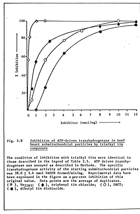
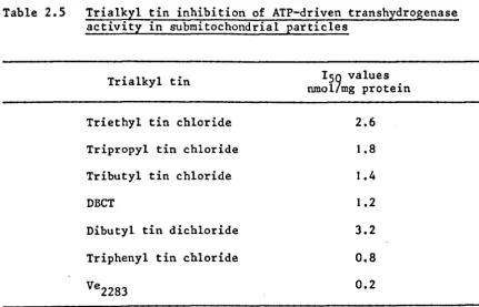
2.8 Inhibition of ATP-driven transhydrogenase in beef heart submitochondrial particles by trialkyl tin compounds
2.10 2.11 2.12 2.13 2.14 2.15 2.16 3.1 3.2 3.3
3.4
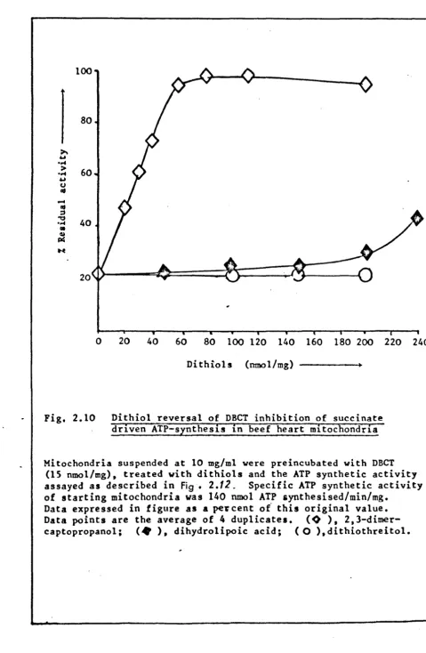
3.5 3.6 3.7 3.8 4.1Dithiol reversal of DBCT inhibition of
succinate driven ATP synthesis in beef heart mitochondria
Dithiol reversal of DBCT inhibition of
succinate driven ATP synthesis in beef heart submitochondrial particles
Dithiol reversal of DBCT inhibited OS-ATPase activity in beef heart submitochondrial particles
Dithiol reversal of DBCT inhibition of ATP driven transhydrogenase activity in submitochondrial particles
Dithiol reversal of dibutyl tin dichloride inhibited ATP driven transhydrogenase activity in beef heart submitochondrial particles
Dithiol reversal of tripheny~ tin inhibition of ATP driven transhydrogenase in beef heart
submitochondrial particles
Proposed structure for trialkyl tin binding in haemoglobin, pyruvate kinase and OS-ATPase
[3H]-DBCT binding to beef heart submitochondrial particles as a function of increasing concentra-tions of [3H]-DBCT
Effect of various trialkyl tins on [3H]-DBCT binding to submitochondrial particles
Scatchard plot of [3H]-DBCT binding to beef heart submitochondrial particles
Dixon plot of the inhibition of the ATPase activity of submitochondrial particles by [3H]-DBCT
Thin layer chromatography of purified chloroform! methanol extract of DBCT labelled submitochondrial
particles
Mass spectra of DBCT-X
Mass spectra of lipoic acid (A) and DBCT (B)
Mass spectra of DBCT-lipoic acid complex
Transmembrane transport of protons by dihydrolipoic acid
r : '."'
5. 1 5.2 5.3 5.4 5.5 5.6 5.7 5.8 5.9 6.1 6.2 6.3 6.4 6.5 6.6 6.7
The oleoyl cycle of oxidative phosphorylation
Inhibition of succinate driven oxidative phos-phorylation in beef heart mitochondria by fatty acids
Fatty acid/derivatives inhibition of succinate driven oxidative phosphorylation in submito-chondrial particles
Effects of fatty acids/derivatives on ATP-drive trans hydrogenase
Stimulation of ATPase activity in mitochondria and submitochondrial particles by oleic acid
Stimulation of ATPase activity in beef heart submitochondrial particles by fatty acids
Stimulation of the FI-ATPase activity by oleic acid
Reversal of nBCT and oligomycin inhibited ATPase activity of submitochondrial particles by oleic acid
Effect of bovine serum albumin on oleic acid reversal of DBCT inhibited ATPase activity in submitochondrial particles
Structure of lipoic acid
Reaction mechanism of pyruvate dehydrogenase complex of E. coli
Proposed mechanism for the catalytic reduction of 5,5-dithiobis(2-nitrobenzoic acid) to
thionitrobenzoic acid by NADH
Typical spectrophotometric tracings obtained during reduction of DTNB by catalytic
quantities of lipoic acid and lipoamide in the standard assay system containing NADH and pig heart lipoamide dehydrogenase
Dependence of the rate of DTNB reduction in the presence of catalytic amounts of lipoic acid on the concentration of the components of the standard assay system.
Calibration curve for lipoic acid
Calibration curve for lipoamide
6.9
6.10
Reduction of DTNB in the standard assay system with dihydrolipoic acid as a catalyst
Reduction of limiting amounts of DTNB in the standard assay system
(xi)
165
1 • 1
2. J
2.2 2.3 2.4 2.5 2.6 2.7 2.8 2.9 3.1 3.2 4.1 4.2 4.3
Subunit composition of the FIFO-ATPase complex
Trialkyl tin inhibition of OS-ATPase activity in mitochondria and submito-chondrial particles
Comparison of the ISO values of trialkyl tins of various ATPase preparations
Inhibition of succinate driven ATP synthesis in mitochondria and submito-chondrial particles by trialkyl tins
Differential sensitivity of the ATP synthetic and ATP hydrolysing activities
in mitochondria and submitochondrial particles to DBCT
Trialkyl tin inhibition of ATP-driven
transhydrogenase activity in submitochondrial particles
Dithiol reversal of DBCT and DBT inhibition of oxidative phosphorylation in mitochondria
Dithiol reversal of DBCT and DBT inhibition of OS-ATPase activity in mitochondria and submitochondrial particles
Dithiol reversal of DBCT and DBT inhibition of ATP-driven transhydrogenase in submito-chondrial particles
Sensitivity of dithiol reversed DBCT and DBT inhibition of succinate driven ATP
synthesis and ATP-dependent reactions in SMP's to inhibitors and uncouplers of oxidative phosphorylation
Effect of trialkyl tin compounds on [3H]-DBCT binding to beef heart submitochondrial particles
Chloroform/methanol extraction of DBCT binding component
Dihydrolipoic acid driven ATP synthesis in beef heart mitochondria
ATP synthesis in submitochondrial particles
4.5 4.6 4.7 4.8 4.9 5. I 5.2 5.3 5.4 5.5 6. I 6.2 6.3 6.4
Inhibitor sensitivity of dihydrolipoic acid driven ATP synthesis in mitochondria ATP synthesis in mitochondria driven by various substrates
Ability of various substrates to drive ATP synthesis catalysed by submitochondrial particles
ATP synthesis catalysed by complex V Effect of dihydrolipoic acid and certain acyl-esters on energy-linked transhydrogenase activity in submitochondrial particles
Effect of fatty acids (derivatives) on succinate driven oxidative phosphorylation in mitochondria
Effect of fatty acids/derivatives on
succinate driven oxidative phosphorylation in submitochondrial particles
Effect of fatty acids/deriva~ives on ATP-driven and succinate driven transhydrogenase activity of submitochondrial particles
The effect of fatty acids/derivatives on 32p_ATP exchange catalysed by submitochondrial particles sensitivity of the ATPase activity of oleate treated submitochondrial particles to ATPase inhibitors
Standard assay system
The effect of monothiols and dithiols on the rate of DTNB reduction
Lipoic acid content of selected mammalian tissues and organelle
Comparison of the lipoate content of rat
liver, determined by different assay procedures
I • J GENERAL INTRODUCTION
Elucidation of the mechanism by which energy liberated
in cellular electron transport is conserved and utilised for ATP
synthesis, has been a major challenge in biochemistry ever since
the occurrence of phosphorylation in the respiring cell was first
recognised in the 1930s [see (I, 2, 3) for reviews]. Today, it is
well established that the electron transport system associated with
the inner membrane of mitochondria, the thylakoid membra~ of
chloroplasts and the plasma membrane of respiring and photosynthetic
prokaryotes are functionally linked to an ATP-synthesising system
by a mechanism that is fundamentally similar in all living cells.
It is also well established that the energy deriving from the oxidation
of reducing equivalents by the respiratory chain is converted to ATP
by the ATP-synthase complex. Before discussing the mechanisms
involved in this process, the structure and function of the respiratory
chain will be briefly outlined [see (6, 7, 8) for more in depth
reviews] •
1.2 THE RESPIRATORY CHAIN
Current ideas of the component composition and s~q~~nce of
the electron transport chain are shown, in Fig. 1.1 (4). This scheme
has emerged from the confluence of several lines of investigation.
They include measurement of the redox potential of individual
components (6), kinetic determination of the reaction sequence
(6, 7), studies on the donor and acceptor specificity of isolated
components (6) and the interaction of electron transfer inhibitors
a-Ketoglutarate
::::tr /
\.
Isoc~trate
\\lJ
NADH :> IFMN
Glutjt~
3-Hydroxyacyl-CoA
I
(Fe-S)N_lb
(Fe-S)N_3 .
Rotenone
(Fe-S)N_Ja (Fe-S)N_4 (Fe-S)N-2J~
. I
(Fe-S)N_5 I
I
j
Fattyacyl-CoA·
Glycerolphosphate
--- .> FP:
--- •. FP4
III
Antimycin A
I
UQ.cyt.bK.b
T Cyt.ct(Fe-S)Cyt.c
IV
KCN
-+
[c
yt.a.a3]+
02Cu.Cu I I
Fig. 1.1 The electron transfer components of the respiratory chain arranged as a continuous sequence from NADH to
oxygen. The points of entry of electrons from various substrates are shown, as well as the sites of inhibition of electron transport. The symbol FP designates flavoproteins; FAD, flavin adenine dinucleotide; FMN,
flavin mononucleotide; (Fe-S), iron-sulphur centres where the subscript N or S indicates NADH and succinate. UQ is ubiquinone (coenzyme Q) and Cyt. the cytochromes bK,
br,
C,C}, aanda3,Cu copper. The bracketedportions list the total component in each complex. [Adopted from Ohnishi 1975 (4»).
r
I N
[image:19.843.25.794.71.451.2]studies by Hatefi et ale have shown that the respiratory chain can be split into four complexes, designated complex I, II, III and IV according to the classification of D. E. Green (9-11). The complexes catalyse the partial reactions shown in Fig. ].2 and can be
recombined stoichiometrically to give a reconstituted respiratory chain which behaves in a similar manner in its responses to electron transfer inhibitors, as the respiratory chain found in intact
mitochondria (9-1]).
Redox potential measurements on both 'in situ' and isolated components of the respiratory chain have enabled the presentation of the electron carriers on a potential diagram (Fig. 1.3) (12). In Fig. ].3 it is shown, that when the components associated with three
levels of mid-potentials are classed or grouped together, there are three spans or gaps in the chain in whi~h relatively large decreases in free energy occurs, each sufficient to provide the energy for the formation of ATP from ADP and inorganic phosphate. The sites are designated Site I, II and III and represents the span between NADH and coenzyme
Q;
cyt.b and cyt.c and cyt.a and oxygenrespectively. Chance, in his review of ]972 (12), suggested that the three groups of electron carriers having fixed mid-potentials (or isopotential pool) were linked with one another by means of transducing carriers of variable potential. The energy-transducing carriers of site I, II and III are proposed to be
(Fe-S)N2' cyt.bT and cyt.a3 respectively, based on the findings that their mid-potentials were ATP and pH dependent (4, 12, 13). The isopotential pools functions as isopotential redox ballasts
NADH
Succinate
I
I I
..
I I I
..
IV
..
Fig. J. 2
Complex I
)
I
,
FMN FeS )Cyt.b Cu~
--.
-
~-- a a3 -- --+--°2
FAD
) Cyt,c 1 FeS
Cu
Complex II Complex III Complex IV
NADH-ubiquinone reductase complex which catalyses the reaction: NADH + Q + H + ~ NAD + + QH2
Succinic Ubiquinone reductase complex which catalyses the reaction: Succinate + Q ~ Fumarate + QH2
Ubiquinol cytochrome c reductase complex which catalyses the reaction: QH2 + 2 ferricyt. c ~ Q + 2 ferrocyt. c
Cytochrome c oxidase complex which catalyses the
reaction: 2 ferrocyt. c + 2H+ + 102 ~ 2 ferricyt. c + H 20
[image:21.558.45.523.66.836.2]NAD/NADH
~
Succinate/FumarateF:(
Fig. 1.3
FpDH
(Fe-S)
~ (Fe-S)S
t
(Fe-S)
(Fe-S)
(Fe-S)
(Fe-S)
(Fe-S)N2""-" UQ
ATP
Site I
EH(mV)
,
-300
r
Cyt.b
K
1
Cyt. bT.J.c!t,C1ATP Fe-S
t
Site I I Cyt.c++cyt.a Site I I I
Cu2+
~Cyt.~
(Cu)~\ O2
ATP
o +600
The electron carriers of the respiratory chain arranged as a group of fixed potential (or isopotential 'pools') and individual components of variable mid-potentials
(bT, (a3) Bnd
Z).
The other components are the same as in Fig. 1.1, from [Modified from Chance 1972 (12)]. [image:22.563.65.520.58.793.2]operates through a series of quasi-equilibrium steps. In the case of cyt.bT for example, there are four species; oxidised high mid-potential, oxidised low mid-potential, reduced high mid-potential and reduced low mid-potential. bT in its low mid-potential form can only react with group II carriers and in
its high mid-potential form can only react with group III carriers. The actual site of ATP synthesis is now known to
reside on the multi-enzyme ATP synthase complex, which although located in the inner membrane of the mitochondria, is not directly linked to the respitatory chain. It is therefore important, to consider the problem of how energy from the respiratory chain is made available to the ATP synthase in such a form that it drives ATP synthesis.
1.3 MECHANISMS OF ENERGY TRANSDUCTION
Currently, the theories available to describe the phenomenon of oxidative phosphorylation, falls mainly into three broad categories:
(a) chemical,
(b) conformational, and (c) protonic.
1.4 THE CHEMICAL HYPOTHESIS
The chemical hypothesis as originally proposed [Slater
1953 (14)] was based on a mechanism of substrate level phosphorylation
(15). The central feature of the hypothesis is that the respiratory
chain generates high energy chemical intermediates which can be
used to drive ATP synthesis or other energy-linked reactions. Using
the notation of Chance and Williams (16), the hypothesis can be
represented by the following sequence of reactions:
1 • AH2 + B + I
*
A'"
I + BH2 2. A'"
I + X*
A + X'"
·13. X
'"
I + Pi ~ X'"
P + I4. X
'"
P + ADP*
X + ATPAH
Z and B are two adjacent respiratory carriers and '",' refers to a high energy bond (probably anhydride or thioester bond). The
nature of X and I is unknown. X '" I is regarded as the common energy
transducing intermediate. This is considered as being the target
of action of uncoupling agents, which, directly or indirectly,
catalyse its hydrolysis (19, 20). Oligomycin is thought to block
reaction 3, and transhydrogenation and ion transport are both driven
by utilisation of X '" I, produced either by respiration or by ATP
(17-20). Evidence for the chemical hypothesis is largely based on the
actions of inhibitors like oligomycin, and on the fact that partial
reactions such as transhydrogenation and reversed electron transfer
can be driven either by energy deriving from respiration or ATP •
• However, there are a number of disquieting features in the chemical
hypothesis. It provides no explanation for:
(a) the apparent dependence of oxidative phosphorylation
structure; and
(b) the fact that a wide range of molecules of wid~ly different structures will uncouple respiration from phosphorylation.
The inability to isolate and identify the proposed high energy
intermediate adds considerable complication to the chemical hypothesis (21, 22).
1.5 THE CHEMIOSMOTIC HYPOTHESIS
The chemiosmotic hypothesis, proposed by Mitchell in 1961
(23) is based on the following four main postulates which are summarised in Fig. 1.4:
1. The ATP synthase is a chemiosmotic membrane-located reversible protonmotive ATPase, having characteristic
+/ " h"
H P st01C 10metry.
2. The respiratory chain is a membrane located vectorial metabolic proton trans locating system, having a
characteristic H+/2e stoichiometry and having the same polarity of proton translocation across the membrane for normal forward redox activity as
the ATPase for ATP hydrolysis.
3. There are proton (or hydroxyl) linked solute porter systems for osmotic stabilisation and metabolite transport.
Fig. 1.4 Fundamental postulates of chemiosmosis (23-28)
ADP + POH
+ +
(+ H /P)H ~
ATP + H 2O
AH2 + B
+
(+H/e)H ( 2. Respiratory chain
A ) cation linked ~ A
C+~ porters
Cytosol 4. Membrane Matrix
+ .
H+---~l - -il leak- - -- ---~) H (stimulated + by uncouplers)
Mitchell pointed out that, since the basis of oxidative
phosphorylation was a dehydration reaction to form an acid anhydride
link, a system which causes the removal of water from the active site
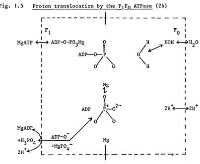
of the ATPase would favour the formation of ATP (Fig. 1.5). The
system Mitchell sur.gested for causing this dehydration consisted of
an anisotropically organised respiratory chain consisting of
alternating electron carriers and hydrogen carriers (Fig. 1.6)
and an anisotropic membrane ATPase. He proposed that phosphorylation
was linked to electron transport by a protonic electrical potential
set up across the coupling membrane by the action of the respiratory
chain which pump protons from one side of the membrane to the other.
+
The protonic electrical potential cause the removal of (H
20 as) H
[image:26.579.121.534.120.429.2]centre of the ATPase (Fig. 1.5). The protonic electrical potential
or protonmotice force (~p) is the sum of the pH gradient (~pH) and
the membrane potential (~~) according to the equation (23-28):
z~pH •
Fig. 1.5 Proton translocation by the FIFo ATPase (26)
r'- - -- - - -
-t
---I
- ,
I
I F 1 I
MgATP ~ ADP-O-P03Mg
Fa I I
/H~HOHiH20
o
I"H
I
ADP 2H " +~"'--I~" 1 + 2H
ADP-O
Mg
I
---+-
_ _ _ _ _ _ _ --. _ ..JWhen the membrane is not leaky and most of the proton
current generated by the redox system passes back through the
reversible ATPase, there will be stoichiometric coupling between
oxidation-reduction and phosphorylation. The P/O (P/2e-) quotient of
the overall process is given by the ~H+/2e- quotient of the redox
+
system divided by the ~H /p quotient of the ATPase, that is:
+ +
(~H /2e)/ (~ll /p).
A short circuiting proton pathway across the membrane [as provided
by uncouplers (23-28)] would,collapse ~p, uncouple phosphorylation,
[image:27.574.87.491.191.522.2]Fig. 1.6 Proton translocating redox loops (26)
SH2
NAD (NAD-linked)
2H+ S LooE
Fe(S)
2H+ FMN
SH2
(FAD-linked)
2H+ S LooE 2
Cyt.b
2H+ CoQ
2H+
Cyt'C 1
1
1°2 + 2H+ LooE 3Cyt.c
~
Cyt.a/a3/
Cu
'. '. - H2O
Detailed arrangement of the respiratory carriers according to Mitchell. The scheme provides for the translocation of 6H+/O for the oxidation NAD~linked substrates and 4H+/O for succinate oxidation (29).
With the chemiosmotic hypothesis, Mitchell, circumvented two of the major objections to the chemical hypothesis. Firstly, the integrity of the coupling membrane is essential to maintain a membrane potential and pH gradient. Secondly, there is no need to postulate the existence of energy-rich intermediates to couple respiration and phosphorylation since the ATPase transferspi directly to ADP.
[image:28.574.120.493.121.466.2]particles, and reconstituted vesicles, summarised below. 1.
2.
3.
4.
5.
Vectorial, transmembrane movement of H+ and other ions accompany both respiration and ATP hydrolysis (30-35). Most if not all the various types of energy transducing
systems can generate a proton gradient and/or a membrane potential across the membrane in which they are located
(35) •
Energy transfer between energy-transducing systems located in the same membrane can take place yia a proton gradient and/or membrane potential (35).
The action of some uncoupler's depends on their ability to dissipate the transmembrane proton gradient, since almost all are lipophilic weak acids (35-38).
Artificially generated electrochemical proton gradients are kinetically competent in driving ATP synthesis
(39, 40).
Although the'chemiosmotic hypothesis offer a generally acceptable framework for energy transduction, the proposed mechanisms for H+ translocation (Fig. 1.6) and ATP synthesis (Fig. 1.5) are no less hypothetical than those involving high energy intermediate
(14). Mitchell'~ suggestion that the prolon gradient is generated by the action of H+ translocating 'redox loops', formed by
alternating hydrogen carriers and electron carriers of the respiratory chain (Fig. 1.6), has been refuted by a number of workers (30).
Harmon et al. have pointed out, that although complexes I and III contain both hydrogen (NAD, FMN, CoQ) and electron carriers
(Fe-S, cyt.b and c), (unlike complex IV, which contains only
to propose his 'protonmotive ubiquinone cycle' (Q cycle) for H+ translocation [see (42, 43) for reviews]. However, the 'Q cycle' requires the movement of Q and QH2 across the membrane, and a certain distribution of b cytochromes across the membrane. Available evidence suggests that these situations are unlikely
(30). In the case of the ATPase system, much evidence has been compiled, which indicates that the FOF) ATPase complex may
+
function as a H trans locator (44, 45). However, the direct interaction of pi and ADP, with the intermediate formation of a
phosphonium ion [P03]+' to yield ATP at the catalytic site (Fig. 1.5) proposed by Mitchell (46, 47) has been questioned on theoretical grounds by Boyer and Williams. (48, 49).
The mechanisms by which the respiratory chain and the ATPase complex transport H+ and the H+/site quotient are matters of
considerable controversy. In both cases the problem concerns whether proton translocation proceeds directly, involving the catalytic site(s) of the enzyme, or whether it proceeds indirectly via a 'proton pump'. Information deriving from investigation of the membrane topology of the catalysts involved and measurement of the
+ +
~H /2e and ~H /ATP ratios, support an indirect mechanism involving proton pumps (50). Direct coupling redox loops would require
a stoichiometry of two H+ translocated per two electron translocated through each coupling site (i.e., ~H+/site
=
2). Similarly,workers have obtained significantly higher ~H+/site ratios, often exceeding 3, for both the electron transport and the ATPase
systems (35, 5J-54). These stoichiometries (~H+/site ratio ~ 3)
are not compatible with the proposed 'redox loop' and 'proton well' mechanisms, but can be readily accommodated by a mechanism involving proton pumps (32). The reported transmembrane location of Fe.S centres 1 and 2 in complex I (55-56) or the two b cytochromes in the case of complex III (55-58) is equally compatible with the
occurrence of transmembrane proton pumps driven by nonvectorial electron transfer catalyst. However, the mechanism by which protons are translocated through the 'pumps' is not defined.
1.6 LOCALISED PROTON HYPOTHESES
Two alternative hypothesis envisaging proton coupling have been put forward to explain the inadequacies of the chemiosmotic hypothesis. They are the 'Localised proton hypothesis' of Williams
(59-60) and the 'Electrodic hyPothesis' of Kell (61). Only a summary of these hypotheses will be presented, because they have been presented, only as logical models for discussion [for in depth reviews see
ref. (219-22 J )).
In the 'localised proton hypothesis', Williams proposes that the protons produced by the respiratory chain, and which are
responsible for ATP synthesis, are not pumped across the membrane as proposed by Mitchell (23-28), but remain in the membrane where they lower the activity of the "solvent- : 'water" in the ATPase region (Fig. J. 7) • The energy of the redox reactions is thus transferred
I
which is connected to the ATPase (Fig. 1.8).
Fig. 1.7 The localised proton model for ATP synthesis (59)
Aqueous (A) Aqueous (C)
ADP
Organic (B)
~---2e---
,..---•
I
f
---~
ATPase
, I
The solid lines (box) represent a very hydrophobic particle within which ADP and pi are condensed to ATP by the ATPase.
Thus during the synthesis of ATP from ADP and pi the H+ moves so
as to pull H
20 molecules from the polyphosphate condensation site
into the diffusion channel [Fig. 1.8 (60-63)].
The obvious advantages of Williams' hypothesis over
the chemiosmotic hypothesis are that it offers:
(a) more efficient energy utilisation,
(b) better control, and
(c) discriminatory coupling of energy to different
processes.
However, although a certain degree of localisation of H+ takes place
between the electron transport and ATPase system (64-65), Mitchell
has pointed out that Williams' hypothesis is very limiting and not
experimentally falsifiable (28). In particular, it lacks explanation
for transport, and because it lacks stoichiometry, it cannot
be used to explain the experimentally observed
plo
quotients.In his 'electrodic hypothesis', Kell proposes that the
Fig. 1.8 Indirect localised proton coupling model (63)
A
Redox system
Boun~H
- +
e H
c
H 2O
H2O
H 2O H 2O H 20 H 0+
3 H 20 H 20 H 20 ATPase
-
----I IP-OH I I IHO-P I
.'
r -I I I I IP-O-P B
....
-+OH + H30 ~ 2H20 + P-O-P
An open channel model is shown so that it could in principle be linked to chemiosmotic gradient, although this is not required in the hypothesis.
the membrane from one S-phase (SR-phase) (membrane-liquid interphase)
to the other (SL-phase) by the electron transfer complex (Fig. 1.9).
The protons then flow along the SL-phase to the ATPase through which
they re-enter the original SR-phase completing the protonic circuit.
The flow of protons along the S-phase is 'conducted' by 'structured
+
water molecules' (or membrane water) which allows H tunnelling
(66, 67). The movement of protons between the S-phases results in
charging the membrane surfaces relative to the bulk aqueous phases,
and it is the potential drop across the membrane/solution (electrode/
Fig. 1.9 Electrodic hyPothesis (61)
I
ETC
I I
I
if I
,
H+ H+
I H+ H
+
,
I .Ir-.
I
I
1;'
ADP+PiI
... , ... 1\
VI
I
I ATP
- I
I
."I
I I
I I
I
I
L SL M SR R
ATPase
A typical protic circuit. The diagram represents a phospholipid membrane phase (M) separating two aqueous phases Land R
repsectively. Interphase SL and SR exist between the M phase and the two aqueous phases. The M phase contains the protonmotive electron transport complex (FTC) and protonmotive ATPase of the appropriate polarities, and the radial and lateral flow of the proton current, i.e., a two-dimensional proton current flow, between them is indicated by the arrows. Adopted from Kell
Whilst the postulates of Kell (61) are in broad agreement with the proposals of Williams (59-63), the electrodic hypothesis is
chemiosmotic because a transmembrane-phase H+ gradient is involved. Kell has layed particular emphasis on localised interphase
phenomena based on generally accepted physical, chemical and
electrochemical principles, and ha's provided explanations for -~
ion transport, and the action of uncouplers (61, 68, 69). However, like the chemiosmotic and 'localised proton' hypotheses, it lacks detailed molecular mechanisms for H+ translocation and ATP
synthesis.
1.7 CONFORMATIONAL HYPOTHESIS
The conformational hypothesis in its original form [Boyer 1965 (72)] proposed that the energy generated by electron transport, was conserved in a 'high-energy' conformational state of the respiratory carrier. The energy inherent in this conformation being used to drive ATP synthesis:
A*
AH2
+ ADP
+ B ~ A*
+ Pi =t. A
+
+ ATP +
Support for this hypothesis is obtained from Green (73) and Hackenbrock
(74) who observed that the structure of the mitochondria was energy
dependent)and from the observations that electron flow in bacteria, chloroplast and mitochondria is accompanied by changes in the
conformation of their FI ATPase (70, 75-77). However, little compelling evidence has been found to support direct interaction between the ATPase and the redox centres. Mitchell (28) has pointed out that such an interaction does not occur.
proposed that ATP synthesis occursat the active site of the ATPase complex prior to the input of energy; which is necessary, only, to bring about the release of ATP from the active site, via an energy dependent conformational change [Fig. 1.10, (70, 79)]. This hypothesis was based on the finding that the exchangeability of adenine nucleotides bound to FI was dependent on the flow of electrons (70, 79) and on the insensitivity of the anhydride formotion to uncouplers of oxidative phosphorylation (80). These
findings although intimately correlating with the suggested molecular explanation for exchange reactions; namely that they result from the dynamic reversal of ATP formation, are difficult to reconcile with Mitchell's proposal that the protons are directly involved in the anbydride formation (28).
Fig. 1.10 New conformational couplirtg model (70)
)
energy
~
ADP+Pi
1.8 THE MITOCHONDRIAL ATPase COMPLEX
The mitochondrial ATPase complex appears to be one of
the most complex enzyme systems known to date, performing 'in situ'
a number of reactions such as ATP-pi exchange, ATP synthesis and
hydrolysis. These reactions are affected by a variety of inhibitors
~
including oligomycin, DCCD, venturicidin and trialkyltins. The
inhibitor studies have proved to be very useful in trying to
determine the subunit/activity relationships in the complex, and
in some cases, have proved to be instrumental in the proposal of
new mechanisms for coupling ATP synthesis and respiration. Inhibitor
studies have also indicated that the ATPase complex is at least
bifunctional. Of interest are those experiments which show that
some inhibitors such as AMP-P-NP are capable of either activating
or inhibiting ATP hydrolysis without altering ATP synthesis (81).
Work presented in Chapter 2 of this thesis has shown that such a
differential effect on ATP synthesis and ATP hydrolysis can be
demon-strated with trialkyltin compounds in beef heart mitochondria. A
brief description of the ATPase will now be presented. For more
in depth reviews see references (81-88).
The mitochondrial ATPase complex is located exclusively
in the inner membrane where it protrudes into the matrix space
(82). In general, it accounts for about 2% of the mitochondrial
mass (81). The 'complete' ATPase complex (Fig. 1.IIA) is depicted
by many authors as consisting of four major components: a headpiece
called Fl which is water soluble, catalyses the hydrolysis of nucleoside
triphosphates and binds the inhibitor aurovertin; a basepiece or
membrane sector, which is detergent soluble and binds the inhibitors
oligomycin and DCCD; a"stalk connecting the headpiece and basepiece;
Peptide inhibitor
..
Headpiece...
Stalkl
..
Basepiece
'-Membrane section
J
Fig. I.IIA Schematic re£resentation of the mitochondrial ATPase complex (81)
F I
FO
Fig. I.IIB Plan view Fig.I.IIC Side view
Three dimensional structure of FIFo-ATPase (109)
The stalk and basepiece are referred to collectively as FO.
The FIFO ATPase complex (or complex V) of beef heart
mitochondria has been prepared by a variety of methods (81, 87-89),
all of which involves solubilisation of the inner mitochondrial
membrane with chaotrophic detergents such as Triton-X-IOO and
deoxycholic acid. These treatments result in severe delipidation
of the final purified form of the FIFO ATPase complex, and the
expression of its maximal enzymic activities is dependent on the
presence of exogenous lipids (81). The purified complex has an
estimated molecular weight of 460-480,000 and contains 8-12
different subunits (84, 88-90). Hatefi etal. have recently
shown that the FIFO ATPase complex of beef heart mitochondria
contained at least 12 subunits, most of which have been tentatively
described on a functional basis in Tabl~ 1.1 (84).
The purified FIFO ATPase complex catalyses ATP
hydrolysis and ATP-pi exchange; however maximal catalytic
activity is dependent on the addition of exogenous phospholipids
(81). Both the ATP-pi exchange and ATP hydrolytic activities are
sensitive to inhibitors such as oligomycin, venturicidin, trialkyltins
and DCCD. The FIFO ATPase complex appears to be divided into two
functionally dissimilar portions: the hydrophylic FIATPase which
protrudes from the inner membrane into the matrix; and the F O
portion which is hydrophobic in nature and is buried in the inner
membrane, both of which, have been isolated in purified form
(92, 93, 104, 105).
The FI ATPase has been isolated from the inner mitochondrial
membrane by a variety of methods, including sonication (92, 93) and
chloroform treatment (94). The enzyme prepared from beef heart
mitochondria has a molecular weight of 347-360,000 and has been
Table 1.1
Subunit M.W.
53,000
50,000
33,000
30,000
23,000
22,000
15,000
12,000
8,000
11-13,000
6,000
Identity of subunits of the FIFO ATPase Complex (84)
a subunit Fl ATPase
a
subunit Fl ATPasey subunit FI ATPase
uncoupler binding protein
Fa
component; pantotheine binding protein (86) oligomycin sensitivity conferring protein (OSCP)~ subunit FI ATPase
DceD
binding proteincoupling factor F6 (85)
ATPase inhibitor or coupling factor B (87, 91)
E subunit FI ATPase
Numbers in brackets are references; M.W.
=
molecular weight.subunits (84, 93). These subunits are designated a,
a,
y, ~ and Ein Table 1.1. Measurement of the subunit stoichiometry, using
dye staining and amino acid frequency methods, indicate that there are
at least 9-10 polypeptide components per FI ATPase molecule (92, 93).
Senior has suggested a subunit stoichiometry of a3~3YI~El for the beef heart enzyme (92) and a similar stoichiometry has been proposed
for the rat liver enzyme by Catterall and Pedersen (95) and for the
yeast enzyme, by Tzagoloff and Meagher (90). An additional 10,000
molecular weight subunit found in certain preparations of FI ATPase
has been proposed to be the 'ATPase inhibitor protein' which regulates the
ATPase activity 'in vivo' (96).
and is insensitive to inhibitors such as oligomycin, DCCD,
venturicidin and trialky1tin compounds. However, F1 ATPase is
inhibited by aurovertin and efrapeptin. The true substrate
of the enzyme is the Mg ATP complex; however, inosine and uridine
triphosphate can also act as substrates (97). 'In situ' the F1 ATPase
catalyse ATP synthesis, hydrolysis and ATP-dependent reactions such
as transhydrogenation. This has been demonstrated by a series of
reconstitution experiments, in which F1 ATPase was shown to couple'
ATP synthesis and ATP-dependent activities in F]-ATPase depleted
membrane vesicles (81).
1.9 NUCLEOTIDE BINDING SITES
The binding of nucleotides to FI has been studied
extensively using many different techniques (98-101). Slater et ale
have suggested that there are at least seven ATP and ADP binding
sites on each FI ATPase molecule and that these could characterise
into four types, designated type I, II, III and IV (99). Types I
and II bind ATP and ADP respectively, very strongly and are thought
to be involved in ATP synthesis, since their binding affinities
are weakened in membrane bound F I , when the membrane is energised by electron flow through the respiratory chain (99, 102, 103).
There are two type III binding sites, one on each B-subunit, and
these are proposed to be the catalytic site for ATP hydrolysis.
One type IV binding site is found on each a-subunit and these sites
are proposed to be allosteric anion binding sites (99, 101). Based
on these binding studies, Slater et ale (99) have suggested that
there are at least two catalytic sites on the FI ATPase, one
ATP-dependent reactions. Support of this suggestion comes from the
differential effects of aurovertin (124), AMP-PNP (125),
quercetin (126) and the inhibitor protein of Pullman and Munroy
(127) on oxidative phosphorylation andATP dependent reactions.
It is well known that a ten-fold greater concentration
of aurovertin is required to inhibit ATPase than to inhibit
oxidative phosphorylation. Chang and Penefsky (128) have suggested
that there are two aurovertin binding sites, one involved in the
inhibition of oxidative phosphorylation, and the other in the
inhibition of the ATPase.
The FO component of the F}FO ATPase complex unlike
the F} ATPase component is not catalytically active. However,
it is required for the expression of oligomycin sensitivity and
ATP-dependent reactions. The FO sector. is supposed to be the
region in which the hi~h energy state '~', generated by coupled
electron transport, is transduced to activate inorganic phosphate.
In the chemiosmotic hypothesis the FO component is considered to
be a proton conducting well spanning the inner membrane, through
which protons flow to the active site of the FI ATPase. This
view is supported by the findings that the increased proton
per-meability in submitochondrial particles depleted of FI can be
inhibited by oligomycin, which binds to the FO component (}04, 105).
The FO component contains 5-7 different subunits, the most
well characterised of which are, the DCCD binding protein, the
oligomycin sensitivity conferring protein (OSCP) and coupling
factor F6 (see Table 1.1). The OSCP binds directly to F} (in
a 1:1 ratio) and to the membrane (106, 107). F6 appears to
facilitate the binding of FI to the membrane by binding OSCP
shown in Fig. 1.11.
Although the distribution of subunits between the Fl
and FO portions of the FIFO ATPase complex has been known since
1973 (92), little is known about the spatial arrangement of these
subunits in the FI or FIFO ATPase complexes. However, Enns and
Criddle have recently investigated the 3-dimensional arrangement
of these subunits in the FI and FIFO ATPase complexes, using
cleavable and non-cleavable cross-linking reagents (e.g.
mercaptobutyriimidate) and 2-dimensional SDS-gel electrophoresis
(109). From these investigations Enns and Criddle found that the
following pattern of cross-linking occurs;
8-e,
a-a, a-B, y-€.The B and a subunits were also found to link with each of the
other subunits. From the results of these studies Enns and Criddle
proposed the structure presented in Fig. I.IIB, and I.IIC as a
diagramatic representation of the 3-dimensional structure of the
FIFO ATPase complex (109).
I. 10 A GENETIC APPROACH TO OXIDATIVE PHOSPHORYLATION
As outlined in the preceding sections of this Chapter,
the chemical transformations occurring in oxidative phosphorylation
have been difficult to resolve with the usual biochemical techniques.
The fundamental problem is that the catalytic units are often an
integral part of the membrane structure. This observation has
prompted a number of laboratories to tackle the problem of energy
conservation by means of a mutant approach. Much of the work to
date has been carried out on the eucaryote, SQcchc:lrGmycE's cerevisiae
Kovac has reviewed the advantages of yeast as suitable
organisms for biochemical genetic studies of oxidative phosphorylation
1 •
2.
3.
it possesses mitochondria with properties very similar to mammalian mitochondria;
its biochemistry, genetics and cytology are known in great detail;
and it can survive major genetic lesions affecting the mitochondria, since it can grow on fermentable
substrates.
He concluded that the most suitable yeast for these studies is the strain Saccharomyces cerevisiae, largely because its biochemistry, genetics and cytology are the most well documented.
The basic approach has been to make use of the fact that mitochondria are semi-autonomous organelles possessing their own
DNA (mtDNA) and protein synthesising systems. Moreover, mitochondrial protein synthesis can be specifically iuhibited by chloramphenicol and erythromycin but not by cycloheximide which inhibits cytoplasmic protein synthesis (Ill, 112). This has enabled Schatz et a1. (113) and Tzago10ff et a1. (114) to produce results which indicate that three subunits of cytochrome oxidase, two subunits of ubiquinone
-cytochrome c reductase and four subunits of the ATPase system are mitochondrial1y synthesised.
The first type of mutant to be actively investigated were the resp~ratory deficient mutants known as petites. Petites
can form spontaneously, but they are usually induced by the action interca1.ating drugs such as ethidium bromide. They are usually characterised by the fact that they form smaller than normal size colonies on media containing glucose as energy source, their
mitochondrial protein synthesis (116). Schatz (113) and Tzagoloff
and Meagher (90) have shown that the ATPase present in petite
mitochondria is F1-ATPase and that the FO components were absent. Mitochondria of petite mutants do not catalyse an ATP-Pi exchange
reaction and are not energised by ATP as is shown by the lack of
fluorescence with ANS (110). The conclusion from these experiments
is that the FO subunits are necessary for the ATPase to participate
in energy conservation and transduction.
An alternative approach to the isolation of mutants
with altered components of the energy conservation system has
been adopted by Griffiths (117-121). Mutants are selected for
growth on non-fermentable substrate in the presence of inhibitors
of oxidative phosphorylation. Any such mutants not due to cell or
mitochondrial membrane impermeability OP detoxification may be due
to a modified ATPase complex. Mutanffiresistant to oligomycin (1]8),
triethyltin (119, 121) and venturicidin (117, 120) have been isolated.
The oligomycin-resistant mutants can be grouped into two classes
(Class I and Class II) on the basis of cross resistance to other
mitochondrial drugs (118). Class I mutants show cross resistance
to aurovertin, Dio-9, venturicidin, triethyltin, uncouplers and
other mitochondrial drugs. In contrast, Class II mutants
are specifically resistant to oligomycin and structurally related
antibiotics and shows no cross resistance to venturicidin,
triethyltin, DCCD or uncoupling agents (122). All the Class II
mutants exhibit tyPical cytoplasmic inheritance and the resistance
determinants are located on the mtDNA. Genetic analysis indicates
that two loci (OLI and OLII) located on independent cistrons on
mtDNA are involved (118). Biochemical studies (121, 123) on the
stimulated respiration in isolated mitochondria were all less
sensitive to oligomycin. Class II triethyl tin mutants have also been shown to be cytoplasmic and map at the T locus (121). Decreased
sensitivity to triethyl tin is found in the ATPase and ADP stimulated respiration of Class II mutants (121).
Mitochondrial venturicidin resistant mutanGmap at two loci, those exhibiting cross-resistance to oligomycin mapping at the OLIII locus which is closely linked to OLI, and those exhibiting cross-resistance to triethyl tin mapping at the VI locus (120). Venturicidin mutants mapping at VI are very similar to the triethyl tin mutants mapping at T{ and it seems likely that Ti and VI are identical. All the available evidence suggests that OLI, OLII and VI are located on separate cistrons
(120).
The resistance phenomenon may be explained by a binding of oligomycin at three attachment points, modification of which arise by mutation of OLI, OLII and OLIII. Venturicidin is complexed by two attachment points, one of which it has in common with
2. 1
ENERGY-LINKED FUNCTIONS OF BEEF HEART
MITOCHONDRIA AND SUBMITOCHONDRIAL PARTICLES
. INTRODUCTION
Trialkyl tin compounds have been shown to be potent
inhibitors of mitochondrial oxidative phosphorylation, oligomycin
sensitive ATPase (OS-ATPase) and some energy-linked reactions,
e.g. 32pi_ATP exchange (135-138). The mechanism by which the
organotin compounds cause inhibition of these energy-linked reactions
is not known. However, Aldridge·et ·al. have proposed rhat inhibition
is brought about by anyone of/or a combination of the following three
mechanisms [summarised in Fig. 2.1 (140)]:
1. binding to a component of the energy conservation system
to produce an oligomycin-like response;
2. uncoupling, due to their abllity to mediate the exchange
of halide for hydroxyl ions across membranes; and
3. uncoupling, due to induced gross swelling of the
mitochondria.
Support for an oligomycin-like action of trialkyl tin
compounds is obtained from the demonstration by Dawson and Selwyn
(141) and Cain (142), that tributyl tin chloride and dibutyl-chloromethyl
tin chloride (DBCT) block ADP stimulated respiration in mitochondria
and lower the proton permeability of F
1-ATPase depleted ("stripped")
submitochondrial particles (141). Studies on the binding of
trialkyl tin compounds to submitochondrial particles of rat
liver (143), beef heart (14J) and yeast (145) have indicated
the presence of one high affinity and one low affinity binding site for
trialkyl tins. Aldridgeet·al. (143) have reported that the dissociation
• -8
M
\
constant of the hlgh affinity site of triethyl tin (k
D ~ 10 ~.~as
OB
Cl
~---~~~_~
__
~,~r--- OH: TAT:
..
_-
..
-'
---+--...:.-...:.-+----~ Cl
FO component
ADP + pi
Fig. 2.1 Two mechanism by which trialkyl tins may inhibit oxidative phosphorylation. An oligomycin type inhibition; direct binding of trialkyl tin (TAT) to FIFO ATPase complex; and uncoupler type action; catalysis of OH-/Cl- exchange across the coupling membrane; M, (140).
oxidative phosphorylation. Biochemical genetic studies by Griffiths
and Lancashire (121) and binding studies by Cain et a1. (144)
have indicated that the high affinity binding sites of trialkyl tins
were localized in the FIFO ATPase complex. However, genetic studies
on tria1ky1 tin resistant mutants of yeast by Griffiths et ale (111)
and extraction of the 'trialkyl tin-X' complex (X - tin binding
component) by Cain (142) (see Chapter 3 of this thesis), have shown
that the high affinity sites are not localized on any of the protein
components of the ATP synthesising system. These findings make it
difficult to relate the mechanism of trialkyl tin inhibition to
that of oligomycin or dicyc10hexy1carbodiirnide.
Tria1ky1 tins have also been proposed by Stockdale et a1.
phosphorylation. These authors showed that tria1ky1 tins cata1yse
an anion/hydroxyl (C1-/0H-) exchange across the inner membrane of
mitochondria and chloroplasts, which results in the breakdown of
the proton gradient responsible for driving ATP synthesis. An
uncoup1er mode of action has also been proposed by Selwyn et a1.
(146, 147) who have shown that tria1kyl tins facilitate the swelling
of mitochondria in C1- containing media. However, it is not known if
the C1-/0H- exchange system in mitochondria represents a specific
property of the tria1ky1 tin compounds, or whether it is the result
of interaction with specific sites in the mitochondrial membrane (e.g.
components of the FIFO ATPase complex).
Although an uncoupling mode of action might be used to
explain tria1kyl tin inhibition of oxidative phosphorylation, it cannot
be used to explain the inhibition of mitochondrial membrane ATPase
activity (145-147). It is thus, clearly important to establish the
molecular basis for trialky1 tin inhibition of energy-linked reactions.
Two approaches have been made:
(a) a study of the chemical specificity of trialkyl tins
for the few proteins to which they bind (148), and
(b) the use of dibutylchloromethyl tin chloride (DBCT) as
a covalent inhibitor of mitochondrial ATP synthase,
followed by the isolation of the component(s) to which
it is attached (142, 149).
The 'simple' chemical nature of trialkyl tin compounds, combined with
their high biological activity and limited chemical reactivity, in
addition to the fact that they appear to specifically inhibit oxidative
phosphorylation and energy-linked reactions, make tria1ky1 tins useful
probes in the investigation of the molecular mechanism of these
The present chapter describes the inhibitory properties
of a number of trialkyl tin compounds; including
dibutylchloro-methyl tin chloride (DBCT) and a penta-coordinate tin compound
{2-[(dimethylamino)methylJphenyl}diethyl tin bromide (Ve
2283)
on mitochondrial energy-linked reactions. In contrast to other trialkyl
tin compounds, DBCT has been proposed to act as a specific covalent
inhibitor of OS-ATPase activity and oxidative phosphorylation (150).
Preliminary results showing that the OS-ATPase activity was 6-10 times more sensitive to all trialkyl tin compounds than oxidative
phosphory-lation was investigated more thoroughly using DBCT.
2.2 MATERIALS
All chemicals were of AnalaR or similar grade. Organic
solvents of AnalaR grade were redistilled before use. Trialkyl tin
chlorides and dicyclohexylcarbodiimide (DCCD) were obtained from BDH
Chemicals Ltd., Poole, Dorset. Dibutylchloromethyl tin chloride was
synthesised in this laboratory by Dr. D. E.Griffiths.
{2-[(dimethyl-amino)methylJphenyl}diethyl tin bromide (Ve
2283) and
4,S,6,7-tetra-chloro-2-trifluoro-rnethylbenzimidazole (TTFB), were gifts from
Professor B. Beechey, Shell Research Ltd., Sittingbourne, Kent.
Oligomycin, valinomycin, gramicidin D, ATP, ADP hexokinase, glucose,
rotenone, carbonyl-cyanide-rn-chlorophenyl hydrazone (CCCP),
dihydrolipoic acid, ~-lipoic acid, glutathione 2,3-dimercaptopropanol, dithiothreitol and B-rnercaptoethanol were obtained from Sigma
Chemical Company. Antimycin A was obtained from Calbiochem, San Diego,
2.3 METHODS
Beef heart mitochondria and submitochondrial particles
were prepared by the method of Low and Vallin (153). Freshly
excised bovine hearts were brought on ice from a nearby slaughter
house; all subsequent operations, preparation of submitochondrial
particles and enzymes, were carried out at 4°C unless otherwise
stated. Fat, connective tissue and ligaments were carefully trimmed
from the muscle tissue which was then cut up into 3 on cubes and
passed through a meat grinder. The resulting minced tissue was
resuspended in 2 volumes ice cold 0.25 M sucrose, the pH adjusted
to pH 7.0-7.5 by addition of M Tris base, and the suspension
homogenised in a Waring tissue blender for 40 seconds at maximum
speed. The suspension was then pH'd to a stable pH 7.0-7.5 by the
addition of I M Tris base with rapid stirring; care being taken at
this stage to ensure that the pH is stabilised and is not above
7.5. The suspension was then centrifuged at 2,000 r.p.m. in a
Mistral 6 L centrifuge 4 x 1.25 L rotor for 30 minutes at 4°C.
The supernatant was passed through 4 layers of muslin and the pH
adjusted to 7.5 with I M Tris base before being centrifuged at
10,000 r.p.m. for 20 minutes in a SorvallRC-2B centrifuge G.S.A. rotor,
o
pre-cooled to 4 C. The supernatant from this spin was carefully
removed by aspiration, as was the top light layer of the pellet.
Any fat lining the centrifuge bottle was then carefully removed by
wiping with a tissue. The dark brown mitochondrial pellet was then
resuspended and homogenised in a glass homogeniser fitted with a
teflon pestle in 0.25 M sucrose; )0 roM Tris-CI, pH 7.5, diluted to
0.5-1.0 litres in the same buffer and recentrifuged at )0,000 r.p.m.