The Size of Metastasis in the Sentinel Node Is a Predictor
of Additional Non-Sentinel Node Positivity
Todd A. Baker1, Nitin Wadhwani2, Prabha Rajan2, Sharfi Sarker1, Gerard Aranha1, Margo Shoup1, Holly Mattix-Kramer3, Constantine Godellas1*
1Department of Surgery, Stritch School of Medicine, Loyola University Chicago, Maywood, USA; 2Department of Pathology, Stritch
School of Medicine, Loyola University Chicago, Maywood, USA; 3Department of Preventative Medicine and Epidemiology, Stritch
School of Medicine, Loyola University Chicago, Maywood, USA. Email: *[email protected]
Received August 29th,2012; revised September 30th, 2012; accepted October 8th, 2012
ABSTRACT
Background: The need for axillary lymph node dissection (ALND) when sentinel lymph nodes (SLN) contain micro-metastasis is controversial. The purpose of this study was to determine if the size of tumor in the SLN corresponds with additional positive non-sentinel lymph nodes (non-SLN) in pT1 breast cancer. Methods: This retrospective review of 483 patients with pT1 breast cancer identified 96 patients with tumor positive SLN biopsies between June 1999 and February 2010. The size of SLN metastasis and the number of tumor positive non-SLN were recorded using AJCC cri-teria. Receiver operating characteristic analysis was used to discriminate the SLN size with the optimal sensitivity, specificity and likelihood ratios (LR) for additional positive non-SLN. Results: Among 96 patients with a tumor posi-tive SLN, 41% (n = 39) had micrometastasis, and 59% (n = 57) had macrometastasis. A posiposi-tive non-SLN was identi-fied after ALND among 18% (n = 7 of 39) with micrometastasis compared with 39% (n = 22 of 57) with macrometasta-sis (p = 0.04). The size of the SLN metastamacrometasta-sis and presence of additional tumor positive non-SLNs corresponds to a positive likelihood ratio of 1.1 for micrometastasis and 1.6 for macrometastasis (95%CI: 0.56 - 0.74). Conclusions: Increased size of tumor in SLN is associated with greater likelihood of non-SLN positivity and should be considered for more aggressive follow-up and therapy.
Keywords: Sentinel Node; Axillary Lymph Node Dissection (ALND)
1. Introduction
Sentinel lymph node (SLN) biopsy is the standard method for assessment of nodal involvement in clinically nega- tive early breast lesions whereby only SLNs containing metastasis require subsequent axillary lymph node dis- section (ALND) [1]. Identification of the SLN allows greater scrutiny of lymphatic tissue most likely to contain metastasis via serial sectioning and immunohistochemi- cal (IHC) analysis, improving the identification of small tumor foci [2]. Improved pathologic methodology has resulted in a greater than seven fold increase in the diag- nosis of sentinel node micrometastasis (SNMM) over the last decade [3]. To prevent stage migration, the classifi- cation of “isolated tumor cells” was introduced as sepa- rate category distinct from micrometastasis [4]. Isolated tumor cells (ITC) are classified as node negative for treat- ment and prognostic purposes whereas SNMM are con- sidered positive and may necessitate additional axillary surgery.
nodal positivity; the majority of studies are pooled co- horts of early and advanced lesions combined in a single analysis. Patients with small tumors and limited nodal involvement may be ideal candidates for more limited axillary interventions. With this in mind, we hypothe- sized that size of SLN metastasis among early pathologic T1 (pT1) cases will accurately predict additional non- SLN positivity and may be useful in differentiating which subgroups would benefit from more aggressive axillary clearance strategies.
2. Materials and Methods
Patients and Tumor Characteristics. After approval of the institutional review board for human research sub-jects, a retrospective review of a prospectively collected database identified patients who underwent sentinel node biopsy for pT1 invasive breast cancer at Loyola Univer-sity Medical Center between June 1999 and February 2010. Patients with primary tumors larger than 2 cm or with pathology limited to ductal carcinoma in-situ were excluded.
Electronic medical records were reviewed for the fol-lowing patient characteristics: age, sex, extent of breast excision, size of primary tumor, Nottingham tumor grade, tumor sub-classification, estrogen and progesterone re-ceptor status, number of sentinel nodes (SN) removed, frequency of SN containing tumor, size of SN metastasis, presence of SN extranodal tumor extension, number of non-SLNs removed, frequency of non-SLNs containing tumor and peritumoral lymphovascular invasion.
Evaluation of Lymph Nodes. Sentinel nodes were identified by preoperative injection of 99mTechnetium
labeled sulfur colloid and intradermal subareolar injec-tion of vital blue dye (isosulfan or methylene blue). Sen-tinel nodes were intraoperatively identified by visual blue staining and using a hand held gamma counter. Subclas-sification of SN size was according to AJCC Cancer Staging Manual 7th ed.: small cell clusters of tumors up
to 0.2 mm or nonconfluent cells containing less than 200 cells per section were classified as isolated tumor cells (ITC), metastasis greater than 0.2 mm and less than or equal to 2.0 mm as micrometastasis, and nodal involve-ment greater than 2.0 mm as macrometastasis [6,7]. Sen-tinel nodes identified as ITC or micrometastasis were independently confirmed by a single breast-trained pa-thologist (P.R.).
The protocol for sectioning and evaluation of sentinel nodes has been previously described. In brief, a 4 μm section from sentinel node is taken for frozen section and for permanent section the SN specimen is fixed in 10% formalin, embedded in paraffin and stained with conven-tional and eosin stain (H & E). Permanent sections that do not show metastasis with H & E are serially sectioned for three additional H&E staining and one
immunohisto-chemical (IHC) staining using a pankeratin antibody. Up to four H & E sections and one IHC section are evaluated before reporting a SLN as negative; any foci of metasta-sis is measured and classified according to AJCC guide-lines. When multiple foci of tumor are present the largest width is used for staging purposes. Tumor histologic grade was assessed using the Nottingham grading system [8].
Statistical Analysis. Continuous variables were de-scribed as medians and interquartile ranges (25th - 75th percentile). Normal distribution was assessed with the D’Agostino-Pearson omnibus K2 test; because not all data sets passed the normality test (α = 0.05), the Mann-Whitney U test was used for continuous variables. Fischer’s exact test and Pearson’s chi-square test were used for dichotomous categorical variables. A two-tailed p < 0.05 was considered significant. Receiver operating characteristic (ROC) curves were created to determine the SLN size with the optimal sensitivity and specificity for additional non-SLN positivity. Sensitivity and speci-ficity were used to calculate the positive likelihood ratio (+LR) for additional non-SLN positivity (+LR = sensi-tivity/1-specificity = true positives/false positives). The +LR estimates how a positive test result (SLN exceeds a particular threshold) changes the odds of having addi-tional positive non-SLNs. The higher the +LR, the higher the increase in odds of disease given a positive test. Sta-tistical analyses were calculated with Stata/IC 11.0 for Mac OS X (StataCorp) and GraphPad Prism 5 for Mac OS X (GraphPad Software Inc.).
3. Results
Table 1. Patient data and lymph node features among pa-tients with positive sentinel lymph nodes.
Micrometastasis Macrometastasis p value
N = 39 (%) N = 57 (%)
Median age, yrs (IQR) 58 (47 - 71) 56 (47 - 67) 0.71
Age < 50 years 14 (35.9) 17 (29.8) 0.27
Age > 70 years 9 (23.1) 11 (19.3%) 0.80
Procedure
Partial mastectomy 28 (71.8) 39 (68.4) 0.82
Mastectomy 11 (28.2) 18 (31.6) 0.72
Sentinel lymph nodes
Median no. SLN
removed, (IQR) 2 (1 - 3) 2 (1 - 3) 0.25
Median size SLN
metastasis, cm (IQR) 0.14 (0.1 - 0.2) 0.7 (0.25 - 1.1) <0.0001* Detection by H & E
stain 24 (61.5) 52 (91.2) 0.0007*
Detection by pankeratin
IHC 23 (59.0) 7 (12.3) <0.0001*
Extrahilar nodal
extenstion 1 (2.6) 23 (40.4) <0.0001*
Axillary lymph node dissection
Median no. non-SLN
removed, (IQR) 11 (2 - 17) 14 (10 - 18) 0.06
Median no. non-SLN
with tumor, (IQR) 1 (1 - 3) 2 (1 - 5) 0.02*
Freq. non-SLN positve
on ALND 7 (17.9) 22 (38.6) 0.04*
*statistically significant p < 0.05; IQR: Interquartile Range (25th - 75th
[image:3.595.58.287.117.467.2]percentile); SLN: Sentinel Lymph Nodes; H & E: Hematoxylin & Eosin; IHC: Immunohistochemistry; ALND: Axillary Lymph Node Dissection.
Figure 1. Frequency of non-sentinel lymph nodes (non-SLN) containing metastasis stratified according to the size of tu-mor in the sentinel node biopsy. Additional positive non- SLNs identified in SLN with micrometastasis 17.9% (7/39) versus 38.6% (22/57) in SLN containing macrometastasis (*p = 0.04).
Features of the primary tumors in both subsets were also compared as shown in Table 2. Among the 96 pa-tients with a positive SLN biopsy there was no difference in breast pathology between groups. The median size of the primary tumor was significantly larger among cases of macrometastasis. No significant differences were ob-served between classifications of SLN metastasis for Nottingham grade, estrogen and progesterone receptor status, as well as peritumoral lymphovascular invasion.
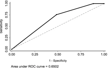
The results of Table 3 and Figure 2 summarize the association between increasing SLN tumor size and the likelihood of an additional positive non-SLN. The posi-tive likelihood ratio for additional non-SLN metastasis is approximately 1.1 for micrometastasis and 1.6 in the case of micrometastasis (95% CI 0.55 - 0.74). The ROC curve shows that increasing the size of SLN metastasis im-proves the specificity for identifying positive non-SLN while maintaining sensitivity corresponding to an area under the curve (AUC) of 0.6502.
4. Discussion
[image:3.595.307.536.444.714.2]In the present study we demonstrate an association be-tween the size of metastasis in the SLN and the presence of additional positive non-SLN in pT1 invasive breast cancer. Both SLN micro and macrometastasis have an increased likelihood of additional positive non-SLNs.
Table 2. Features of primary tumors with sentinel nodes containing metastasis.
Micrometastasis Macrometastasis p value
N = 39 (%) N = 57 (%)
Primary tumor
Median size, cm (IQR) 1.2 (0.7 - 1.5) 1.4 (0.1 - 2.0) 0.01*
Median Nottingham
grade, (gr. 1,2,3) 2 (1 - 2) 2 (1 - 2) 0.51
Pathology
Infiltrating ductal 29 (74.4) 40 (70.2) 0.82
Infiltrating lobular 6 (15.4) 9 (15.8) 0.95
Other 4 (10.3) 8 (14.0) 0.58
Receptor status
ER+/PR+ 30 (76.9) 42 (73.7) 0.81
ER+/PR- 1 (2.6) 7 (12.3) 0.14
ER−/PR+ 3 (7.7) 2 (3.5) 0.39
ER−/PR− 5 (12.8) 6 (10.5) 0.75
Lymphovascular
invasion, no. (%) 10 (25.6) 17 (29.8) 0.82
*statistically significant p < 0.05; IQR: Interquartile Range (25th - 75th
[image:3.595.58.287.509.667.2]Table 3. Receiver operating characteristic analysis of non- sentinel lymph nodes.
Status of Non-SLN Size SLN
Metastasis Negative Positive Sensitivity Specificity + L.R. Isolated Tumor
Cells 8 0 100% 0% 1.00
Micrometastasis 7 32 100% 10.0% 1.13
Macrometastasis 22 35 75.9% 51.4% 1.56
ROC Area 0.6502
Standard Error 0.048
95% CI 0.56 - 0.74*
*statistically significant; ROC: Receiver operating characteristic; Non-SLN:
Non-Sentinel Lymph Nodes.
Figure 2. Nonparametric receiver operating characteristic curve identifies the threshold of a positive screening test that optimizes sensitivity and specificity as expressed by the area under curve (AUC). Increasing the size of SLN metas-tasis improves the specificity for identifying positive non- SLN with an AUC = 0.6502 (S.E. 0.048. 95% CI 0.56 - 074).
Increasing the size of tumor in SLNs corresponds to a greater than two fold increase in the prevalence of non-SLN metastasis, from 17.9% among micrometastasis to 38.6% with macrometastasis. A positive SLN biopsy will increase the odds of additional non-SLN metastasis by a factor of 1.1 in micrometastasis and by nearly 1.6 when macrometastasis is present.
The size of the primary tumor within the pT1 subclass was also associated with non-SLNs containing metastasis. Extranodal tumor extension found predominantly in SLN macrometastasis was also associated with positive non- SLNs on univariate analysis. The association between extranodal tumor extension, macrometastasis and axillary nodal dissemination is demonstrative of the increased metastatic potential of larger neoplasms. Previous studies have shown more aggressive patterns of tumor spread when peritumoral lymphovascular invasion is present [9,10]. The present study did not find differences in the
occurrence of peritumoral lymphovascular invasion nor a tendency for nodal spread among cases of SLN micro and macrometastasis.
Both the size of the primary lesion and the extent of regional metastasis are mutually reflective of the tumor’s growth potential. Turner, et al identified a similar in-crease in non-SLN positivity among pT1-T4 lesions with larger primary tumors and increasing SLN tumor burden. In this study, SLN micrometastasis corresponded to a 26% incidence of non-sentinel nodal positivity and a 63% occurrence for SLN containing macrometastasis [9]. In other case series limited to pT1-T2 neoplasms, the frequency of non-SLN positivity ranges from 6% - 22% for SLN with micrometastasis and 44% - 55% for mac-rometastasis [10-12]. In a meta-analysis of 25 articles reporting non-SLN spread concurrent with small volume SLN metastasis, Cserni, et al report a risk of additional axillary disease ranging from 10% - 15% [13]. The trend towards decreased incidence of non-SLN metastasis cor-responding to smaller primary tumors was also reflected in the present study.
There are several explanations for the variability that is noted among retrospective reviews attempting to quan-tify and correlate sizing of SLN metastasis. Studies were routine ALND was performed regardless of SLN status may have higher estimates of non-SLN positivity com-pared to those study protocols allowing omission of sur-gical axillary clearance. [13] side from selection biases, the method of pathologic detection will affect the sensi-tivity-specifically the utilization of H & E versus IHC. As noted in the present study, H & E was more often util-ized for identifying macrometastasis whereas pankeratin IHC was more frequently utilized for micrometastasis.
[image:4.595.59.289.290.437.2]ad-ministration of adjuvant systemic therapy known to duce locoregional recurrence. Patients who do not re-ceive radiation and adjuvant systemic therapy may not achieve the same results as in the Z0011 trial and may still benefit from additional scrutiny of their axillary nodal status.
5. Conclusion
The present study demonstrates a significant correlation between the size of tumor in the SLN and axillary me-tastasis to non-SLN in patients with pT1 breast cancer. The size of the SLN metastasis is an important risk factor even among early breast lesions. Although the strength of association is strongest for patients with macrometastasis, our data suggests that patients with either micro or mac-rometastasis in their SLN biopsies have an increased likelihood of additional nodal positivity. This has impor-tant therapeutic implications given the recent trend to-wards minimizing axillary interventions in perceived low risk subpopulations. The findings of this study suggest patients with pT1 lesions and large volumes of SLN tu-mor should be considered for closer follow-up and tu-more aggressive axillary management strategies.
REFERENCES
[1] G. H. Lyman, A. E. Giuliano, M. R. Somerfield, A. B. Benson, D. C. Bodurka, H. J. Burstein, A. J. Cochran, et al., “American Society of Clinical Oncology Guideline Recommendations for Sentinel Lymph Node Biopsy in Early-Stage Breast Cancer,” Journal of Clinical Oncology, Vol. 23, No. 30, 2005, pp. 7703-7720.
doi:10.1200/JCO.2005.08.001
[2] A. E. Giuliano, P. S. Dale, R. R. Turner, D. L. Morton, S. W. Evans and D. L. Krasne, “Improved Axillary Staging of Breast Cancer with Sentinel Lymphadenectomy,” An- nals of Surgery, Vol. 222, No. 3, 1995, pp. 399-401. doi:10.1097/00000658-199509000-00016
[3] N. Wasif, M. A. Maggard, C. Y. Ko and A. E. Giuliano, “Underuse of Axillary Dissection for the Management of Sentinel Node Micrometastases in Breast Cancer,” Ar- chives of Surgery, Vol. 145, No. 2, 2010, pp. 161-166. doi:10.1001/archsurg.2009.269
[4] D. L. Weaver, “Sentinel Lymph Nodes and Breast Carci- noma: Which Micrometastases Are Clinically Signifi- cant?” The American Journal of Surgical Pathology, Vol. 27, No. 6, 2003, pp. 842-845.
doi:10.1097/00000478-200306000-00018
[5] N. Wasif, X. Ye and A. E. Giuliano, “Survey of ASCO Members on Management of Sentinel Node Micrometas- tases in Breast Cancer: Variation in Treatment Recom- mendations According to Specialty,” Annals of Surgical Oncology, Vol. 16, No. 9, 2009, pp. 2442-2449.
doi:10.1245/s10434-009-0549-7
[6] S. D. Edge, D. R. Byrd, C. C. Compton, A. G. Fritz, F. L. Greene, A. Trotti, et al., “AJCC Cancer Staging Hand-
book, from the AJCC Cancer Staging Manual,” 7th Edi-tion, Springer, New York, 2009.
[7] M. Hulvat, P. Rajan, E. Rajan, S. Sarker, C. Schermer, G. Aranha, et al., “Histopathologic Characteristics of the Primary Tumor in Breast Cancer Patients with Isolated Tumor Cells of the Sentinel Node,” Surgery, Vol. 144, No. 4, 2008, pp. 518-524. doi:10.1016/j.surg.2008.06.006 [8] C. W. Elston, “The Assessment of Histological Differen-
tiation in Breast Cancer,” Australian and New Zealand Journal of Surgery, Vol. 54, No. 1, 1984, pp. 11-15. doi:10.1111/j.1445-2197.1984.tb06677.x
[9] R. R. Turner, K. U. Chu, K. Qi, L. E. Botnick, N. M. Han- sen, E. C. Glass and A. E. Giuliano, “Pathologic Features Associated with Nonsentinel Lymph Node Metastases in Patients with Metastatic Breast Carcinoma in a Sentinel Lymph Node,” Cancer, Vol. 89, No. 3, 2000, pp. 574-581. doi:10.1002/1097-0142(20000801)89:3<574::AID-CNCR 12>3.0.CO;2-Y
[10] M. R Weiser, L. L. Montgomery, L. K. Tan, B. Susnik, D. Y. Leung, P. I. Borgen, et al., “Lymphovascular Invasion Enhances the Prediction of Non-Sentinel Node Metasta- ses in Breast Cancer Patients with Positive Sentinel Nodes,” Annals of Surgical Oncology, Vol. 8, No. 2, 2001, pp. 145-149. doi:10.1007/s10434-001-0145-y
[11] K. U. Chu, R. R. Turner, N. M. Hansen, M. B. Brennan, A. Bilchik and A. E. Giuliano, “Do All Patients with Sen- tinel Node Metastasis from Breast Carcinoma Need Com- plete Axillary Node Dissection?” Annals of Surgery, Vol. 229, No. 4, 1999, pp. 536-541.
doi:10.1097/00000658-199904000-00013
[12] G. Viale, E. Maiorano, G. Mazzarol, S. Zurrida, V. Galimberti, A. Luini, et al., “Histologic Detection and Clinical Implications of Micrometastases in Axillary Sen-tinel Lymph Nodes for Patients with Breast Carcinoma,”
Cancer, Vol. 92, No. 6, 2001, pp. 1378-1384.
doi:10.1002/1097-0142(20010915)92:6<1378::AID-CNC R1460>3.0.CO;2-Y
[13] G. Cserni, D. Gregori, F. Merletti, A. Sapino, M. P. Mano, A. Ponti, et al., “Meta-Analysis of Non-Sentinel Node Metastases Associated with Micrometastatic Sentinel Nodes in Breast Cancer,” British Journal of Surgery, Vol. 91, No. 10, 2004, pp. 1245-1252. doi:10.1002/bjs.4725 [14] A. E. Giuliano, L. Mccall, P. Beitsch, P. Whitworth, P.
Blumencranz, A. Leitch, et al., “Locoregional Recurrence after Sentinel Lymph Node Dissection with or Without Axillary Dissection in Patients with Sentinel Lymph Node Metastases,” Annals of Surgery, Vol. 252, No. 3, 2010, pp. 426-433.
[15] C. Aristei, F. Chionne, A. R. Marsella, M. Alessandro, A. Rulli, A. Lemmi, et al., “Evaluation of Level I and II Axillary Nodes Included in the Standard Breast Tangen- tial Fields and Calculation of the Administered Dose, Results of a Prospective Study,” International Journal of Radiation Oncology, Biology and Physics, Vol. 51, No. 1, 2001, pp. 69-73. doi:10.1016/S0360-3016(01)01595-4 [16] P. J. Schlembach, T. A. Buchholz, M. I. Ross, S. M.
nal of Radiation Oncology, Biology and Physics, Vol. 51, No. 3, 2001, pp. 671-678.
doi:10.1016/S0360-3016(01)01684-4
[17] J. S. Wong and J. R. Harris, “Postmastectomy Radiation