Original Article
Correlation analysis of integrin αvβ3, T-cadherin, and
VEGF expression in gastric cancer tissue with
microangiogenesis
Qing Deng1*, Li Xiong2*, Shuying Shen3, Jun Li4
Departments of 1General Surgery, 3Central Sterile Supply, The First Hospital of Jingzhou (The First Affiliated Hos-pital of Yangze University), Jingzhou, China; 2Department of Infectious Disease, Clinical Medical College, Hubei University of Science and Technology, Xianning, Hubei, China; 4Department of Surgery, Clinical Medical College, Hubei University of Science and Technology (Hubei University of Science and Technology National Demonstration Center for Experimental General Medicine Education), Xianning, Hubei, China. *Equal contributors.
Received September 6, 2017; Accepted January 4, 2018; Epub April 15, 2018; Published April 30, 2018
Abstract: This study was designed to analyze the relationship between integrin αvβ3, T-cadherin, and VEGF expres -sion with microvessel density (MVD) and clinicopathological characteristics of gastric cancer. A total of 68 gastric cancer patients and 30 healthy volunteers were enrolled. Serum T-cadherin and VEGF levels were tested by ELISA. Integrin αvβ3, T-cadherin, VEGF, and CD34 expression in gastric cancer tissues and adjacent normal tissues was detected by immunohistochemistry. The relationship between integrin αvβ3, T-cadherin, VEGF, and CD34 expres -sion with MVD and clinicopathological characteristics was analyzed. Serum VEGF level was significantly higher, while T-cadherin was lower in gastric cancer patients compared to those in the normal control group (P < 0.05). Positive rates of integrin αvβ3 and VEGF were higher, MVD counting was larger, and T-cadherin positive rate was lower in gastric cancer tissues than those in the adjacent normal controls (P < 0.05). αvβ3 positive expression was corre -lated with differentiation, infiltration depth, lymph node metastasis, and TNM staging. VEGF positive expression was related to lymph node metastasis and TNM staging. T-cadherin and CD34 positive expression was associated with differentiation, lymph node metastasis, and TNM staging (P < 0.05). αvβ3 and VEGF expression showed a positive correlation (r = 0.53 and 0.69, respectively; P < 0.05), whereas T-cadherin exhibited a negative correlation with MVD (r = -0.51, P < 0.05). VEGF was positively correlated with αvβ3 (r = 0.58, P < 0.05), while it was negatively correlated with T-cadherin (r = -0.49, P < 0.05). Abnormal expression of integrin αvβ3, T-cadherin, and VEGF was associated with gastric cancer progression and microangiogenesis.
Keywords: Gastric cancer, integrin αvβ3, T-cadherin, VEGF
Introduction
Gastric cancer is a common clinical malignancy
with high mortality and morbidity. Due to
hid-den clinical symptoms, early screening for gas -tric cancer is poor. Most patients are diagnosed in the late stage, which is combined with
dis-tant metastasis [1, 2]. Disdis-tant metastasis of
gastric cancer includes liver and extrahepatic metastasis. Liver metastasis is a multi-step
complex process involving multiple factors,
such as angiogenesis, extracellular matrix metalloproteinase, adhesion molecules, and
other factors [3, 4]. At present, the etiology and pathogenesis of gastric cancer have not been fully elucidated. Angiogenesis plays an impor
-tant role in the development and progress of malignant tumors. The balance of angiostatin and angiogenic factors is important in the regu
-lation of angiogenesis. Tumor angiogenesis is a
complex interactive process between vascular endothelial cells and tumor cells, including new
basement membrane and vascular ring forma -tion, endothelial matrix membrane dissolu-tion,
and endothelial cell migration and proliferation
[5, 6].
The initiation of tumor angiogenesis is the deg
matrix structural protein. Vascular endothelial growth factor (VEGF) plays a critical role in vas
-cular regulatory factors that can specifically stimulate the proliferation of vascular endothe
-lial cells. VEGF overexpression in a variety of
tumor tissues is associated with tumor inva-sion and metastasis. Endostatin suppresses tumor angiogenesis by inhibiting endothelial cell growth and migration [7, 8]. It has been shown that the adhesion molecule E-cadherin can induce tumor angiogenesis, whereas the
role of T-cadherin in gastric cancer invasion,
metastasis, and tumor angiogenesis is scar- cely explored [9, 10]. Integrins, are important adhesion molecules that mediate the interac-tion between cells and extracellular matrix. Integrins play an important role in tumor
angio-genesis by regulating cell adhesion, prolifera -tion, and migration. They are also involved in
the process of invasion and metastasis of vari -ous malignant tumors. In vitro and in vivo
experiments show that anti-αvβ3 monoclonal antibody can effectively inhibit angiogenesis and inhibit tumor proliferation [11, 12]. This
study was intended to analyze the relationship
between integrin αvβ3, T-cadherin, and VEGF expression with microvessel density (MVD) and clinicopathological characteristics of gastric
cancer.
Materials and methods
General information
A total of 68 gastric cancer patients between Jan 2015 and Oct 2016 in the First Hospital of Jingzhou (Hubei, China) were enrolled, including 32 males and 36 females with mean age at
59.1 ± 3.3 (30-76) years old. All the patients were diagnosed by pathology. The gastric tumor was removed by surgery, and the adjacent
nor-mal tissue was obtained from the place with more than 5 cm from the tumor edge. No
patients received chemotherapy or
radiothera-py before operation. Another 30 cases of
healthy volunteers that received physical exam-ination in the corresponding period were
select-and the study was approved by the ethics
com-mittee in First Hospital of Jingzhou (Hubei,
China).
Reagents and instruments
VEGF and T-cadherin ELISA kits were provided
by Jiancheng Bioengineering Institute (Nan-
jing, China). VEGF, αvβ3, and T-cadherin anti
-bodies, SP immunoassay kit, CD34 monoclo-nal antibody, and protein quantification kit
were supplied by Boster (Wuhan, China). In-
verted microscope was obtained from Olympus (Japan). Tissue embedder was purchased fr-om SAKURA (Japan). Slicer was bought frfr-om Leica (Germany). Oscillator was obtained from Jinghong (Shanghai, China). High-temperature resistance plastic dyeing frame was obtained from Maxim (Fuzhou, China). Computer image analysis system was provided by Hp (USA).
Methods
ELISA: The venous blood was extracted and
centrifuged to obtain the supernatant. Serum T-cadherin and VEGF contents were tested by
ELISA. The plate was read at 450 nm to ob-tain the absorbance value. A linear regression equation was established to calculate concen-tration.
Immunohistochemistry: The tissues were fixed by formalin and embedded after dehydra
-tion and waxing. Next, paraffin sec-tions were toasted and repaired after dehydration. Then the sections were blocked by goat serum and incubated in 50 μL primary antibodies (1:100,
1:200, 1:200, respectively) at room
tempera-ture for 1 h. Next, the sections were added with 50 μL secondary antibody (1:100, 1:200, 1:200, respectively) for 10 min. The sections were treated by 50 μL streptavidin-peroxidase for 10 min. After coloration, redyeing, and dif
-ferentiation, the sections were observed under
the microscope. CD34 was used to stain new blood vessel endothelial cells by SP method.
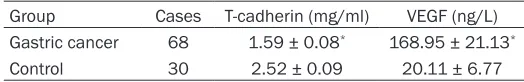
MVD value was calculated upon one blood ves -Table 1. Serum T-cadherin and VEGF comparison
Group Cases T-cadherin (mg/ml) VEGF (ng/L)
Gastric cancer 68 1.59 ± 0.08* 168.95 ± 21.13*
Control 30 2.52 ± 0.09 20.11 ± 6.77
*P < 0.05, compared with control.
ed as controls, including 15 males
and 15 females with mean age at
60.2 ± 4.2 (30-75) years old. No
sta-tistical significance was observed
on gender, age, and weight between the two groups (P > 0.05). All the
[image:2.612.88.350.84.125.2]sel counting by claybank staining of
endothe-lial cell.
Positive judgement: The immunohistochemi- stry result was scored by using a
semi-quanti-tative method. Positive cells were defined as those with claybank particles in the cell mem -brane or cytoplasm. Positive cell percentage: Score 0, no positive cells; Score 1, positive cells < 25%; Score 2, positive cells between 25% and 50%; Score 3, positive cells between
50% and 75%; Score 4, positive cells ≥ 75%.
Staining intensity: score 0, no staining; Score 1,
light yellow; Score 2, claybank; Score 3, sepia.
Staining score = cell staining intensity ×
posi-tive cell percentage. Negaposi-tive, score 0-3. Weak
positive (+), score 4-8. Positive (++), score 9-12.
Statistical analysis
All data analyses were performed on SPSS 19.0 software. The enumeration data were
analyzed by chi-square test. Measurement data were presented as mean ± standard
devi-ation and compared by ANOVA. Correldevi-ation analysis was performed by Spearman. P < 0.05 was depicted as statistical significance.
Results
Serum T-cadherin and VEGF levels in the gas -tric cancer patients
Serum VEGF level was significantly higher, while
T-cadherin was lower in gastric cancer patients than those in the normal control (P < 0.05, Table 1).
αvβ3, T-cadherin, and VEGF positive expres
-sion in gastric cancer tissue and MVD counting
The positive rates of integrin αvβ3 and VEGF were higher, MVD counting was larger, and
T-cadherin positive rate was lower in gastric cancer tissue than those in the adjacent
[image:3.612.91.527.72.301.2]nor-Figure 1.αvβ3, T-cadherin, and VEGF positive expression in gastric cancer tissue (× 100). A. αvβ3; B. T-cadherin; C. VEGF; D. CD34; 1, adjacent normal tissue; 2, gastric cancer tissue.
Table 2.αvβ3, T-cadherin, and VEGF positive expression and MVD count in gastric cancer
Tissue αvβ3 (%, n) T-cadherin (%, n) VEGF (%, n) MVD (/HPF)
Gastric cancer tissue 58.82 (40/68)* 44.12 (30/68)* 67.65 (46/68)* 18.25 ± 6.01*
Adjacent normal tissue 20.59 (14/68) 61.76 (42/68) 27.94 (19/68) 7.31 ± 5.07
χ2/t value 20.762 4.250 21.483 23.277
P value 0.000 0.032 0.000 0.000
[image:3.612.92.524.360.429.2]Table 3. Correlation analysis of αvβ3, T-cadherin, and VEGF positive expression with clinicopathologi -cal characteristics in gastric cancer
Item N αvβ3 positive rate T-cadherin positive rate VEGF positive rate MVD (/HPF) Gender
Male 32 19 (59.37) 14 (43.75) 22 (68.75) 18.44 ± 5.21
Female 36 21 (58.33) 16 (44.44) 24 (66.67) 18.08 ± 4.66
χ2/t value 0.070 0.033 0.036 0.380
P value > 0.05 > 0.05 > 0.05 > 0.05
Age
< 60 46 27 (58.69) 18 (39.13) 31 (67.39) 18.05 ± 4.99
≥ 60 22 13 (59.09) 12 (54.54) 15 (68.18) 18.67 ± 5.12
χ2/t value 0.012 1.434 0.042 0.782
P value > 0.05 > 0.05 > 0.05 > 0.05
Tumor size (cm)
< 5 25 14 (56.00) 10 (40.00) 14 (56.00) 17.25 ± 4.78
≥ 5 43 26 (60.47) 20 (46.51) 32 (74.42) 18.83 ± 5.17
χ2/t value 0.113 1.809 2.450 1.056
P value > 0.05 > 0.05 > 0.05 > 0.05
Pathology type
Adenocarcinoma 49 30 (61.22) 23 (46.94) 34 (69.39) 18.56 ± 4.78
Other type 19 10 (52.63) 7 (36.84) 12 (63.16) 17.45 ± 4.55
χ2/t value 0.417 0.824 0.243 1.172
P value > 0.05 > 0.05 > 0.05 > 0.05
Differentiation
Poor 47 33 (70.21) 25 (53.19) 34 (72.34) 21.34 ± 5.16
Well-moderate 21 7 (33.33) 5 (23.81) 12 (57.14) 11.33 ± 4.11
χ2/t value 9.192 5.083 1.532 10.558
P value < 0.05 < 0.05 > 0.05 < 0.05
Infiltration depth
Non-serosa infiltration 18 2 (11.11) 4 (22.22) 9 (50.00) 16.63 ± 4.08
Serosa infiltration 50 38 (76.00) 26 (52.00) 37 (74.00) 18.83 ± 5.11
χ2/t value 19.903 3.118 3.483 1.708
P value < 0.05 > 0.05 > 0.05 > 0.05
Lymph node metastasis
No 16 1 (6.25) 1 (6.25) 5 (31.25) 9.15 ± 3.24
Yes 52 39 (75.00) 29 (55.77) 41 (78.85) 21.05 ± 6.02
χ2/t value 12.169 12.665 12.570
P value < 0.05 < 0.05 < 0.05 < 0.05
TNM staging
I+II 36 13 (36.11) 6 (16.67) 19 (52.78) 13.63 ± 4.11
III+IV 32 27 (84.37) 24 (75.00) 27 (84.37) 23.45 ± 6.07
χ2/t value 16.292 23.382 7.728 10.558
P value < 0.05 < 0.05 < 0.05 < 0.05
mal control (P < 0.05). αvβ3 is mainly express-ed on the inner side of the cell membrane. VEGF is mainly located in the cytoplasm.
T-cadherin was on the cell membrane. CD34 is mostly expressed in the cell membrane and cytop-lasm (Figure 1 and Table 2).
Correlation analysis of αvβ3, T-cadherin, and VEGF positive expression in gastric cancer tis -sues with clinicopathological characteristics
metastasis, and TNM staging. VEGF positive
expression was related to lymph node metasta-sis and TNM staging. T-cadherin and CD34
positive expression were associated with differ -entiation, lymph node metastasis, and TNM staging (P < 0.05) but not tumor size, age, and gender (Table 3).
Correlation analysis of αvβ3, T-cadherin, and VEGF positive expression in gastric cancer tis
-sue with MVD
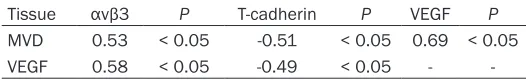
αvβ3 and VEGF expression showed positive
correlation (r = 0.53, 0.69, P < 0.05), whereas T-cadherin exhibited negative correlation with
MVD (r = -0.51, P < 0.05). VEGF was positively correlated with αvβ3 (r = 0.58, P < 0.05), while negatively correlated with T-cadherin (r = -0.49, P < 0.05) (Table 4).
Discussion
The invasion and migration of gastric cancer
and other malignant tumors depend on tumor angiogenesis. Tumor angiogenesis processes include extracellular matrix degradation,
cellu-lar proliferation, and multiple other steps. The proportion of angiogenesis inhibiting factors and promoting factors affects the tumor angio
-genesis process. Endostatin is a highly specific
endogenous angiogenesis inhibitor, which inhibits tumor angiogenesis by inhibiting eNOS
activation to block the VEGF signaling pathway. VEGF can promote vascular endothelial cell division by specifically binding the receptor on
the cell membrane to promote angiogenesis
and regulate endothelial cell proliferation. It has been suggested that VEGF overexpression in a variety of tumor cell lines and tumor tis -sues, is associated with tumor invasion, met-
astasis, and prognosis [13, 14]. VEGF can also
promote tumor distant metastasis by promot-ing lymphatic hyperplasia around the tumor
tis-sue. The recurrence rate of VEGF positive patients is higher than that of negative expres
-sion. VEGF blockade therapy significantly
reduces the primary tumor volume and
decreas-sis, and invasion depth. It has been shown that
αvβ3 is highly expressed in esophageal and
colorectal cancers, and is positively correlated
with tumor infiltration. Blocking αvβ3 in colorec
-tal cancer decreased tumor cell invasion. αvβ6 may affect gastric cancer distant metastasis through the VEGF and PI3K/AKT pathways [17, 18]. The role of αvβ3 expression in gastric can -cer has not been investigated.
T-cadherin is different from the classical cad -herin molecule. Since there is no transbrane region, T-cadherin attaches on cell mem-brane through glycosylphosphatidylinositol molecules, thus playing an important role in the
process of tumor angiogenesis and distant
migration. It is downregulated in various tumor tissues, such as pancreatic cancer and colorec-tal cancer, and is closely related to lymph node
metastasis [19, 20]. However, studies docu -menting T-cadherin expression in gastric
can-cer have been relatively few. In this study, we
investigated the correlation between the
expression of αvβ3, T-cadherin, and VEGF in gastric carcinoma, and MVD and clinicopatho -logical characteristics, aiming to provide a
basis for the clinical treatment and prognosis evaluation of gastric cancer. Serum VEGF level was significantly higher, while T-cadherin was
lower in gastric cancer patients compared with
normal controls, suggesting that integrin αvβ3, T-cadherin, and VEGF are abnormally expressed
in gastric cancer tissue. It was reported that
integrin αvβ6 positive expression was related to tumor differentiation, infiltration depth,
lymph node metastasis, and TNM staging.
αvβ3 is mainly involved in tumor cell invasion
and metastasis, and mediates cell adhesion
and migration. It is significantly expressed in
malignant tumors and is positively correlated with tumor cell invasive ability through
mediat-ing the TGF-β1 signalmediat-ing pathway. The positive expression of VEGF is associated with lymph
node metastasis and TNM staging, which is
similar to the results of this study [21, 22], sug
[image:5.612.89.352.97.137.2]-gesting that integrin αvβ3, T-cadherin, and VEGF may promote angiogenesis, invasion, and
Table 4. Correlation analysis of αvβ3, T-cadherin, and VEGF positive expression with MVD count
Tissue αvβ3 P T-cadherin P VEGF P
MVD 0.53 < 0.05 -0.51 < 0.05 0.69 < 0.05
VEGF 0.58 < 0.05 -0.49 < 0.05 -
-es the incidence of distant metas
-tasis [15, 16]. αvβ3 is expressed in
most malignant tumor tissues, su- ch as colorectal cancer and
metasta-distant metastasis. T-cadherin is an endoge-nous negative regulator that can inhibit cancer
cell metastasis. The inactivation of T-cadherin
gene in malignant tumors is related to promo-
ter methylation. The specific mechanism of
T-cadherin expression in gastric cancer is still unclear. Limited by the sample size, this study only investigated the relationship between
the expression of integrin αvβ3, T-cadherin, and VEGF with the progression and angiogene
-sis of gastric cancer. The related indexes of
angiogenesis in gastric cancer still need to be
further explored.
Abnormal expression of integrin αvβ3, T-cad-herin, and VEGF were associated with gastric
cancer progression and microangiogenesis. Combined detection could be used to evaluate gastric cancer treatment and prognosis. Acknowledgements
This work was supported by the National Natural Science Foundation of China (NO.
81641154).
Disclosure of conflict of interest
None.
Address correspondence to: Dr. Jun Li, Department of Surgery, Clinical Medical College, Hubei University of Science and Technology, 88 Xianning Road, Xianning 437100, Hubei, China. Tel: +86-715-8270-912; Fax: +86-715-8270+86-715-8270-912; E-mail: junlierr@ aliyun.com
References
[1] Song A, Zhang X, Yu F, Li D, Shao W, Zhou Y. Surgical resection for hepatic metastasis from gastric cancer: a multi-institution study. Onco-target 2017; 8: 71147-71153.
[2] Fu Y, Li H, Hao X. The self-renewal signaling pathways utilized by gastric cancer stem cells. Tumour Biol 2017; 39: 1010428317697577. [3] Macedo F, Ladeira K, Longatto-Filho A, Martins
SF. Gastric cancer and angiogenesis: is VEGF a useful biomarker to assess progression and remission? J Gastric Cancer 2017; 17: 1-10. [4] Li JH, Shen WZ, Gu XQ, Hong WK, Wang ZQ.
Prognostic value of EUS combined with MSCT in predicting the recurrence and metastasis of patients with gastric cancer. Jpn J Clin Oncol 2017; 47: 487-493.
[5] Ouyang S, Zhu G, Ouyang L, Luo Y, Zhou R, Pan C, Bin J, Liao Y, Liao W. Bapx1 mediates
trans-forming growth factor-beta-induced epithelial-mesenchymal transition and promotes a ma-lignancy phenotype of gastric cancer cells. Biochem Biophys Res Commun 2017; 486: 285-292.
[6] Xu L, Zhou R, Yuan L, Wang S, Li X, Ma H, Zhou M, Pan C, Zhang J, Huang N, Shi M, Bin J, Liao Y, Liao W. IGF1/IGF1R/STAT3 signaling-induc -ible IFITM2 promotes gastric cancer growth and metastasis. Cancer Lett 2017; 393: 76-85.
[7] Wang D, Xin Y, Tian Y, Li W, Sun D, Yang Y. Pseudolaric acid B inhibits gastric cancer cell metastasis in vitro and in haematogenous dis-semination model through PI3K/AKT, ERK1/2 and mitochondria-mediated apoptosis path-ways. Exp Cell Res 2017; 352: 34-44.
[8] Shi A, Shi H, Dong L, Xu S, Jia M, Guo X, Wang T. CXCR7 as a chemokine receptor for SDF-1 promotes gastric cancer progression via MAPK pathways. Scand J Gastroenterol 2017; 52: 745-753.
[9] Dai Y, Jiang J, Wang Y, Jin Z, Hu S. The correla -tion and clinical implica-tion of VEGF-C expres -sion in microvascular density and lymph node metastasis of gastric carcinoma. Am J Transl Res 2016; 8: 5741-5747.
[10] Ma DM, Luo DX, Zhang J. SDF-1/CXCR7 axis regulates the proliferation, invasion, adhesion, and angiogenesis of gastric cancer cells. World J Surg Oncol 2016; 14: 256.
[11] Wang L, Chang Y, Xu J, Zhang Q. Predictive sig-nificance of serum level of vascular endothelial growth factor in gastric cancer patients. Biomed Res Int 2016; 2016: 8103019. [12] Hu L, Zang MD, Wang HX, Li JF, Su LP, Yan M, Li
C, Yang QM, Liu BY, Zhu ZG. Biglycan stimu -lates VEGF expression in endothelial cells by activating the TLR signaling pathway. Mol On-col 2016; 10: 1473-1484.
[13] Watanabe K, Hirata M, Tominari T, Matsumoto C, Fujita H, Yonekura K, Murphy G, Nagase H, Miyaura C, Inada M. The MET/vascular endo-thelial growth factor receptor (VEGFR)-targeted tyrosine kinase inhibitor also attenuates FMS-dependent osteoclast differentiation and bone destruction induced by prostate cancer. J Biol Chem 2016; 291: 20891-20899.
[14] Wu ZZ, Chen LS, Zhou R, Bin JP, Liao YL, Liao WJ. Metastasis-associated in colon cancer-1 in gastric cancer: beyond metastasis. World J Gastroenterol 2016; 22: 6629-6637.
[15] Zhuang K, Yan Y, Zhang X, Zhang J, Zhang L, Han K. Gastrin promotes the metastasis of gastric carcinoma through the beta-catenin/ TCF-4 pathway. Oncol Rep 2016; 36: 1369-1376.
-herin and vascular endothelial growth factor and the prognosis of patients with gastric can -cer. Mol Med Rep 2015; 12: 2075-2081. [17] Tang Y, Dai Y, Huo J. Decreased expression of
T-cadherin is associated with gastric cancer prognosis. Hepatogastroenterology 2012; 59: 1294-1298.
[18] Li L, Jiang X, Zhang Q, Dong X, Gao Y, He Y, Qiao H, Xie F, Xie X, Sun X. Neuropilin-1 is associat -ed with clinicopathology of gastric cancer and contributes to cell proliferation and migration as multifunctional co-receptors. J Exp Clin Can -cer Res 2016; 35: 16.
[19] Akagi M, Kawaguchi M, Liu W, McCarty MF, Takeda A, Fan F, Stoeltzing O, Parikh AA, Jung YD, Bucana CD, Mansfield PF, Hicklin DJ, Ellis LM. Induction of neuropilin-1 and vascular en -dothelial growth factor by epidermal growth factor in human gastric cancer cells. Br J Can -cer 2003; 88: 796-802.
[20] Yang XW, Gao F, Chen YJ, Teng FM. The clinical study of urokinase-type plasminogen activator and vascular endothelial growth factor in gas -tric cancer. Cell Biochem Biophys 2015; 72: 649-652.
[21] Hafez NH, Tahoun NS. Expression of cyclooxy -genase 2 and vascular endothelial growth fac -tor in gastric carcinoma: relationship with clini-copathological parameters. J Egypt Natl Canc Inst 2016; 28: 149-156.