Original Article
Icariin administration is associated with enhanced
homing of bone marrow-derived progenitor cells,
increased levels of CXCR-4/SDF-1α homing factors,
and attenuated brain injury in a rat stroke model
Sen Liang1*, Ying-Hao Pei2*, Jia-Cheng Wu3, Cheng-Cheng Xu1, Ming-Hua Wu1
Departments of 1Neurology, 2Intensive Care Unit, Jiangsu Province Hospital of Traditional Chinese Medicine, The
Affiliated Hospital of Nanjing University of Traditional Chinese Medicine, Nanjing, China; 3Kangda College, Nanjing
Medical University, Nanjing, China. *Equal contributors.
Received March 9, 2018; Accepted July 14, 2018; Epub November 15, 2018; Published November 30, 2018
Abstract: The goal of this study was to investigate the mechanisms of neuroprotection by Icariin (ICA) treatment against brain ischemia in a rat stroke model via enhanced homing of bone marrow-derived progenitor cells and modulation of the CXCR-4/SDF-1α homing factor axis. Infarct size, brain water content, and neurological deficits by neurobehavioral scoring were evaluated in a middle cerebral artery occlusion model in rats treated with ICA or saline as a control. mRNA expression and protein levels of SDF-1α and CXCR-4 were investigated by quantitative reverse transcription polymerase chain reaction and Western blot analysis. Fluorescence-activated cell sorting analysis was performed to determine the effects of ICA on bone marrow cells. Neurological scores, infarct size, and brain edema all significantly improved following ICA treatment (P < 0.05). In comparison with the saline control group, CD45+/
CD34+, CD45+/CD34+/CD31+, and CD45+/CD34+/c-kit+ bone marrow-derived stem cells were increased in the
pe-ripheral blood (P < 0.05), while CD45+/CD34+ and CD45+/CD34+/c-kit+ stem cells were elevated in bone marrow in
the ICA treatment group (P < 0.05). CD45+/CD34+/CD31+ cells showed only a slight, but not significant, increase in
bone marrow in ICA-treated rats. Compared with the control group, protein, and mRNA levels of the homing factors, SDF-1α and CXCR-4, were higher with ICA treatment (P < 0.05), peaking after seven days and remaining elevated for at least 14 days following stroke. ICA protects against brain ischemic injury likely by enhancing the number of mobilized bone marrow-derived progenitor cells, which may play a key role in the repair of ischemic brain damage, and by elevating CXCR-4/SDF-1α homing factor levels.
Keywords: Icariin, bone marrow-derived stem cells, CXCR-4/SDF-1α homing factors, neuroprotection, ischemic stroke
Introduction
Ischemic stroke has become a leading cause of long-term disability and mortality [1]. Currently, pharmacological treatment is not available to prevent neuron death and degeneration trig-gered by ischemia and reperfusion. Admini- stration of bone marrow-derived mesenchymal stem cells (BMSCs) can improve the functional outcome for stroke in rats [2]. Studies using granulocyte-colony stimulating factor (G-CSF) in combination with BMSCs revealed an impro- vement in neurological function and survival in a stroke model in rats. This was accompanied by robust angiogenesis in the infarct core and
the surrounding region [3]. However, G-CSF in combination with BMSC treatment failed to impact spatial reference-memory or infarct vol-ume in this study.
Stromal cell-derived factor-1α (SDF-1α) and its
cellular receptor CXCR4 have been demonstrat-ed to be pivotal elements for stem-cell
mobili-zation and homing in many species and tissue types [4, 5]. SDF-1α/CXCR4 plays a key role in
the development of the cerebellum [6], the cerebral cortex [7], the dentate gyrus [8], and motor axons [9]. In some hypoxic conditions, such as ischemic cardiomyopathy [10] and
and CXCR4 are significantly elevated. These results raise the possibility that the SDF-1α/
CXCR4 axis might play a crucial role in regulat-ing BMSC migration in the ischemic brain. Icariin (ICA) (2-(4’-methoxylphenyl)-3-rhamno- sido-5-hydroxyl-7-glucosido-8-(3’methyl-2-but- ylenyl)-4-chromanone) (Figure 1), a flavonol iso -lated from Epimedii herba, is considered the primary active component of Epimedium ex- tracts. Epimedium is a traditional Chinese herb, which has been extensively used for more than one thousand years in China. Because of its
effect on increasing cerebral blood flow, ICA
has been widely used for the treatment of stroke in Chinese traditional medicine [12]. ICA has the ability to improve spatial learning and memory abilities in rats with brain dysfunction due to decreased expression of TNF-alpha, IL-1 and COX-2 in the hippocampus [13]. However, it has not been investigated whether the neuro-protective ability of ICA is associated with enh-
anced mobilization and homing of BMSCs via the CXCR-4/SDF-1α axis.
Therefore, in this study, rats were treated with ICA before performing middle cerebral artery occlusion (MCAO) in rats, and effects on infarct
size, brain water content and neurological defi -cits by neurobehavioral scoring, as well as on
the number of mobilized BMSCs and CXCR-4/
SDF-1 expression were evaluated. Materials and methods
MCAO model for stroke in rats
Adult male Sprague-Dawley rats (250-300 g) were purchased from the Shanghai Laboratory Animal Center, Chinese Academy of Sciences. The animals were housed in groups of eight
and maintained in controlled temperature (22 ± 2°C) and humidity (60-70%), and under a 12 hour light-dark cycle (lights on 07:00 AM). Food and water were available ad libitum. The MCAO model for stroke was established as described
[14]. Briefly, rats were anesthetized with pento -barbital (1%, 50 mg/kg), and the middle cere-bral artery was occluded permanently using a
piece of 6-0 monofilament nylon suture. Succ-essful occlusion was confirmed by an 87-90% reduction in cerebral blood flow, as measured by Laser-Doppler flowmetry. Mortality was
aro-und 10%. In all experimental protocols, the ani-mals were monitored twice daily for signals of severe distress. The criteria adopted to decide on euthanasia included the occurrence of con-vulsions, and/or severe abdominal distension. All procedures were performed in accordance with the guidelines of the Institutional Animal Care and Use the National Research Council’s Guide for the Humane Care and Use of Laboratory Animals. The experimental protocol was approved by the Ethics Committee of Jiangsu Province Hospital of Traditional Chinese Medicine, China.
Drug treatment with ICA
In accordance with previous reports [13, 15], three doses of ICA (30, 60 and 120 mg/kg) were chosen for preliminary experiments. The results showed that based on the infarction
size of the brain tissue, 120 mg/kg ICA was the
optimal dose for protection against brain isch-emia injury. In the following study, 81 rats were
equally and randomly divided into three groups:
sham group, MCAO with ICA group, and MCAO with vehicle group. Rats in the ICA group were treated with ICA (purity > 98%; Sulang Phar- maceutic Technology Co., LTD, Nanjing, China) through intragastric administration at a dose of 120 mg/kg per day for 2 weeks before MCAO. Physiological saline solution was used as the vehicle treatment. For further analysis, nine
rats of each group were deeply anesthetized
with pentobarbital (1%, 50 mg/kg) and
eutha-nized by decapitation days 3, 7, or 14 post
injury.
Infarct size measurement in MCAO rats
Nine rats of each group were selected for
infarct size measurement. The infarct size after
MCAO was determined by 2, 3,
[image:2.612.90.289.73.179.2]5-Triphenyl-2H-tetrazolium chloride (TTC) staining (Sigma, USA)
at days 3, 7 and 14 as described previously [16]. TTC stained the normal brain tissue deep red, while the infarct area was stained a pale gray color. Infarction area and hemispheric areas of each slice were photographed and
analyzed using an image-analysis software
Osiris (version 4.19, University Hospital of
Geneva, Switzerland). The infarct volume in all
slices was expressed as a percentage of the contralateral hemisphere after correcting for edema [17].
Nissl staining
Nissl staining was performed 14 days after MCAO. Sections (4 mm) were hydrated in 1%
toluidine blue at 50°C for 20 min. After rinsing with double distilled water, they were dehydrat-ed and mountdehydrat-ed with Permount. Images of the cortex and CA1 area of the hippocampus from each animal were captured and
Imaging-Pro-Plus software (Leika DMLB, Leica, Wetzlar, Germany) was used to perform quantitative
analysis of cell numbers.
Neurobehavioral evaluation of MCAO rats
To evaluate the neurological damage in MCAO rats, the performance of each rat was rated
according to neurological deficit scores, which
were described previously [18]. Brain water content in MCAO rats
Animals were anesthetized and sacrificed by
decapitation at different time points (days 3, 7
and 14). The brains were quickly removed,
weighed to obtain the wet weight, dried at 100°C for 24 h, and then weighed on an ana-lytical balance (Sartorius AG, Goettingen, Ger- many) to obtain the brain water content. The formula for calculating the water content was the following: water content (%) = 100 × (wet weight-dry weight)/wet weight.
Western blot analysis
Proteins were extracted from the cortex accord-ing to procedures described previously [11]. Membranes were probed with primary
antibod-ies against SDF-1α and CXCR4 (1:1000, Santa Cruz Biotech, Santa Cruz, CA, USA) followed by
horseradish peroxidase-conjugated anti-rabbit or anti-mouse secondary antibodies. Immun-
ostained protein bands were visualized using
the enhanced chemiluminescence method with an ECL kit (Amersham Pharmacia Biotech, Piscataway, NJ). The intensity of protein bands
was quantified by densitometry using Image J
software (NIH, Bethesda, MA). The loading
con-trol was β-actin, and relative expression of pro
-teins was normalized to β-actin levels.
Real-time quantitative RT-PCR
Total cellular RNAs from rats’ brain cortex were
extracted using TRIzol reagent (GIBCO-BRL,
Waltham, MA, USA) and reversed into cDNA using AMV reverse transcriptase (Invitrogen,
Carlsbad, CA, USA). Real-time quantitative
RT-PCR (qRT-PCR) was performed with the ABI Prism 7300 sequence detection system
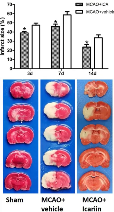
(App-Figure 2. Effect of ICA treatment on the infarction size in MCAO rats. Three rats of each group were used for infarction size measurement at each time point. The infarction size in the ICA treatment group was signifi-cantly decreased compared with the vehicle-treated group at all time points measured (day 3: 38.98% ± 1.62 vs. 47.43% ± 2.32; day 7: 46.12% ± 2.46 vs.
[image:3.612.92.284.70.426.2]lied Biosystems, Foster City, CA, USA) using
Taqman probes (Shanghai Shinegene
Mole-cular Biotechnology Co. Ltd, Shanghai, China).
β-actin was used as housekeeping gene for internal normalization. The transcript copy
number of the target genes was determined on the basis of their Ct values. The fold change in mRNA expression was obtained using the 2TΔΔCt
method. Flow cytometry
Flow cytometry was used to measure the levels of BMSCs in peripheral blood, bone marrow, and brain tissue. At days 3, 7, and 14 after MCAO, 1 ml of peripheral blood was harvested from the caudal vein of the animals in each group. Bone marrow cells were obtained by
flushing the tibiae and femurs from euthanized
rats. Mononuclear cells were separated by den-sity-gradient centrifugation using 1.077 g/ml
way analysis of variance (ANOVA). Comparisons between two groups were performed using the
Student’s t test. P < 0.05 was considered as statistically significant.
Results
Protective effect of ICA treatment in the MCAO model of stroke in rats
The visual evaluation of the infarction size (grey
areas) in the TTC-stained coronal sections reve-
aled a noticeably reduced size in ICA-treated
rats compared with sham-operated and vehi-cle-treated rats (Figure 2). The results of image
quantification showed that the infarction size in the ICA treatment group was significantly
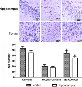
[image:4.612.93.369.72.363.2]decreased compared with the vehicle-treated group at all of the time points measured (Figure 2, day 3: 38.98% ± 1.62 vs. 47.43% ± 2.32; day 7: 46.12% ± 2.46 vs. 58.62% ± 3.52; day 14: Figure 3. Effect of ICA treatment on brain cell damage in MCAO rats. In
the sham and ICA group, the cell outline was clear and cell structure was compact. Cells were large and had abundant cytoplasm and Nissl body staining. In the vehicle group, cells were arranged sparsely and the cell outline was fuzzy. The number of cells with eumorphism in the ICA group was significantly reduced. More Nissl-stained cells were observed in the ICA group compared with the vehicle-treated group on day 14 after MCAO (cortex: 44.2 ± 5.6 vs. 20.1 ± 3.3; hippocampus: 35.6 ± 3.2 vs. 17.7 ± 2.4; P < 0.05). *P < 0.05 vs. vehicle group.
Histopaque solution
(Sigma-Aldrich, St. Louis, MO, USA),
purified, and re-suspended in
phosphate buffered saline con-taining 1% bovine serum albu-min. Cells were incubated for 40 minutes in the dark at 4°C
with fluorescein isothiocyanate
(FI-TC), phycoerythrin (PE), or peri-dinin chlorophyll-protein (Per- CP) conjugated monoclonal an- tibodies CD45-PerCP, CD34-FITC, CD31-PE and c-kit-PE (all from BD Biosciences Phar- mingen, San Diego, CA, USA). Matching isotype antibodies (BD Biosciences Pharmingen) served as controls. Cells were
analyzed by three-color fluores -cence-activated cell sorting (FACS) using a Coulter Epics
XLMCLTM flow cytometer
(Be-ckman Coulter, Fullerton, CA, USA). Each analysis included 50000 events.
Statistical analysis
The data are expressed as mean ± standard deviation (SD)
and were analyzed using the
one-23.55% ± 2.65 vs. 33.67% ± 3.18; P < 0.05).
Extensive neuronal changes were noticed in the cortex and CA1 region of the hippocampus by Nissl staining with features, such as consid-erable dark, pyknotic neurons, in the vehicle-treated group. More Nissl-stained cells were observed in the ICA group compared with the vehicle-treated group on day 14 after MCAO (Figure 3, cortex: 44.2 ± 5.6 vs. 20.1 ± 3.3;
hip-pocampus: 35.6 ± 3.2 vs. 17.7 ± 2.4; P < 0.05).
The scores ranged from the lowest score of three to a maximum score of 18. In vehicle-treated MCAO rats, the neurological score decreased to a score of 3.2 after three days, and gradually improved to a score of 6.9 after 14 days. The neurological scores for MCAO rats
treated with ICA significantly improved com -pared to vehicle-treated rats (Figure 4, day 3:
5.4 ± 0.3 vs. 3.2 ± 0.6; day 7: 7.3 ± 0.5 vs. 5.5
± 0.6; day 14: 11.2 ± 0.7 vs. 6.9 ± 0.4; P < 0.05
for all conditions). The cerebral water content, which is another measure of stroke-associated brain damage, was increased in vehicle-treated MCAO rats compared with the sham-operated controls (Figure 5). ICA-treatment led to a
sig-nificant reduction of the brain water content
compared with the vehicle group at day 7 and 14 after stroke induction (Figure 5, day 3: 83.2% ± 1.2 vs. 84.1% ± 1.4; day 7: 81.1% ± 1.5 vs. 84.7% ± 1.6; day 14: 79.0% ± 1.4 vs.
81.9% ± 1.3; P < 0.05 at day 7 and 14).
ICA-treatment up-regulates SDF-1α and CXCR4
protein and mRNA levels
The vehicle-treated group had a marked incr-
ease in SDF-1α and CXCR4 mRNA levels at days
3, 7 and 14 (Figure 6). However, ICA
[image:5.612.90.291.72.174.2]adminis-tration significantly increased their expression
Figure 5. Effect of ICA treatment on brain water content in MCAO rats. Quantification of the cerebral water content was less in the ICA group compared with the vehicle group (day 3: 83.2% ± 1.2 vs. 84.1% ± 1.4; day 7: 81.1% ± 1.5 vs. 84.7% ± 1.6; day 14: 79.0% ± 1.4 vs. 81.9% ± 1.3; P < 0.05 at day 7 and 14). *P < 0.05 vs. vehicle group.
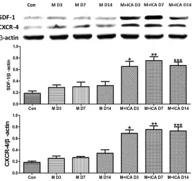
Figure 6. Effect of ICA treatment on mRNA expres-sion of SDF-1 and CXCR4 evaluated by qRT-PCR in MCAO rats. ICA administration significantly increased the mRNA expression of SDF-1α and CXCR4 in MCAO rats at all time points (1.8, 1.4, and 1.9-fold relative to sham control versus 1.6, 1.7, and 1.6-fold in the MCAO + vehicle group at days 3, 7, and 14, respec-tively, P < 0.05 compared with MCAO + vehicle). *P <
[image:5.612.322.520.74.352.2]0.05 vs. MCAO + vehicle group (n = 9). Figure 4. Effect of ICA treatment on neurological
deficits in MCAO rats. Ischemia-induced neurological deficits were significantly ameliorated in rats that re-ceived ICA treatment compared with vehicle-treated rats at days 3, 7, and 14 after MCAO (day 3: 5.4 ± 0.3 vs. 3.2 ± 0.6; day 7: 7.3 ± 0.5 vs. 5.5 ± 0.6; day 14: 11.2 ± 0.7 vs. 6.9 ± 0.4; P < 0.05 for all conditions).
[image:5.612.90.284.286.392.2]over that of the vehicle-treated group (1.8, 1.4, and 1.9-fold relative to sham control versus 1.6, 1.7, and 1.6-fold in the MCAO + vehicle group at days 3, 7, and 14, respectively, P <
0.05 compared with MCAO + vehicle).
Western blot analysis revealed that the protein
levels of SDF-1α and CXCR4 were both
incr-eased in the vehicle-treated control group com-pared to the sham-operated group, and ICA treatment resulted in further elevated protein levels (Figure 7). Densitometric quantification using β-actin normalized values showed a 1.96,
1.86, and 1.69-fold increase at days 3, 7, and 14, respectively, in the ICA group compared to the vehicle treatment group. The CXCR4 protein levels were 3.28, 2.15, and 2.38-fold increased after ICA treatment at the same time points as
compared with vehicle-treated controls (P <
0.05).
In bone marrow, the number of CD45+/CD34+
cells significantly increased 1.4, 1.3, and
1.5-fold, and CD45+/CD34+/c-Kit+ cells increased
numbers by 1.5, 1.4, and 1.2-fold at days 3, 7,
and 14, respectively (P < 0.05 for all condi -tions) (Figure 8).
In brain tissue, CD45+/CD34+ cells displayed a
6.5, 4.8, and 4.1-fold increase, CD45+/CD34+/
CD31+ cells had a 4.8, 4.9, and 5.8-fold
increase, and CD45+/CD34+/c-kit cells had a
2.4, 3.3, and 3.5-fold increase at days 3, 7, and
14 after ICA treatment, respectively (P < 0.05
for all conditions) (Figure 8). Discussion
In the present study, the protective effects of ICA in the preclinical MCAO rat model for stroke were examined. We particularly focused on
[image:6.612.94.372.69.331.2]homing mechanisms for mobilized BMSCs. The
Figure 7. Effect of ICA treatment on SDF-1 and CXCR4 protein levels in MCAO rats. Quantification of the β-actin-normalized densities of the immu-noreactive bands revealed significantly increased expression of SDF-1α and CXCR4 in MCAO rats at all of the time points measured (D3, D7, and D14) compared to the vehicle group. Densitometric quantification using β-actin normalized values showed a 1.96, 1.86, and 1.69-fold increase at days 3, 7, and 14, respectively, in the ICA group compared to the vehicle treatment group. CXCR4 protein levels were 3.28, 2.15, and 2.38-fold increased after ICA treatment at the same time points as compared with vehicle-treated controls (P < 0.05). *P < 0.05, **P < 0.01, and *P < 0.001 vs. vehicle group
(n = 9).
ICA-treatment increases the number of BMSCs in periph-eral blood, bone marrow and brain tissue of MCAO rats
To investigate the effect of ICA
on the mobilization of BMSCs
in peripheral blood, bone mar-row and brain tissue, we
per-formed flow cytometry using
different immune markers for subpopulations of mononucle-ar cells (Figure 8). We found a
significant increase in the
nu-mbers of different subtypes of CD45+/CD34+ cells in the pe-
ripheral blood after ICA admin-istration. CD45+/CD34+ cells
displayed a 3.7, 4.2, and 4.1-fold increase, CD45+/CD34+/
CD31+ cells had a 3.3, 5.1, and
5.5-fold increase, and CD45+/
CD34+/c-kit cells had a 1.4,
1.7, and 2.2-fold increase at day 3, 7, and 14, respective-
ly (P < 0.05 for all conditions)
(Figure 8). A representative
FACS analysis visualized the
difference in the number of CD45+/CD34+ cells in
main findings indicate 1) ICA was protective
against ischemic brain injury as revealed by the
size of infarcted tissue, scoring neurological
deficits and measuring the cerebral water con -tent; 2) ICA increased the expression of the
homing factors CXCR-4 and SDF-1; 3) mobiliza -Figure 8. Effect of ICA treatment on the number of CD45+/CD34+, CD45+/CD34+/CD31+ and CD45+/CD34+/c-Kit+
cells at days 3, 7, and 14 in MCAO rats. A. A significant increase of different subtypes of CD45+/CD34+ (3.7, 4.2,
and 4.1-fold increase), CD45+/CD34+/CD31+ (3.3, 5.1, and 5.5-fold increase), and CD45+/CD34+/c-Kit+ (1.4, 1.7,
and 2.2-fold increase) cells in peripheral blood (PB) at days 3, 7, and 14 after MCAO. In bone marrow (BM), CD45+/
CD34+ (1.4, 1.3, and 1.5-fold increase), and CD45+/CD34+/c-Kit+ (1.5, 1.4, and 1.2-fold increase) cells were
signifi-cantly increased after ICA administration. In brain tissue (BT), CD45+/CD34+ cells displayed a 6.5, 4.8, and 4.1-fold
increase, CD45+/CD34+/CD31+ cells had a 4.8, 4.9, and 5.8-fold increase, and CD45+/CD34+/c-Kit cells had a 2.4,
3.3, and 3.5-fold increase at days 3, 7, and 14 after ICA treatment. *P < 0.05 vs. vehicle group (n = 9). B.
[image:7.612.91.524.71.561.2]tion of subpopulations of BMSCs into the peripheral blood occurred without depleting the bone marrow.
The traditional Chinese herb Epimedium has been reported to possess various important pharmacological effects, such as increasing
cerebral blood flow, anti-inflammatory effects,
promoting nerve regeneration, improving blood circulation, regulating immunity, preventing osteoporosis, and anti-aging effects [19, 22]. Recently, Zhu et al. [23] reported that ICA could protect against brain ischemic injury by increas-ing expression of SIRT1 and PGC-1a. For the present study, a dose of 120 mg/kg ICA per day for two weeks before MCAO was chosen based on preliminary experiments. The brain infarc-tion area, brain water content, neurological function scores, and the expression levels of
the homing factors SDF-1α and CXCR-4 and the mobilization of BMSCs into peripheral blood were analyzed to evaluate the effect of ICA at
days 3, 7, 14 after stroke induction. Our results indicate that ICA ameliorates brain injury in MCAO, which resulted in a smaller area of brain infarction, less brain water content, and imp- roved neurological function scores compared to the vehicle group at the three time points examined.
Mobilization of different subpopulations of
BMCs was detected in the peripheral blood in the rats of the ICA group. These BMSC subpop-ulations remained unchanged or increased in the bone marrow, suggesting an effect of ICA on BMSC proliferation within the bone marrow. Activation of BMCSs and bone marrow cell transplantation has protective effects after ischemic brain damage [24]. However, to our knowledge the effect of ICA on the migration of
mobilized BMSCs has not been reported. In the present study, a significant increase in the
number of CD45+/CD34+ cells in the peripheral
blood was observed. Among these bone mar-row-derived progenitor cells, subpopulations additionally expressing the markers CD31+ and
c-kit were increased in the ICA-treated MCAO group. The observed increase in BMSCs was time-dependent, and reached a peak at seven days after stroke induction. Migration of BMSCs depends on homing factors, which mediate attraction, adhesion, and migration of these
cells. SDF-1α, the ligand for CXCR-4, protein
and mRNA levels were up-regulated compared to control. The mRNA and protein levels of CXCR-4, which is the corresponding surface
receptor, also significantly increased in the ICA
treatment group compared to controls. BMSCs can alleviate ischemic brain injury via the
CXCR4/SDF-1α axis [25]. G-CSF is one of the most reliable BMSC mobilizing agents for tis -sue repair. However, it has poor chemotaxis properties for CXCR4+ cells [10]. It is tempting
to speculate that ICA might improve CXCR4+
chemotaxis, improving tissue repair in ischemia after stroke.
The brain contains a reservoir of progenitor cells that may play a key role in the repair of ischemic brain damage [26]. To investigate
these cells, we quantified CD45+/CD34+/c-Kit+
cells. The results revealed a mild increase in CD45+/CD34+/c-Kit+ cells both in the peripheral
blood and in the bone marrow of MCAO rats. One other thing to note is that enhancement of BMSC homing may not be suitable for all stroke patients. MSCs were reported to be recruited and enriched in tumors, such as hepatocellular carcinoma, and MSCs may show trophic effects on tumors [27, 28]. Currently it is unclear whether MSCs have a positive or negative impact on tumor progression. Thus, it remains to be determined whether enhancing BMSC homing is an appropriate treatment strategy in stroke patients with malignant tumors.
A shortcoming of our study is that we only focused on investigating the effects of ICA on
the SDF-1α/CXCR4 chemokine axis and on
BMSC migration. In addition, we did not address a link between the SDF-1/CXR4 axis and BMSC
mobilization, as well as the signaling pathways involved in regulating the SDF-1α/CXCR4 che -mokine axis in the injured brain following ICA treatment.
In summary, our data demonstrate that in vivo administration of ICA can enhance BMSC hom-ing and the expression of homhom-ing factors
CXCR-4 and SDF-1α. Additional studies are needed to
further investigate the effects of ICA and its mechanism of action. Stroke treatment with ICA and other agents, such as G-CSF, may
opti-mize regeneration of an ischemic brain follow -ing a stroke.
Acknowledgements
Medicine for their support of this study (grant no. LJ200912, ZX2016A2), 333 high level tal-ents training project in Jiangsu (grant no. BRA 2016507).
Disclosure of conflict of interest
None.
Address correspondence to: Ming-Hua Wu, Depart- ment of Neurology, Jiangsu Province Hospital of Traditional Chinese Medicine, Nanjing 210029, China. Tel: +86-25-86617141; Fax: +86-25-8655- 8407; E-mail: [email protected]
References
[1] Maldonado NJ, Kazmi SO and Suarez JI. Up-date in the management of acute ischemic stroke. Crit Care Clin 2014; 30: 673-697. [2] Li Y, Chen J, Chen XG, Wang L, Gautam SC, Xu
YX, Katakowski M, Zhang LJ, Lu M, Janakira-man N, Chopp M. HuJanakira-man marrow stromal cell therapy for stroke in rat: neurotrophins and functional recovery. Neurology 2002; 59: 514-523.
[3] Balseanu AT, Buga AM, Catalin B, Wagner DC, Boltze J, Zagrean AM, Reymann K, Schaebitz W and Popa-Wagner A. Multimodal approaches for regenerative stroke therapies: combination of granulocyte colony-stimulating factor with bone marrow mesenchymal stem cells is not superior to g-csf alone. Front Aging Neurosci 2014; 6: 130.
[4] Huang C, Gu H, Zhang W, Manukyan MC, Shou W and Wang M. SDF-1/CXCR4 mediates acute protection of cardiac function through myocar-dial stat3 signaling following global ischemia/ reperfusion injury. Am J Physiol Heart Circ Physiol 2011; 301: H1496-1505.
[5] Lehwald N, Duhme C, Wildner M, Kuhn S, Furst G, Forbes SJ, Jonas S, Robson SC, Knoefel WT, Schmelzle M, Schulte Am Esch J. HGF and SDF-1-mediated mobilization of cd133+ bmsc for hepatic regeneration following extensive liver resection. Liver Int 2014; 34: 89-101. [6] Klein RS, Rubin JB, Gibson HD, DeHaan EN,
Alvarez-Hernandez X, Segal RA and Luster AD. SDF-1 alpha induces chemotaxis and enhanc-es sonic hedgehog-induced proliferation of cerebellar granule cells. Development 2001; 128: 1971-1981.
[7] Kondziolka D, Wechsler L and Achim C. Neural transplantation for stroke. J Clin Neurosci 2002; 9: 225-230.
[8] Shyu WC, Lin SZ, Yen PS, Su CY, Chen DC, Wang HJ and Li H. Stromal cell-derived factor-1 alpha promotes neuroprotection, angiogene-sis, and mobilization/homing of bone
marrow-derived cells in stroke rats. J Pharmacol Exp Ther 2008; 324: 834-849.
[9] Lazarini F, Tham TN, Casanova P, Arenzana-Seisdedos F and Dubois-Dalcq M. Role of the alpha-chemokine stromal cell-derived factor (SDF-1) in the developing and mature central nervous system. Glia 2003; 42: 139-148. [10] Zaruba MM and Franz WM. Role of the
sdf-1-CXCR4 axis in stem cell-based therapies for ischemic cardiomyopathy. Expert Opin Biol Ther 2010; 10: 321-335.
[11] Liu H, Liu S, Li Y, Wang X, Xue W, Ge G and Luo X. The role of SDF-1-CXCR4/CXCR7 axis in the therapeutic effects of hypoxia-preconditioned mesenchymal stem cells for renal ischemia/ reperfusion injury. PLoS One 2012; 7: e34608. [12] Wang X, Li J, Qian L, Zang XF, Zhang SY, Wang
XY, Jin JL, Zhu XL, Zhang XB, Wang ZY, Xu Y. Icariin promotes histone acetylation and at-tenuates post-stroke cognitive impairment in the central cholinergic circuits of mice. Neuro-science 2013; 236: 281-288.
[13] Guo J, Li F, Wu Q, Gong Q, Lu Y and Shi J. Pro-tective effects of icariin on brain dysfunction induced by lipopolysaccharide in rats. Phyto-medicine 2010; 17: 950-955.
[14] Jia J, Guan D, Zhu W, Alkayed NJ, Wang MM, Hua Z and Xu Y. Estrogen inhibits fas-mediated apoptosis in experimental stroke. Exp Neurol 2009; 215: 48-52.
[15] Xu RX, Wu Q, Luo Y, Gong QH, Yu LM, Huang XN, Sun AS and Shi JS. Protective effects of icariin on cognitive deficits induced by chronic cerebral hypoperfusion in rats. Clin Exp Phar-macol Physiol 2009; 36: 810-815.
[16] Benedek A, Móricz K, Jurányi Z, Gigler G, Lévay G, Hársing LG Jr, Mátyus P, Szénási G and Al-bert M. Use of ttc staining for the evaluation of tissue injury in the early phases of reperfusion after focal cerebral ischemia in rats. Brain Res 2006; 1116: 159-165.
[17] Yanamoto H, Hong SC, Soleau S, Kassell NF and Lee KS. Mild postischemic hypothermia limits cerebral injury following transient focal ischemia in rat neocortex. Brain Res 1996; 718: 207-211.
[18] Garcia JH, Wagner S, Liu KF and Hu XJ. Neuro-logical deficit and extent of neuronal necrosis attributable to middle cerebral artery occlu-sion in rats. Statistical validation. Stroke 1995; 26: 627-634.
[19] Jin Q, Lee C, Lee JW, Yeon ET, Lee D, Han SB, Hong JT, Kim Y, Lee MK and Hwang BY. 2-Phe- noxychromones and prenylflavonoids from epi-medium koreanum and their inhibitory effects on lps-induced nitric oxide and interleukin-1beta production. J Nat Prod 2014; 77: 1724-1728.
immunity in patients of hemodialysis mainte-nance]. Zhongguo Zhong Xi Yi Jie He Za Zhi 1995; 15: 202-204.
[21] Yan S, Wu B, Lin Z, Jin H, Huang J, Yang Y, Zhang X, Shen Z and Zhang W. Metabonomic characterization of aging and investigation on the anti-aging effects of total flavones of epi-medium. Mol Biosyst 2009; 5: 1204-1213. [22] Wang J, Tian XF, Wu SY, Meng XC and Wen GW.
Accelerated healing by composites containing herb epimedium for osteoinductive regenera-tion. Biomed Mater 2014; 9: 035013.
[23] Zhu HR, Wang ZY, Zhu XL, Wu XX, Li EG and Xu Y. Icariin protects against brain injury by en-hancing sirt1-dependent pgc-1alpha expres-sion in experimental stroke. Neuropharmacol-ogy 2010; 59: 70-76.
[24] Lin MN, Shang DS, Sun W, Li B, Xu X, Fang WG, Zhao WD, Cao L and Chen YH. Involvement of pi3k and rock signaling pathways in migration of bone marrow-derived mesenchymal stem cells through human brain microvascular en-dothelial cell monolayers. Brain Res 2013; 1513: 1-8.
[25] Schonemeier B, Schulz S, Hoellt V and Stumm R. Enhanced expression of the CXCl12/SDF-1 chemokine receptor CXCR7 after cerebral isch-emia in the rat brain. J Neuroimmunol 2008; 198: 39-45.
[26] Zhang SC and Fedoroff S. Expression of stem cell factor and c-kit receptor in neural cells af-ter brain injury. Acta Neuropathol 1999; 97: 393-398.
[27] Hernanda PY, Pedroza-Gonzalez A, van der Laan LJ, Broker ME, Hoogduijn MJ, Ijzermans JN, Bruno MJ, Janssen HL, Peppelenbosch MP and Pan Q. Tumor promotion through the mes-enchymal stem cell compartment in hum- an hepatocellular carcinoma. Carcinogenesis 2013; 34: 2330-2340.