Genetic influence on the kinetics and associated pathology of
the early stage (intestinal-hepatic) migration of
Ascaris suum
in mice
C. DOLD1*, J. P. CASSIDY2, P. STAFFORD1, J. M. BEHNKE3andC. V. HOLLAND1
1
Department of Zoology,School of Natural Sciences,Trinity College Dublin,Dublin 2,Ireland
2
Veterinary Sciences Centre,University College Dublin,Belfield,Dublin 4,Ireland
3
School of Biology,University of Nottingham,University Park,Nottingham NG7 2RD,UK
(Received 25 March 2009; revised 20 May, 1 June and 3 June 2009; accepted 8 June 2009; first published online 21 September 2009)
S U M M A R Y
The generative mechanism(s) of aggregation and predisposition toAscaris lumbricoidesandA. suuminfections in their host population are currently unknown and difficult to elucidate in humans and pigs for ethical/logistical reasons. A recently developed, optimized murine model based on 2 inbred strains, putatively susceptible (C57BL/6j) and resistant (CBA/Ca) to infection, was exploited to elucidate further the basis of the contrasting parasite burdens, most evident at the pulmonary stage. We explored the kinetics of early infection, focusing on the composite lobes of the liver and lung, over the first 8 days in an effort to achieve a more detailed understanding of the larval dispersal over time and the point at which worm burdens diverge. Larval recoveries showed a heterogenous distribution among the lobes of the lungs, being higher in the right lung of both strains, and in the susceptible strain larvae accumulating preferentially in 2 (caudal and middle) of the 4 lobes. Total larval burdens in these 2 lobes were largely responsible for the higher worm burdens in the susceptible strain. While total lung larval recoveries significantly differed between mouse strains, a difference in liver larval burdens was not observed. However, an earlier intense inflammatory response coupled with more rapid tissue repair in the hepatic lobes was observed in CBA/Ca mice, in contrast to C57BL/6j mice, and it is possible that these processes are responsible for restricting onward pulmonary larval migration in the resistant genotype.
Key words :Ascaris suum, mouse model, C57BL/6j, CBA/Ca, susceptibility, migration, liver, lungs, lobar distribution, inflammatory reaction.
I N T R O D U C T I O N
Ascaris lumbricoidesandAscaris suumare important widespread parasitic nematodes that cause infec-tions of humans and pigs respectively (O’ Lorcain and Holland, 2000 ; Crompton, 2001).Ascaris exhi-bits an aggregated frequency distribution in its hosts (Croll and Ghadirian, 1981 ; Boeset al. 1998 ; Holland and Boes, 2002). Furthermore, observations of re-infection patterns post-chemotherapy have indicated that individuals display a degree of pre-disposition to their worm burden status (Haswell-Elkins et al. 1987 ; Holland et al. 1989 ; Keymer and Pagel, 1990 ; Boes et al. 1998). The generative mechanism(s) of these observed phenomena are currently unknown and are difficult to elucidate in humans and pigs for ethical and logistical reasons. Recent studies have proposed that host genetics play a role in predisposition although precisely which genes are involved and what they control is still
largely unknown. One possibility is that genes fluencing immune mechanisms that control the in-tensity of infection play a key role (Holland et al. 1989, 1992 ; Williams-Blangero et al. 2008 ; Nejsum et al. 2009), and conceivably together with physio-logical factors, contribute to the processes that are responsible for predisposition (Mc Sharry et al. 1999 ; Holland and Boes, 2002). Inevitably these processes are likely to be confounded further by the influence of environmental factors such as nutrition and concurrent infections which are known to affect susceptibility to infection (Keymer and Pagel, 1990). Predisposition to Ascaris infection has important implications for transmission, morbidity and con-trol, and it is therefore crucially important to identify the mechanistic basis of resistance/susceptibility (Anderson and May, 1985) and their roles in pre-disposition to infection.
Comparative studies on larval migration have demonstrated that the mouse is a suitable model for investigating the early phase of A. suum infection (Murrell et al. 1997 ; Slotvedet al. 1998). Mitchell et al. (1976) reported that mouse strains vary in their susceptibility toAscarisinfection and therefore provide a convenient model for investigating the * Corresponding author : Department of Zoology, School
of Natural Sciences, Trinity College Dublin, Dublin 2, Ireland. Tel : +353 (0)1 8962194. Fax : +353 (0)1 6778094. E-mail : [email protected]
Parasitology(2010),137, 173–185. fCambridge University Press 2009
genetic basis of variation in migratory kinetics and immune responses to infection. A murine model was recently developed and optimized and 2 inbred strains were identified as putatively susceptible (C57BL/6j) and resistant (CBA/Ca) to infection (Lewiset al. 2006). This was reflected in the distinct, repeatable difference in A. suum larval burden in the lungs on day 7 post-infection (Lewiset al. 2006). The contrast of resistance and susceptibility between CBA and C57BL/6j mouse strains respectively re-presents the extremes of host phenotype displayed in an aggregated distribution.
The underlying mechanism responsible for the loss ofA. suumlarvae during primary infections and its precise location within the body are currently unknown. Recent studies undertaken by Lewiset al. (2006, 2007) revealed that a similar numbers of larvae were detected in the liver of both mouse strains. The majority of the larvae observed in the liver success-fully migrated to the lungs in the C57BL/6j, while far fewer larvae accumulated in the lungs of CBA/Ca mice. Subsequent examination of the leukocyte population in bronchoalveolar lavage (BAL) fluid and lung histopathology indicated that whilst there were clear differences in the intensity of the response between strains, the pulmonary inflammatory re-action was unlikely to play a prominent role in explaining the difference between Ascaris larval burdens in these mouse strains (Lewiset al. 2007). The liver has previously been cited as a key site in the immobilization of migrating Ascarislarvae in mice (Sprent and Chen, 1949 ; Taffs, 1968 ; Mitchellet al. 1976 ; Johnstoneet al. 1978 ; Songet al. 1985), guinea pigs (Kerr, 1938 ; Fallis, 1948) and rabbits (Arean and Crandall, 1962).
The white spot (WS) lesions that form as a result of mechanical injury and inflammatory response in-duced by migratingAscarislarvae in the liver have been suggested to form along the larval migration routes or encapsulate trapped larvae (Rone´us, 1966 ; Copeman and Gaafar, 1972 ; Pere´z et al. 2001). Therefore WS have been proposed to play a role in immunity toA. suuminfection in pigs (Copeman and Gaafar, 1972 ; Eriksen et al. 1980). A. suum larval debris has been detected within WS (Schwartz and Alicata, 1932 ; Sprent and Chen, 1949 ; Copeman and Gaafar, 1972 ; Pere´zet al. 2001).
There has been little focus on the larval distri-bution within the various lobes of the liver and lungs. McCraw and Greenway (1970) recovered A. suum larvae from lung lobes in calves but stated that no significant difference between the pulmonary lobes was detected. The larval and adult intestinal burden has been quantified in pigs (Eriksen et al. 1992 ; Murrellet al. 1997 ; Roepstorffet al. 1997 ; Jungersen et al. 1999) and in model organisms such as mice and guinea pigs (Jenkins, 1968 ; Keittivuti, 1974). Furthermore, larvae ofA. suumhave been observed in the wall and lumen of large intestinal segments, the
caecum, colon and rectum in mice (Jenkins, 1968 ; Douvres and Tromba, 1971 ; Keittivuti, 1974 ; Lewis et al. 2007) and pigs (Murrellet al. 1997).
The main aim of the present study was to deter-mine if the liver plays a significant role in parasite attrition in resistant mice. In order to assess the precise times and locations of larval attrition and accumulation, during the tissue-migratory phase of Ascarisinfection in C57BL/6j and CBA/Ca mice, the distribution of larval numbers was quantified in the small and large intestines and in individual hepatic and pulmonary lobes. Moreover, the role of hepatic inflammation in explaining variation in susceptibility between the two inbred mouse strains was deter-mined by semi-quantitative scoring of the histo-pathological changes within the liver.
M A T E R I A L S A N D M E T H O D S
Experimental animals
A total of 180 (90 C57BL/6j and 90 CBA/Ca) male inbred mice was purchased from Harlan UK Ltd at 7 weeks of age. They were allowed a week to accli-matize to animal house conditions before infection at 8 weeks of age. Mice were housed in an animal maintenance room in the Bioresources unit, Trinity College Dublin for the duration of the experimental procedure. The room was maintained at approxi-mately 22xC with a daily 12 h light/dark photo-periodicity. Water and commercial pelleted food were suppliedad libitum, and cages were cleaned on a re-gular basis. The mice were individually weighed on arrival, and randomly assigned, within each strain, to groups of 4 per cage.
Parasite
Approximately 5 000 000 embryonatedAscaris suum ova (batch no AP 04.04) were supplied by the Danish Centre for Experimental Parasitology (CEP), Copenhagen. Individual doses were adjusted in or-der to contain 1000 fully embryonated ova in 0.1M
H2SO4as described previously (Lewis et al. 2006). Placebo doses for control mice consisted of 0.9 % saline only.
Infection and post-mortem of mice
liver. InfectiveA. suumova and placebo doses were administered by gastric intubation to experimental and control mice respectively. Post-mortems were conducted at 6 h and on days 1–8 post-infection (p.i.). At each post-mortem time-point 8 exper-imental mice (5 for larval counts and 3 for histo-pathological examination) and 2 control mice of each strain were euthanized by cervical dislocation. Prior to euthanasia, mice were individually weighed.
Larval counts
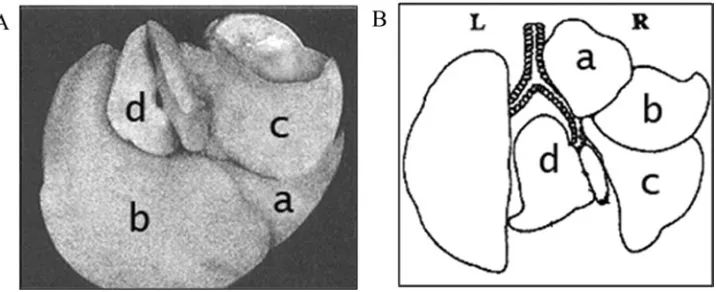
Five mice of each strain were euthanized at each post-mortem time-point and viable larvae were en-umerated in the large and small intestines, liver and lungs. The small intestine was separated into 7 cm segments (1–7 cm, 8–14 cm, 15–21 cm and the re-mainder, 22-x cm) from the pylorus of the stomach. The large intestine was also divided into the caecum and colon. The liver was separated into its multiple lobes, the median, caudate, left and right lobes. Similarly, the right lung was separated into its composite lobes, the cranial, middle, caudal and ac-cessory lobes. The left lung cannot be further sep-arated (Fig. 1). All lobes and intestinal segments were evaluated as separate entities.
Living larvae were recovered from each lobe and segment by means of the modified Baermann tech-nique (Lewis et al. 2006). A pellet of the isolated viable larvae was suspended in a 5 ml solution of 0.9 % saline and 6 % formalin. Prior to larval counts the 5 ml solutions were agitated to ensure a homo-genous distribution of larvae within the sample. Larval counts were conducted on 1 ml of each sam-ple. In order to enumerate the live larvae in the liver, large and small intestinal samples, twenty 50ml, ten 100ml and five 200ml aliquots respectively were screened underr40 magnification. When recording the larval burden in the lung samples, 2 ml were pipetted into the chamber of a nematode counting
slide (Chalex Corporation). The number of larvae present in the grid area, which represents 1 ml, was counted under r40 magnification. The number of larvae in a 1 ml solution was multiplied by total volume in order to estimate the number of larvae in the tissue sample.
Histopathological examination
Three experimental and 2 control mice of each strain were euthanized at each post-mortem time-point. The liver was separated into individual lobes as outlined above, fixed in 10 % buffered formalin for 24 h. Following paraffin embedding, 5mm sections of each lobe were cut, stained with haematoxylin and eosin, and examined. Hepatic injury and inflam-mation was scored semi-quantitatively ranging from +/x(no inflammation) and + (very mild inflam-mation) to ++++ (severe inflammation). Exam-ining sections of each hepatic lobe from each animal reduced the margin of error in scoring that could have resulted from a localized response. Histo-pathological examination was performed without prior knowledge of sample identity.
To observe changes in the hepatic inflammatory reaction an initial histopathological section from each hepatic lobe from each animal sampled at 6 h and days 1–8 p.i. was examined and graded. A further 3 histopathological sections were taken at a fixed distance of 100mm apart from each lobe from each animal euthanised on days 2–5 p.i. The scores allot-ted to the 3 further serial histopathological sections examined were subjected to statistical analysis.
Statistical analysis
[image:3.595.113.471.63.209.2]All statistical analysis was carried out at the 95 % confidence limit. Larval recovery data were assessed for normality and were subsequently log trans-formed. Intestinal larval numbers were not subjected Fig. 1. The composite lobes of the (A) murine liver (a, median lobe ; b, left lobe ; c, right lobe ; d, caudate lobe) and (B) murine left (L) and right (R) lungs (a, cranial lobe ; b, middle lobe ; c, caudal lobe ; d, accessory lobe) (adapted from Maronpotet al. 1999).
to statistical analysis as larvae were almost exclus-ively recovered on a single post-mortem time-point. The influence of the factors, time and strain on the mean larval recoveries from the liver and lungs were investigated using a 2-way GLM (SPSS 15.0). To investigate the influence of the factors, time, strain and the distribution of larvae within organs on the mean larval recoveries in the lobes of the liver and the lungs, a MANOVA (SPSS 15.0) was conducted. In order to gauge whether a larval predilection for a specific lobe in an organ exists, a MANOVA was undertaken on the proportion of total larvae in each mouse recovered from each hepatic and pulmonary lobe. Proportional data were arcsine, square root transformed prior to analysis. Analysis of the semi-quantitative pathological scores (converted into a scale ranging from zero (+/x), to 4 (++++) in the liver was carried out using a mixed-GLM model, with time and strain as fixed factors, mouse as a random factor nested within the 2-way interaction of time *strain and liver lobe as a fixed effect. The model was therefore time+strain+time *strain+ mouse (time *strain)+liver lobe, with mouse as a random factor. We also explored models involving interactions between liver lobe and time and strain, but none of these proved significant. Appropriate statistics are reported in the text.
R E S U L T S
Ascaris suummigration in the intestines, liver and lungs of C57BL/6j and CBA/Ca mice at 6 h and 1 to 8 days p.i.
Larvae were detected at 6 h and on days 1–8 p.i. in the intestines, liver and lungs of both mouse strains, but larval burdens varied over time and differed be-tween mouse strains.
The highest presence of larvae in the intestines of both mouse strains was recorded at 6 h p.i. At this time-point, the majority of larvae was recovered from the large intestine and had a strong predilection for the caecum as can be seen in Table 1. Mean larval burdens were higher in the resistant CBA mice. The mean larval numbers in the large intestine then de-creased by day 1 p.i. in both strains and no consistent pattern distinguishing the two strains was evident (Table 1).
Ascaris suumlarvae were recovered from the liver at every post-mortem time-point in each mouse strain. Appreciable numbers were recovered in this organ at 6 h and marked decreases were observed on days 4 and 5 p.i. in C57BL/6j and CBA/Ca mice respect-ively. A 2-way GLM demonstrated that the total numbers of larvae observed in the liver did not differ significantly between the two strains (main effect of strain,F1,71=3.060,P=0.085). However, recoveries of larvae differed significantly over post-mortem time-points (main effect of time, F8,71=23.785,
P<0.001). A significant strain *time interaction was found (F8,71=2.745, P=0.011) as the larvae recovered from each strain displayed different migratory patterns through this organ (Table 2).
Larvae were recovered in each liver lobe as early as 6 h p.i. In C57BL/6j the mean number of larvae peaked on days 1–3 p.i. but was dependent on the hepatic lobe and subsequently declined to compara-tively low larval numbers on days 7 and 8 p.i. (Fig. 2). As in C57BL/6j mice, low larval numbers were ob-served on the later days of infection in the CBA/Ca liver lobes (Fig. 2).
The results from a 2-way MANOVA on the pro-portion of total larvae recovered in the 4 hepatic lobes as opposed to the absolute mean numbers revealed that a significant strain effect was detected in the median (F1,71=5.519, P=0.022) and right (F1,71= 4.238,P=0.043) lobes. The proportion of total liver larvae recovered in the median lobe of C57BL/6j mice increased to a peak on day 2 p.i. (0.26¡0.07) and then fluctuated until days 7 and 8 p.i. at which point no larvae were recovered in this lobe. In CBA/Ca mice the proportion recovered in the me-dian lobe fluctuated consistently with the lowest proportion recovered on day 4 p.i. (0.04¡0.03) and the highest on day 3 p.i. (0.42¡0.10). With the ex-ception of days 2 and 4 p.i. a higher proportion of larvae was recorded in the median lobe of CBA/Ca mice. In the right lobe, the proportion of total liver larvae observed in C57BL/6j mice fluctuated con-sistently, peaking on day 8 p.i. (0.61¡0.42). How-ever in CBA/Ca mice the proportion of total larvae in the liver recovered in the right lobe increased to a peak on day 4 p.i. (0.46¡0.10) and then decreased steadily.
The strain effect was approaching significance in the caudate lobe (F1,71=3.732, P=0.057), in which the proportion of total liver larvae was seen to fluctuate slightly in both mouse strains. However, a noteworthy decrease in the proportion of larvae recovered in the caudate lobe in CBA/Ca mice was observed on day 7 p.i. (0.00) followed by a con-siderable increase on day 8 p.i. (0.43¡0.17). The
proportion of total liver larvae recovered in the right and caudate lobe significantly changed over time (right lobe : F8,71=2.950, P=0.007, caudate lobe : F8,71=2.437, P=0.022). A significant time *strain interaction was observed in the median (F8,71=2.091,
P=0.048) and right (F8,71=3.085,P=0.005) lobes.
The larval migratory kinetics observed in the liver and lungs of C57BL/6j and CBA/Ca indicated a reduction of larvae in the liver coinciding with the arrival of larvae in the lungs on days 4 and 5 p.i. In C57BL/6j mice, larvae accumulated in the lungs to a peak on day 7 p.i. (125¡21.7) and subsequently fell by day 8 p.i. (50¡21.5). Larvae consistently increased to a maximum on day 8 p.i. (31¡7.5) in CBA/Ca mice. A 2-way GLM conducted on larval numbers in the lungs on days 5 to 8 p.i. indicated a highly significant effect of strain (F1,32=16.164,
P<0.001) and time (F3,32=9.025, P<0.001). No
strain *time interaction was detected (F3,32=2.507,
P=0.077) as the pattern of larval migration in this
organ was relatively similar in both strains.
Appreciable numbers of larvae entered the left and right lungs on day 5 p.i. In C57BL/6j mice larvae in each lung increased to a peak on day 7 p.i. and sub-sequently declined on day 8 p.i. Larvae recovered in the left lung of CBA/Ca mice increased to a maxi-mum on day 8 p.i. A similar burden was recovered on days 7 and 8 p.i. in the right lung of CBA/CA mice (day 7 p.i. : 23¡4.9, day 8 p.i. : 22¡6.0) (Fig. 3).
A 2-way MANOVA conducted on proportional larval burdens on days 5–8 p.i. in the left and right lungs indicated that a strain effect was not detected in either the left (F1,32=0.198, P=0.659) or right (F1,32=2.124, P=0.155) lungs. The proportion of larvae in the lungs, which were recovered in the left and right lungs varied over time (left lung : F3,32= 4.193,P=0.013, right lung :F3,32=5.994,P=0.002). On some post-mortem time-points, the proportion of larvae recovered in the right lung differed signifi-cantly between C57BL/6j and CBA/Ca mice (F3,32= 5.060,P=0.006).
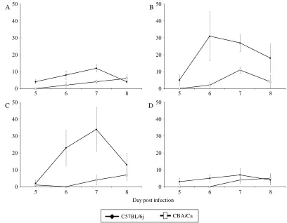
The mean larval burden recovered on days 5–8 p.i. in the right lung lobes is illustrated in Fig. 4. In C57BL/6j larvae increased to a peak on day 7 p.i. in each lobe, which was also observed in the middle lobe in CBA/Ca mice. However, in the cranial, caudal and accessory lobe of CBA/Ca mice, larvae increased to a maximum on day 8 p.i.
[image:5.595.54.280.117.235.2]A 2-way MANOVA conducted on the propor-tional data confirmed the effects of strain (F4,29= 3.576,P=0.014) and time (F12,77=2.565,P=0.007) on lobar distribution in the right lung. A strain *time interaction was observed (F12,77=3.285,P=0.001) as the percentage of larvae in the right lung recovered in the 4 lobes varied between strains at some post-mortem time-points. From the 4 right lung lobes, only the larval burden of the middle lobe changed over time (F3,32=8.546, P<0.001). The migratory pattern of larval burdens differed significantly Table 2. Mean larval numbers (¡S.E.M) recovered
at 6 h and 1–8 days p.i. in the liver of C57BL/6j and CBA/Ca mice, following inoculation with 1000Ascaris suumova
Day p.i. C57BL/6j CBA/Ca
6 h 32¡5.1 63¡14.2
1 98¡15.6 88¡18.5
2 161¡21.9 67¡11.9
3 97.5¡24.4 70¡18.8
4 98¡20.0 57¡10.7
5 51¡17.8 89¡16.3
6 48¡8.7 31¡7.0
7 11¡2.9 3¡2.0
between the two mouse strains in the cranial (F3,32= 3.046,P=0.043) and middle (F3,32=3.410,P=0.029) lobes. The proportional larval burden recovered in the caudal lobe was highly significantly different between C57BL/6j and CBA/Ca mice (F1,32=15.331,
P<0.001). The proportion of larvae in this lobe was
consistently higher in C57BL/6j and the main sites for recovery in this mouse strain were the middle and caudal lobe. The disparity was most pronounced on day 6 p.i., when larvae were absent from the caudal lobe of CBA/Ca mice, and day 7 p.i., at which point 42.5 % and 17.4 % of the larvae were recovered from the caudal lobes of C57BL/6j and CBA/Ca mice re-spectively.
Histopathological examination of the liver
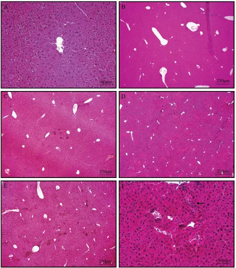
None of the uninfected control animals showed his-topathological changes in the liver (Fig. 5A). Focal accumulations of granulocytes and macrophages were observed in infected animals with a predominant peri-portal and centrilobular distribution in each lobe but were also detected within the intervening hepatic lobular parenchyma (Fig. 5I). Necrotic tracts were widely distributed throughout the tissue with a particular association with portal and perilobular blood vessels (Fig. 5F). Longitudinal and transverse
sections ofA. suumlarvae were frequently identified within blood vessels and sinusoids (Fig. 5F–H).
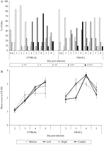
Semi-quantitative grading of sections from liver lobes (Fig. 5B–E) revealed a contrast in timing of the inflammatory reaction in C57BL/6j and CBA/Ca mice (Fig. 6A). A low inflammatory reaction score was recorded in both mouse strains at the initial stages of infection but was relatively greater in the CBA/Ca mice (mixed model as explained in the Materials and Methods section, main effect of strain F1,16=5.0,P=0.04). Although C57BL/6j mice had a higher percentage of lobes with mild inflammation (grade+) than CBA/Ca mice, the C57BL/6j animals had extensive necrotic tracts radiating from blood vessels. Comparatively severe inflammation was ob-served in CBA/Ca mice on day 2 p.i., which became more pronounced on day 3 p.i. On day 4 p.i., more severe inflammation (grade++++) was observed in more hepatic lobes of CBA/Ca animals. A similar response in the hepatic lobes in C57BL/6j mice was not detected until day 6 p.i. Comparatively less severe inflammation was detected on days 7 and 8 p.i. in CBA/Ca mice (Fig. 6A).
The mean semi-quantitative scores allotted to the 3 serial histopathological sections of each hepatic lobe illustrated a similar response in both mouse strains. In C57BL/6j mice the inflammatory reaction
6 h 1 2 3 4 5 6 7 8 6 h 1 2 3 4 5 6 7 8
6 h 1 2 3 4 5 6 7 8 0 6 h 1 2 3 4 5 6 7 8
20 40 60 80 100 0 20 40 60 80 100
0 20 40 60 80 100 0 20 40 60 80 100
Mean larval burden (± S.E.M.)
A B
C D
Day post infection
[image:6.595.73.542.63.397.2]C57BL/6j CBA/Ca
increased steadily over days 2–5 p.i in the left, right and caudate lobes. A similar inflammatory reaction was observed in the median lobe on days 3–5 p.i. In CBA/Ca mice the strong inflammatory reaction on day 4 p.i. and subsequent recovery on day 5 p.i. was observed in each hepatic lobe (Fig 6B).
These changes over time were significant (mixed model, main effect of timeF3,16=8.8,P=0.001), and both strains showed a similar pattern of changes,
if not intensity, over time (2-way interaction time * strain, F3,16=2, P=NS). There was no significant difference between liver lobes (main effect of liver lobe F3,257=1.9, P=NS) but with time, strain, the interaction between time and strain, and the main effect of liver lobe taken into account, there was a highly significant component of variation attributed to between-mouse variation in scores (mouse (time * strain),F16,257=10.4,P<0.001).
M
ea
n
la
rv
al
bur
d
en (± S
.E
.M
.)
A B
Day post infection
5 6 7 8 0
20 40 60 80 100
0 20 40 60 80 100
5 6 7 8
[image:7.595.57.524.63.254.2]C57BL/6j CBA/Ca
Fig. 3. Changes in total mean larval burden in the left (A) and right (B) lungs of C57BL/6j and CBA/Ca mice, following inoculation with 1000Ascaris suumova.
0 10 20 30 40 50
0 10 20 30 40 50
0 10 20 30 40 50
0 10 20 30 40 50
5 6 7 8 5 6 7 8
5 6 7 8 5 6 7 8
M
ea
n
la
rv
al
bur
d
en (± S
.E
.M
.)
A B
C D
Day post infection
C57BL/6j CBA/Ca
[image:7.595.93.501.299.617.2]D I S C U S S I O N
The results of the current study confirm and mark-edly extend earlier work indicating that migration of A. suumlarvae is impeded in the liver or in between the liver and lungs in CBA/Ca mice. During mi-gration in this study larvae were found to pre-ferentially accumulate in particular lobes of critical organs in susceptible mice, thereby contributing to the difference in total larval burdens between strains of mice. Analysis of larval burdens in the organs
through which the parasite migrates, particularly the significant disparity in lung larval numbers between strains and histopathological examination of the liver revealed a significant differential response, adding weight to the idea that the liver of resistant CBA/Ca mice plays an important role in the impediment of onward larval migration.
[image:8.595.70.543.61.597.2]2007). In the current study, the number of larvae in the intestines was monitored over an 8-day period in susceptible and resistant mouse strains yet a high number of larvae was only recovered in the intestines at the 6-h p.i. time-point and was predominantly in the large intestine. The observed burden in C57BL/6j mice at this time in the large intestines was comparable to that of a previous study (Lewiset al. 2007). However, the larval burden detected in the large intestine of CBA/Ca mice was comparatively higher than in the C57BL/6j mice and also relative to the previously recorded larval burden in the large intestine of this strain at 6 h p.i. (Lewiset al. 2007). The observed high caecal numbers at the 6-h p.i. time-point was documented in mice by Keittivuti (1974) and Jenkins (1968), and in both of these studies a predilection for the caecal wall was noted.
Despite higher numbers in the large intestine of CBA/Ca mice at 6 h p.i., equivalent numbers in the liver suggest that a similar number of larvae success-fully migrate from the large intestine to the liver in both mouse strains. This finding is consistent with
[image:9.595.56.529.62.416.2]earlier studies on our mouse model (Lewis et al. 2007) and in immunized and control guinea pigs (Soulsby, 1957). Timing, therefore, may account for the strain difference in the early intestinal burden as suggested by Slotved et al. (1998). These authors noted that individual mice of the same strain show variation in the rate at which larvae passed through the large intestine. While large intestinal A. suum larval burdens have been shown to peak at 6 h p.i. in mice (Keittivuti, 1974), larvae have been recorded in this tissue as early as 2 h p.i. (Keittivuti, 1974 ; Slotved et al. 1998). Furthermore, in the current study, larvae were recovered at 6 h p.i. in liver, in-dicating earlier hatching in the intestines. A more comprehensive examination of larval numbers in the large intestine at hourly intervals p.i. would indicate whether there is a temporal difference in hatching and larval penetration of the large intestinal wall. Nevertheless, if a temporal difference in hatching exists between the two strains, it is confined to a short period of time. This is reflected in the inconsistency displayed in the intestinal larval burdens on days Fig. 5. Photomicrographs of haematoxylin and eosin-stained liver sections. (A) Control uninfected liver lobe (grade
+/x), (B) mild inflammatory reaction (grade+), (C) moderate inflammatory reaction (grade++), (D) strong inflammatory reaction (grade+++), (E) severe inflammatory reaction (++++), (F) Larvae with associated necrotic larval tracts (indicated by black arrow), (G) Longitudinal larva in hepatic parenchyma and nearby granulocytes, (H) Transverse (within blood vessel) (indicated by white arrows) and longitudinal (indicated by black arrow)
1–8 p.i. observed in our mouse model and up to 10 days p.i. in a previous study where the strain of mouse used was not specified (Keittivuti, 1974).
Ascaris suum larvae have been speculated to migrate within the liver of their host, for a period of days, in an attempt to find a suitable blood vessel for onward migration to the lungs. This was reflected in the erratic pattern of larval burden in each hepatic lobe, particularly the right and caudate lobes, in both strains. The pattern of migration within the liver was shown to differ between the two mouse strains in the median and right lobes. Furthermore, a difference in the proportion of total liver larvae was observed in the median and right lobes and was shown to be approaching significance in the caudate lobe. In an
anatomical study of the mouse’s liver, the caudate lobe was found to be the smallest, accounting for 8.1¡1.0 % of the total liver in Balb/c mice, while the left lobe was shown to be the largest (34.4¡2.0 %) (Inderbitzin et al. 2006). However, while no ana-tomical studies have been undertaken on the lobes of C57BL/6j and CBA/Ca mice, the large size of the left lobe does not explain the lack of disparity in the larval burdens in this lobe between the two mouse strains. The higher vascular density of the caudate lobe, observed in Balb/c mice (Inderbitzinet al. 2006) may vary between the two mouse strains exploited in this study, accounting for the increased larval accumu-lation in C57BL/6j mice. However, since Balb/c mice were found to be in the intermediate susceptibility A
0 1 2 3 4
C57BL/6j CBA/Ca
M
ea
n
s
c
o
re
(
±
S.E.M)
Day post infection
Median Left Right Caudate
B
0 10 20 30 40 50 60 70 80 90 100
6 h 1 2 3 4 5
2 3 4 5
2 3 4 5
6 7 8 6 h 1 2 3 4 5 6 7 8
C57BL/6j CBA/Ca
%
of
lo
bes
Day post infection
[image:10.595.136.477.64.539.2]+/- + ++ +++ ++++
category in previous A. suum experiments (Lewis et al. 2006), this explanation seems unlikely.
It is currently not possible to determine if the difference in larval recovery from the hepatic caudate lobe is due to anatomical differences between the two mouse strains. Furthermore, the increased successful migration ofA. suumlarvae in C57BL/6j mice from the liver to the lungs may be explained by a hepatic or post-hepatic anatomical difference between the two strains. Strain-specific imaging of the liver and lungs may indicate barriers to dispersal within organs or between organs.
The histopathological changes induced by the larvae did not differ qualitatively between the two mouse strains. Foci of necrosis and attendant in-flammation were observed in both and the inflam-matory response was comparable to that previously described (Bindseil, 1981). Heavy infiltration of granulocytes and macrophages as well as necrosis was observed in both strains but the kinetics of these hepatic histopathological changes differed between the susceptible and resistant hosts. The earlier severe hepatic inflammatory reaction on day 4 p.i. in the resistant CBA/Ca mice coincided with the low numbers of larvae successfully migrating from the liver to the lungs whereas the more delayed severe inflammatory reaction on day 6 p.i. in the susceptible C57BL/6j mice occurred when the majority of the larvae had already migrated to the lungs. In the CBA/Ca mice, the resolution of the necrosis and in-flammation was more rapid following the reduction of larval numbers.
Therefore, it appears that CBA/Ca mice handled the parasitic insult more effectively and economically in terms of responding earlier to restrict the same degree of larval invasion, and effecting more rapid tissue recovery without triggering additional infla-mmation. Day 4 p.i. has played a prominent role in earlier work onAscarisinfections. Larvae were en-capsulated on day 4 p.i. onwards by inflammatory cells in guinea pigs immunized against A. lumbri-coides (Soulsby, 1957) with consequent reduced larval numbers noted in the lungs (Soulsby, 1957 ; Khouryet al. 1977). Furthermore, neither little in-hibition inA. suum-infected host growth nor a rise of eosinophilia was shown to be evident before this time in guinea pigs (Fallis, 1948 ; Soulsby, 1957). Therefore, day 4 p.i. has previously been considered a key time-point in effecting protection against Ascarisinfection.
While it has been proposed that the day 4 p.i. inflammatory reaction in the liver of guinea pigs is induced by a larval moult in this organ (Taffs, 1968), L3 larvae have been observed inAscarisova (Geenen et al. 1999) and ecdysis has been shown not to occur in the liver (Fagerholm et al. 2000). Excretory-secretory products capable of inducing a response are continuously produced by the metabolic activity of larvae during migration (Guerrero and Silverman,
1969 ; Stromberg, 1979) and have been shown to differ depending on their developmental stage (Kennedy and Qureshi, 1986). The ES-antigens are considered targets for responses during larval migration and the temporal difference observed in hepatic inflammatory response in the current study may be due to restricted responses in the susceptible C57BL/6j to particular ES-antigens secreted early in hepatic tissue invasion. The in vitro response of previously infected NIH mice to A. suum ES-antigens has been observed (Kennedy and Qureshi, 1986) and a lack of responsiveness to the 14 000 MW component of both L2 and L3/L4 ES was noted. Interestingly, the NIH strain has been assigned since to the intermediate susceptibility category when in-fected with A. suum (Lewiset al. 2006). However, whether CBA/Ca mice respond to this particular ES-antigen has yet to be investigated and further inves-tigation of the role of the liver in impeding migration in the mouse model will be required.
Previous evidence for an innate response ac-counting for resistance toA. suuminfection in mice, coupled with a temporal difference in the hepatic cellular response observed in this study, indicate that examining the innate liver response to infection will be a fruitful line of enquiry for future work. As dis-cussed by Holland (2009), we are now confident that we have a model of differential hepatic inflammation that is suitable for examining potential mechanisms of resistance and susceptibility in the liver. Future work will mainly focus on sentinel cells of the hepatic innate immune system and levels of cytokines and chemokines produced during infection, which may explain the temporal difference between strains in the hepatic inflammatory response observed.
The authors would like to thank Dr P. Murphy for comments on earlier drafts of this manuscript. We also thank C. Hearnden, S. Worrell and J. Brady for help with histopathology. This project was funded by the Science Foundation Ireland Research Frontiers Programme (06/RFP/EEB009).
R E F E R E N C E S
Anderson, R. C. and May, R. M.(1985). Helminth
infection of humans : mathematical models, population dynamics and control.Advances in Parasitology24, 1–101.
Arean, V. M. and Crandall, C. A.(1962). The effect of
immunization on the fate of injected stageAscaris
lumbricoideslarvae in the rabbit.American Journal of
Tropical Medicine and Hygiene11, 369–379.
Bindseil, E.(1981). Pathogenesis of liver lesions in mice
following a primary infection withAscaris suum.
Veterinary Pathology18, 804–812.
Boes, J., Medley, G. F., Eriksen, L., Roepstorff, A.
and Nansen, P.(1998). Distribution ofAscaris suum
Copeman, D. B. and Gaafar, S. M.(1972). Sequential development of hepatic lesions of ascaridosis in colostrums-deprived pigs.Australian Veterinary
Journal48, 263–268.
Croll, N. A. and Ghadirian, E.(1981). Wormy persons :
Contributions to the nature and patterns of
overdispersion withAscaris lumbricoides, Ancylostoma
duodenale, Necator americanusandTrichuris trichiura.
Tropical and Geographical Medicine33, 241–248.
Crompton, D. W. T.(2001).Ascarisand ascariasis.
Advances in Parasitology48, 285–375.
Douvres, F. W. and Tromba, F. G.(1971).
Comparative development ofAscaris suumin rabbits, guinea pigs, mice and swine in eleven days.
Proceedings of the Helminthological Society of
Washington38, 246–252.
Eriksen, L., Andersen, S., Nielsen, K., Pedersen, A.
and Nielsen, J.(1980). ExperimentalAscaris suum
infection in pigs. Serological response, eosinophilia in peripheral blood, occurrence of white spots in the liver and worm recovery from the intestine.Nordic Veterinary
Medicine32, 233–242.
Eriksen, L., Nansen, P., Roepstorff, A., Lind, P. and
Nilsson, O.(1992). Response to repeated inoculations
withAscaris suumeggs in pigs during the fattening
period. I. Studies on worm population kinetics.
Parasitology Research78, 241–246.
Fagerholm, H. P., Nansen, P., Roepstorff, A.,
Frandsen, F. and Eriksen, L.(2000). Differentiation of
cuticular structures during the growth of the third-stage larva ofAscaris suum(Nematoda, Ascaridoidea) after emerging from the egg.Journal of Parasitology86, 421–427.
Fallis, M. A.(1948).Ascaris lumbricoidesinfection in
guinea pigs with special reference to eosinophilia and resistance.Canadian Journal of Research, Series D
Zoological Sciences26, 307–327.
Geenen, P. L., Bresciani, J., Boes, J., Pedersen, A., Eriksen, L., Fagerholm, H. P. and Nansen, P. (1999). The morphogenesis ofAscaris suumto the infective third-stage larvae within the egg.Journal of
Parasitology85, 616–622.
Guerrero, J. and Silverman, P. H.(1969).Ascaris suum:
immune reactions in mice. I. Larval metabolic and somatic antigens.Experimental Parasitology26, 272–281.
Haswell-Elkins, M. R., Elkins, D. B. and Anderson,
R. M.(1987). Evidence for predisposition in humans to
infection withAscaris, hookworm,Enterobiusand
Trichurisin a South Indian fishing community.
Parasitology95, 323–337.
Holland, C. V.(2009). Predisposition to ascariasis :
patterns, mechanisms and implications.Parasitology (in the Press).
Holland, C. V. and Boes, J.(2002). Distributions and
predisposition : people and pigs. InThe Geohelminths :
Ascaris, Trichuris and Hookworm(ed. Holland, C. V.
and Kennedy, M. W.), pp. 1–24. Kluwer Academic Publishers, Boston/Dordrecht/London.
Holland, C. V., Asaolu, S. O., Crompton, D. W. T., Stoddard, R. C., Mc Donald, R. and Tormiro,
S. E. A.(1989). The epidemiology ofAscaris lumbricoides
and other soil-transmitted helminths in primary school children from Ile-Ife, Nigeria.Parasitology99, 275–285.
Holland, C. V., Crompton, D. W. T., Asaolu, S. O., Crichton, W. B., Torimiro, S. E. A. and Walters,
D. E.(1992). A possible genetic factor influencing
protection from infection withAscaris lumbricoides in Nigerian children.Journal of Parasitology78, 915–916.
Inderbitzin, D., Studer, P., Sidler, D., Beldi, G.,
Djonov, Y., Keogh, A. and Candinas, D.(2006).
Regenerative capacity of individual liver lobes in the microsurgical mouse model.Microsurgery26, 465–469.
Jenkins, D. C.(1968). Observations of early migration
of larvae ofAscaris suumGoeze, 1782 in white mice.
Parasitology58, 431–440.
Johnstone, C., Leventhal, R. and Soulsby, E. J. L. (1978). The spin method for recovering tissue larvae and its use in evaluating C57BL/6 mice as a model for the study of resistance to infection withAscaris suum.
The Journal of Parasitology64, 1015–1020.
Jungersen, G., Fagerholm, H. P., Nansen, P. and
Eriksen, L.(1999). Development of patent
Ascaris suuminfections in pigs following intravenous
administration of larvae hatchedin vitro.Parasitology 119, 503–508.
Kennedy, M. W. and Qureshi, F.(1986). Stage-specific
secreted antigens of the parasitic larval stages of the nematodeAscaris.Immunology58, 515–522.
Keittivuti, B.(1974). Sites of penetration ofAscaris suum
larvae in experimentally infected mice and swine. Ph.D. thesis. Purdue University, Lafayette, Indiana, USA.
Kerr, K. B.(1938). The cellular response in acquired
resistance in guinea pigs to an infection with pigAscaris.
American Journal of Hygiene27, 28–51.
Keymer, A. E. and Pagel, M.(1990). Predisposition to
helminth infection. InHookworm Disease : Current
Status and New Directions(ed. Schad, G. A. and Warren,
K. S.), pp. 177–209. Taylor and Francis, London and Philadelphia.
Khoury, P. B., Stromberg, B. E. and Soulsby, E. J. L. (1977) Immune mechanisms toAscaris suumin inbred guinea-pigs. I. Passive transfer of immunity by cells or serum.Immunology32, 405–411.
Lewis, R., Behnke, J. M., Stafford, P. and Holland,
C. V.(2006). The development of a mouse model to
explore resistance and susceptibility to earlyAscaris suuminfection.Parasitology132, 289–300.
Lewis, R., Behnke, J. M., Cassidy, J., Stafford, P.,
Murray, N. and Holland, C. V.(2007). The
migration ofAscaris suumlarvae, and the associated pulmonary inflammatory response in susceptible C57BL/6j and resistant CBA/Ca mice.Parasitology 134, 1301–1314.
Maronpot, R. R., Boorman, G. A. and Gaul, B. W. (1999)Pathology of the Mouse : Reference and Atlas. Cache River Press, Vienna, Illinois, USA.
McCraw, B. M. and Greenway, J. A.(1970).Ascaris
suuminfection in calves III. Pathology.Canadian
Journal of Comparative Medicine34, 247–255.
McSharry, C., Xia, Y., Holland, C. V. and Kennedy,
M. W.(1999). Natural immunity toAscaris lumbricoides
associated with immunoglobulin E antibody to ABA-1 allergen and inflammation indicators in children.
Mitchell, G. F., Hogarth-Scott, R. S., Edwards, R. D.,
Lewers, H. M., Cousins, G. and Moore, T.(1976).
Studies on immune response to parasite antigens in mice 1.Ascaris suumlarva numbers and antiphosphorylcholine responses to infected mice of various strains and hypothymic nu/nu mice. International Archives of Allergy and Applied Immunity 52, 64–73.
Murrell, K. D., Eriksen, L., Nansen, P., Slotved, H. C.
and Rasmussen, T.(1997).Ascaris suum: a revision
of its early migratory path and implications for human ascariasis.Journal of Parasitology83, 255–260. Nejsum, P., Roepstorff, A., Jørgensen, C.,
Fredholm, M., Go¨ rring, H. H. H., Anderson, T. J. C.
and Thamsborg, S. M.(2009). High heritability
forAscarisandTrichurisinfection levels in pigs.
Heredity102, 357–364.
O’Lorcain, P. and Holland, C. V.(2000). The public
health importance ofAscaris lumbricoides.Parasitology 121, S51–S71.
Pere´ z, J., Garcia, P. M., Mozos, E., Bautista, M. J.
and Carrasco, L.(2001). Immunohistochemical
characterization of hepatic lesions associated with migrating larvae ofAscaris suumin pigs.Journal of
Comparative Pathology124, 200–206.
Roepstorff, A., Eriksen, L., Slotved, H. C. and
Nansen, P.(1997). ExperimentalAscaris suuminfection
in the pig : worm population kinetics following single inoculations with three doses of infective eggs.
Parasitology115, 443–452.
Rone´us, O.(1966). Studies on aetiology and pathogenesis
of white spots in the liver of pigs. Dr Thesis,Acta
Veterinaria Scandinavica7, 1–112.
Schwartz, B. and Alicata, J. E.(1932).Ascarislarvae as a
cause of liver and lung lesions in swine.Journal of
Parasitology19, 17–24.
Slotved, H. C., Eriksen, L., Murrell, K. D. and
Nansen, P.(1998). EarlyAscaris suummigration in
mice as a model for pigs.The Journal of Parasitology 84, 16–18.
Song, J. S., Kim, J. J., Min, D. Y. and Lee, K. T.(1985).
Studies on the comparative migration patterns ofAscaris suumlarvae between primary and re-infected mice.
Kisaengchunghak Chapchi23, 247–252.
Soulsby, E. J. L.(1957). Immunization againstAscaris
lumbricoidesin the guinea pig.Nature, London179,
783–784.
Sprent, J. F. A. and Chen, H. H.(1949). Immunological
studies in mice infected with the larvae ofAscaris
lumbricoides. I. Criteria of immunity and immunizing
effect of isolated worm tissues.Journal of Infectious
Diseases84, 111–124.
Stromberg, B. E.(1979). The isolation and partial
characterisation of a protective antigen from developing larvae ofAscaris suum.International
Journal for Parasitology9, 307–311.
Taffs, L. F.(1968). Immunological studies on
experimental infection of pigs withAscaris suum Goeze, 1782. VI. The histopathology of the liver and lung.Journal of Helminthology42, 157–172.
Williams-Blangero, S., Vandeberg, J. L., Subedi, J.,
Jha, B., Correa-Oliveira, R. and Blangero, J.(2008).
Localization of multiple quantitative trait loci influencing susceptibility to infection withAscaris
lumbricoides.The Journal of Infectious Diseases197,