J. exp. Biol. (1980), 89, 117-157
With 28 figures Printed in Great Britain
SYNAPTIC PLASTICITY AND THE MODULATION
OF THE Ca
2+CURRENT
BY MARC KLEIN, ELI SHAPIRO AND ERIC R. KANDEL
Division of Neurobiology and Behavior, Departments of Physiology and Psychiatry, College of Physicians and Surgeons, Columbia University, New York, New York 10032
I N T R O D U C T I O N
'Every physiological teaching on the working of the brain based on localization, no matter how excellent, leaves us ignorant about the mechanism of mental activity. These actions are certainly accompanied by molecular modifications in nerve cells and preceded by complex changes in the relationship between neurones. T o understand mental activity it is necessary to understand these molecular modifications and changes in neuronal relationships. One must know, of course, the complete and exact histology of cerebral centres and their tracts. But that is not enough; it will be necessary to know the energetic transformations of the nervous system which accompany perception and thought, conciousness and emotion.'
Ramon y Cajal (1911)
The study of the mechanisms of neuronal plasticity and the attempt to relate these mechanisms to actual instances of learning has accelerated in recent years as a result of the application of the techniques of biophysics and cell biology to central neurones and their interconnexions. Although support for it has been obtained only recently, the idea that learning might involve changes in the effectiveness of the connexions between neurones actually had its origins at the turn of the century. Following an earlier suggestion by E. Tanzi (1893), Ramon y Cajal postulated, in his Croonian Lecture to the Royal Society of London in 1894 (a lecture to which he was invited through the intervention of Charles Sherrington), that learning might involve changes in strength of connexions between neurones. An almost identical idea was put forth by Sigmund Freud also in 1894 in a fragmentary manuscript published only in 1950. A first requirement of this postulate is that some synapses have plastic properties, that they can change their efficacy following simple use or following more complex patterns of stimulation. This basic requirement has now been fully satisfied. A variety of experiments have shown that chemical synapses can undergo changes in effective-ness as a result of activity or inactivity in a given pathway (homosynaptic change) or as a result of activity in other pathways (heterosynaptic change). Some of the best evidence has come from studies of simple synaptic systems such as the synapses between vertebrate motor neurones and skeletal muscle (for reviews, see Katz, 1962; Eccles, 1964; Kandel & Spencer, 1968).
n 8 M. KLEIN, E. SHAPIRO AND E. R. KANDEL
stimulation but persisted for several minutes after the tetanus. Feng called the synaptic. enhancement that persisted after the tetanus post-tetanic potentiation. He also found' that longer tetani produced greater potentiation than did shorter ones. In 1947, Larrabee and Bronk found post-tetanic potentiation at a peripheral neurone-to-neurone synapse between preganglionic and postganglionic cells of the stellate gan-glion. In 1949, Lloyd described similar potentiation in the monosynaptic reflex of the spinal cord, thereby showing that these plastic changes also occur in central neurones. Lloyd found (as had Larrabee and Bronk) that post-tetanic potentiation produced by stimulating one afferent pathway did not increase the response of the postsynaptic cell to synaptic activation via another, unstimulated, afferent pathway. These experi-ments indicated that post-tetanic potentiation is homosynaptic: it is restricted to the stimulated pathway and results from a change in the synapse itself. Lloyd (1949) also described a second type of plastic change when he found that low frequencies of stimulation produced a decrease in synaptic effectiveness. This form of plastic change
he called low-frequency or homosynaptic depresssion.
Analysis remained at this stage for a number of years because it was difficult to determine whether these changes were due to a presynaptic mechanism (a change in transmitter release) or to a postsynaptic mechanism (a change in receptor sensitivity). The solution to this problem was facilitated in 1954 when del Castillo & Katz (1954a) demonstrated that release of acetylcholine at the nerve muscle synapse is not graded but quantized. An action potential releases about 200 multimolecular packets of transmitter - called quanta - and each quantum contains several thousand molecules of ACh. Quantal transmission was soon found to be the general mode of transmitter release at chemical synapses (see Dudel & Kuffler, 1961a; Eccles, 1964). The dis-covery of quantal transmission not only established important new insights into the nature of transmitter release from the terminals but also provided a method for analys-ing the relative contribution to synaptic transmission of changes in presynaptic and postsynaptic mechanisms. Del Castillo & Katz (1954a, b), Liley (1956a, b) and sub-sequently others, analysed alterations in synaptic effectiveness in terms of quantal transmission and found that both homosynaptic depression and post-tetanic poten-tiation represented a presynaptic alteration in the number of transmitter quanta released per impulse. The sensitivity of the receptor seemed not to be affected.
This work was extended in a major direction in 1961 when, following the earlier suggestion of Frank & Fuortes (1957), Dudel and Kuffler described the first clear instance of a heterosynaptic interaction: presynaptic inhibition at the crayfish nerve-muscle synapse. Dudel & Kuffler (1961a) found that in the crayfish the inhibitory axon to muscle has a double function: (1) it produces an inhibitory postsynaptic potential in the muscle, and (2) it depresses the excitatory postsynaptic potential produced by the excitatory axon. By applying a quantal analysis Dudel & Kuffler (19616) found that presynaptic inhibition reduces the number of transmitter quanta released by the excitatory axon without affecting the sensitivity of the receptor mole-cules. These experiments provided the first evidence that the membrane of the presyn-aptic terminals contains receptors to transmitter molecules, and that these receptors can control transmitter release. Subsequently, Kandel & Tauc (1964), Epstein & Tauc
Synaptic plasticity and the modulation of the Ca
2+current 119
n based upon a quantal analysis, Castellucci & Kandel (1976) provided direct vidence for a presynaptic mechanism.
In 1967 Katz and Miledi advanced the study of synaptic plasticity still one import-ant step further by finding that transmitter release is dependent on the influx of Ca2+ that occurs with each action potential. They proposed that Ca2+ allows the synaptic vesicles, the subcellular organelles that store transmitter (and which are thought to represent individual quanta), to bind to release sites. In addition, Katz & Miledi (1967 a, b) found that the presynaptic terminal contained a high density of voltage-gated Ca2+ channels.
These findings suggested to Katz & Miledi (1968) and to Rahamimoff (1968) that changes in the intracellular level of free Ca2+ might be important for short-term synaptic plasticity. Rahamimoff (1968; Alnaes & Rahamimoff, 1975) proposed that the intracellular concentration of Ca2+ is controlled by intracellular organelles that buffer Caa+ - the mitochondria and endoplasmic reticulum - and that aspects of synaptic plasticity might depend upon this control. In support of this idea, Raham-imoff found that short-term homosynaptic facilitation seems to be due to residual Ca2+,
the Ca2+ that remains in the terminal following a series of action potentials, and that is taken up slowly by the buffering organelles. A similar mechanism also seems to be operative in Aplysia (Kretz, Shapiro & Kandel, 1980).
Another factor that controls the free Ca2+ levels in the terminals is the Ca2+ influx. It seemed to us (as it had earlier to Zucker (1974) and to Stinnakre & Tauc (1973)) that influx might not be constant but might be modulated. In the past, however, this idea has proven difficult to examine in central neurones showing plastic changes, since the most direct test of the hypothesis would require recording of Ca2+ current in presynaptic terminals simultaneous with monitoring of transmitter release. This problem can be overcome, to a certain degree, in Aplysia neurones. First, the mem-brane of the cell body of Aplysia neurones contains Ca2* channels whose properties seem to resemble those of the terminal membrane (Geduldig & Junge, 1968; Geduldig & Gruener, 1970; Stinnakre & Tauc, 1973; Llinas, Steinberg & Walton, 1976). Moreover, in certain cases changes in the calcium current of the cell body parallel the changes in transmitter release at the terminals (Klein & Kandel, 1978). In addition, in some of these neurones the presynaptic terminals controlling transmitter release appear to be sufficiently close to the cell body electrically so that it is possible to modify transmitter release from the terminals by injecting current into the cell body (Shimahara & Tauc, 1975; Shimahara & Peretz, 1978; Shapiro, Castellucci & Kandel,
1980a). The observations suggested to us that we might be able to examine the rela-tionships between transmitter release and specific ionic currents of the presynaptic membrane. To this end we have combined analysis of ionic currents of the cell body of the presynaptic neurone, using voltage-clamp analysis and pharmacological block-ade of specific ion channels, with assay of transmitter release from the presynaptic cell, using intracellular recordings of the synaptic potential in the postsynaptic cell (Fig. 1). These combined techniques provide a powerful method for studying changes in specific ionic conductances associated with various presynaptic mechanisms for synaptic plasticity.
1 2 0 M. KLEIN, E. SHAPIRO AND E. R. KANDEL
r
' Bath'
1.
Current monitor
Chamber virtual ground
Fig. i. Diagram of experimental arrangement for analysing both pre- and postaynaptic aspects of the four types of synaptic plasticity examined in this review. (A) Voltage clamp of the presynaptic neurone allows analysis of the ionic currents controlling transmitter release. Simultaneous recording of synaptic potentials from the postsynaptic cell allows examination of the consequences of the presynaptic currents for synaptic transmission. (B) Voltage clamp used for sensory neurone experiments was a Dagan Instruments 8500 pre-amp-voltage damp. The voltage-clamp circuit described in Byrne et al. 1979, was used for Lio experiments. Virtual ground and current monitor were Ag-Agcl wires in the bath. Two independent intra-cellular electrodes of 1-3 MCI resistance for cell Lio and 5-10 MO resistance for sensory neurones were used for voltage recording and current passing. The electrodes were filled with 2 M-K citrate.
cholinergic cell Lio and its identified follower cells, and (2) the connexions between the mechanoreceptor sensory neurones of the gill-withdrawal reflex and their follower cells.
Synoptic plasticity and the modulation of the Ca
i+current 121
A
lm
y 1.1
L,»
-28 -18
mV
B,
3-0-1
2-5-
2-0-3
1-5-a,
a. 1-OH
TTX
H.P. = - 5 3 mV
.'2
- 5 0 - 4 0 - 3 0 - 2 0 - 1 0 0 +10 +20 Km(L1 0)mV
TTX H.P. = - 3 8 mV Step to - 3 mV
9
8-
64
-
2- 0-/
3 / ' 2
/
i—•—>~ . . . i /
10 20 30 40 50 100 Presynaptic pulse duration (ms)
200
I 2 m V
_ ] 200 nA 400 ms
[image:5.451.57.404.50.480.2]1 2 2 M. KLEIN, E. SHAPIRO AND E. R. KANDEL
+20
0
- 2 0
- 4 0
- 6 0
-80*
-100
HP
.J
20
16 £ E
12 .J
8 I
a.
4 £
-50 - 4 0 \ - 3 0 -20 -10 0 +10 +20
20
16
12
8
4
/1
[image:6.451.51.381.64.448.2]0 20 40 60 80 100 120 140 Peak inward current (nA)
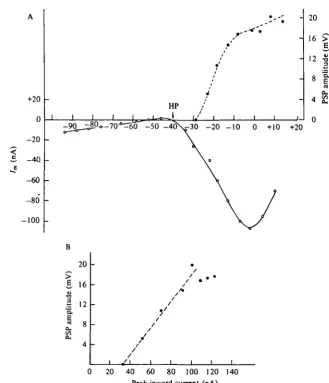
Fig. 3. Transmitter release and presynaptic Cal + current. (A) Cell L i o was voltage clamped at a holding potential of —40 mV in a sea water solution containing high-divalent cation. Transmitter release was evoked by 200 ms duration depolarizing clamp steps. The size of the PSP recorded in cell L5 was plotted against step depolarization. The preparation was then treated with 4-AP (10 mM), TEA"1" (25 ITIM) and extracellular Ca'+ was replaced by Ba'+ ions, to block K+ currents. The peak inward current evoked by 200 ms step depolarizations from
a holding potential (HP) of — 40 mV to various voltages is plotted in the lower part of the figure. (B) Plot of the relationship between peak inward current (corrected for leakage) and PSP amplitude for this cell.
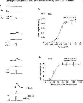
duration of the depolarizing step (Fig. 2B2). Voltage control can be further improved by adding pharmacological agents that block each of the three known outward K+ currents: tetraethylammonium ions (TEA+), which block the delayed K+ channel; 4-aminopyridine (4-AP), which blocks the early K+ channel; and substitution of Ba^ ions for Ca^, which block the Ca^-dependent K+ channel (see Adams, Smith & Thompson, 1980; Shapiro, Castellucci & Kandel, 19800,6). Blocking these K+ channels also lengthens the effective space constant of the neurone. A
Synaptic plasticity and the modulation of the Ca
2+current 123
between transmitter release and presynaptic Ca2+ current (Fig. 3 A). We found that Tn the range of depolarizing voltages in which the Ca2+ current was increasing,trans-mitter release increased in a linear fashion (Fig. 3B). These results suggest that in this voltage range (from about — 40 to zero mV) we could control transmitter release from the terminals with voltage clamp of Lio's cell body.
Although in many cases (particularly in the studies of the connexions made by sensory neurones), we lacked ideal voltage control of the terminals, these procedures nonetheless allowed sufficient control to study transmitter release while at the same time examining ionic currents in the soma of the presynaptic neurone. Moreover, we repeated all experiments except those involving presynaptic inhibition (where the transmitter is not known) in mechanically isolated presynaptic cell bodies where optimal space clamp control can be achieved.
Using this approach, we have examined four types of synaptic plasticity, including two directly involved in simple nonassociative forms of learning: (1) the control of transmitter release in spike generating neurones by the membrane potential of the presynaptic neurones; (2) presynaptic inhibition; (3) homosynaptic depression (the mechanism underlying short-term habituation); and (4) presynaptic facilitation (the mechanism of behavioural sensitization). We have found that each of these involves modulation of the Ca2+ current, although the details of the mechanisms differ in each case.
The control of transmitter release by the membrane potential of the presynaptic cell In 1975 Shimahara and Tauc described a simple form of synaptic plasticity whereby the membrane potential of the presynaptic neurone exerts a powerful influence over the effectiveness of the connexions made by that neurone. Hyperpolarizing the pre-synaptic cell decreased the pre-synaptic potential elicited by the prepre-synaptic action poten-tial whereas depolarizing the presynaptic cell enhanced the synaptic action. Although this effect was opposite to that described in the giant synapse of the squid (Hagiwara & Tasaki, 1958; Takeuchi & Takeuchi, 1962) and at first seemed paradoxical, similar potential dependent control of transmitter release by membrane potential has now been described in the leech (Nicholl9 & Wallace, 1978 a). A form of this plasticity occurs between the identified multiaction cholinergic cell Lio and its follower cells (Waziri, 1977; Shapiro, Castellucci & Kandel, 1980a).
Intracellular stimulation of neurone Lio produces EPSPs in some follower cells (R15 and RB cells) and IPSPs in others (Li to L6, LBVC, LDH I, etc.; see Kandel
et al. 1967; Wachtel & Kandel, 1967, Kehoe, 1972; Koester et al. 1974; Koester &
124 M. KLEIN, E. SHAPIRO AND E. R. KANDEL
RB
- 1 0 6
mV-j 2 0 m V 100 ms
RB
4
-57 mV
I 2 m V
—ilOrnV SO mi
20 h
15
•a .1 10
J i L _L
_75 _70 -65 -60 -55 -50 -45 -40 -35 Presynaptic membrane potential (mV)
Synoptic plasticity and the modulation of the Ca
2+current 125
t
we shall discuss below, these alterations account for only part of the effects of embrane voltage on release. Part of the effect of membrane voltage is independent of changes in spike height and duration.Release of transmitter from the terminals can be controlled from the cell body in a graded manner
The ability to alter transmitter release from the terminals by injecting current into the cell body implies that at least some release sites are electrically close to the soma. If the axon of the cell 19 cut close to the ganglion and the preparation is treated with TTX, graded depolarizing steps under voltage clamp lead to graded release of trans-mitter (Figs. 2, 3 and 5). The sigmoid function relating transtrans-mitter release to pre-synaptic depolarization i9 similar to that reported at the squid giant synapse (Katz & Miledi, 1969; Llinas et al. 1976). As is the case with the squid giant synapse the func-tion relating transmitter release to presynaptic depolarizafunc-tion overlaps the voltage sensitivity of the presynaptic Ca2"1" current (Fig. 3). However, in contrast with results in squid, hyperpolarizing the presynaptic cell decreases the size of the synaptic potential elicited by step depolarization to a given level (Fig. 5 B).
Outward currents are decreased by depolarization
At depolarized holding potentials, currents elicited by step depolarizations under voltage clamp are less outward than at hyperpolarized holding potentials (Fig. 5 A and Connor & Stevens, 19716; Adams et al. 1980). This difference in net current presumably accounts for the difference in the configuration of the action potential at the two holding potentials in undamped cells (Fig. 4B). In addition, the finding that the synaptic potentials are graded with the duration of the command pulse (Fig. 2B2) provides further evidence that the K+ currents, which control spike duration and amplitude, could modulate transmitter release. However, the ability of the mem-brane potential to modulate transmitter release under voltage-clamp conditions, in which the duration and amplitude of the presynaptic command pulse is held constant, suggested that mechanisms other than modulation of the K+ current contribute to this form of plasticity. One possible additional mechanism is a change in transient Ca2+ current. To test this idea we blocked the several outward K+ currents which are responsible for alterations in the shape of the spike to see whether the membrane potential was still capable of modulating the transmitter release caused by a depolar-izing command, and whether this modulation was due to a direct action on the trans-ient Ca2+ current.
Transient Cai+ current is not increased by depolarization
Three pharmacologically separable K+ conductances have been described in mol-luscan somata (Thompson, 1977; Adams et al. 1980). Two of these are voltage-dependent; an early, rapidly inactivating K+ current sensitive to 4-AP (Connor & Stevens, 19716; Byrne et al. 1979; Thompson, 1977), and a delayed K+ current sensitive to TEA+ (Connor & Stevens, 1971a; Thompson, 1977; Byrne et al. 1979). A third K+ conductance is thought to be not dependent on voltage but on intracellular Ca*4" concentration, and can be blocked by agents which block the Ca2+ current (e.g.
«
?+, EGTA) and by substituting Ba24" ions for Ca2"1" ions (Meech, 1972; Meech & inden, 1975; Eckert & Lux, 1976; Thompson, 1977; Adams et al. 1980; Shapiro126 M. KLEIN, E. SHAPIRO AND E. R. KANDEL
RB
H.P. = - 4 5 mV- 3 5
- 1 3
200 nA
- 4 2
H.P. = - 6 2 mV
40 nA
I 2 m V
400 nA
l l O m V lsec
5-0 H TTX H.P. = -45 mV
H.P. = -62 mV
_60 -50 -40 -30 -20 -10 0 +10 Presynaptic membrane potential (mV)
Synoptic plasticity and the modulation of the Ca
2+current 127
Na+-free
TEA 4-AP
- 6 0
(mV)
-50 -40 -30 -20 - 1 0
° ' \ -140 -180 -220 -260 +20 ,„ + 10
- 2 0
- 6 0
-100
\
A
\
\ o
+ 2 0
J
•
+30
f 1
K
f
+40 / / / O H.P. = \ H.P. = O
- 6 0 mV
- 4 0 mV (nA)
B,
TTX Ba1* TEA 4-AP
/ ^
-lOmVi
_36 1
~ T
-10
-62
2 m V
_ | 50 nA 200 ms
C,
Na+-free
Ba3t
TEA 4-AP
r
100 nA -27 mV- 6 2 _
1 s
Fig. 6. (A) Transient calcium currents are independent of holding potential. The current-voltage relationship for Ca1+ current of the presynaptic neurone Lio from two holding potentials (— 6o (O) and — 40 ( • ) mV). The experiment was carried out in a Na+-free solution containing 265 mM-TEA+ and 5 mM-4-AP. The peak transient inward current is not in-creased when elicited from depolarized holding potentials. It may be slightly dein-creased, as is the case here, perhaps due to steady-state inactivation or build-up of intracellular calcium. The graphs are corrected for leakage by subtracting the extrapolated outwaid leakage current elicited by small hyperpolarizing steps from — 60 mV.
(B) Amplitude of PSP is still modulated by holding potential after blockage of most K+ currents. Presynaptic neurone Lio is voltage clamped from two holding potentials in a Ca1+-free
solution containing 60 mM-Ba1+, 30 /*M-TTX, 25 mM-TEA+ and 10 mM-4-AP. A 100 ms step depolarization to — 10 mV from holding potential of — 36 elicits a large transient inward cur-rent and a large IPSP. A 100 ms step depolarization to — 10 mV from a holding potential of
— 60 mV elicits a transient inward current as large from — 36 mV but only a small IPSP. The difference in the inward tail currents at the two holding potentials may be due to the slower inactivation of the Ca1+ channels at the depolarized holding potential. (Parts A and B from Shapiro et al. 1980a.)
(C) Steady-state current through calcium channels is relatively unchanged when neurone Lio is stepped from a depolarized ( — 40 mV) or hyperpolarized ( — 62 mV) holding potential to the same level (— 27 mV). Records were not corrected for leakage. Leakage correction makes the inward current elicited from —62 mV larger than the elicited from —40 mV (see part A of this figure).
128 M . K L E I N , E. SHAPIRO AND E. R. KANDEL
et al. 1980a). Blocking the voltage-dependent K+ channels caused peak transie^
inward currents to appear relatively unchanged from different holding potentials (Figi 6 A). With all K+ channels blocked pharmacologically (so that there is no voltage-sensitive outward current with depolarizing command) and in a solution that was free of both Na+ and Ca24" (265 mM-TEA+, 60 mM-Ba2"4", 10 mM-4-AP) the inward current through the Ca24" channels, now carried by Ba24" ions, can be observed directly. When this inward current was activated with a step depolarization it was not increased by changing the holding potential from — 62 to —40 mV (Fig. 6C). At the same time, the synaptic potential elicited by depolarization from the two levels was still affected by membrane potential (Fig. 6B). When high concentrations of TEA4" and 4-AP were utilized together, the peak inward Ca24" current even decreased slightly when evoked from more depolarized holding potentials (Fig. 6 A), perhaps as a result of steady-state inactivation, or because of an increase in the intracellular concentration of Ca24- at depolarized holding potentials.
These results indicate that differences in the transient currents observed with steps from different holding potentials (Fig. 4 A) are due to changes in K+ conduc-tances and not to changes in Caa+ current. The holding potential does not control transmitter release by regulating directly the transient activation of the Ca2+ channel (Fig. 6).
Depolarization activates a steady-state Ca2+ current
Although membrane potential regulation of transmitter release does not result from direct modulation of the transient Ca*+ current, changes in membrane potential could lead to changes in the steady-state activation of the Ca2+ channels (Fig. 6C). Steady-state activation - the contribution of Ca2+ current to the resting current - might be greater at depolarized levels. Such steady-state activation of Ca2+ channels has been described in molluscan somata by Eckert & Lux (1976) and by Akaike, Lee & Brown (1978).
A steady-state activation of Ca2+ could lead to an increased concentration of intra-cellular Ca2+ at more depolarized holding potentials, which would add to the Ca2+ influx during the action potential, resulting in a higher total intracellular concentration of Ca24" available for transmitter release. In addition, increased intracellular Ca24" could cause relative saturation of intracellular Ca2+ buffering systems that are thought to compete with the release process (Alnaes & Rahamimoff, 1975; Rahamimoff, 1968). A third possibility is that increased intracellular Ca2+ concentration causes changes in screening of surface membrane proteins by changing internal membrane surface charges, and transmitter release efficacy (Bass & Moore, 1966; Van der Kloot & Kita, 1973). In each of these possibilities invasion of the terminal by an action potential would provide a more release-effective Ca*+ concentration.
Synoptic plasticity and the modulation of the Co
2-* current 129
A,
L10 HP =
+ TEA++ 4-AP
NSW WASH
1 I
-60
3 NSW + TTX
i NSW + TTX ., + TEA++ 4-AP
•'
- 9 0 - 8 0 .jbO -SO - 4 0 - 3 0 f V-m (mV)
(nA) +70 +60 +50 +40 +30 +20 + 10
HP = - 6 0 o oNSW + TTX • - - - • B a ^ S W + TTX
M A NSW WASH
/m(nA)
- 1 0 - 1 0
- 2 0
- 3 0
1
-90
i 1 ,
V
'"•lit"*** 60 ~ -40 »(mV)
-1
- 3 0
> -\ _ +40 +20 1 -10 - 2 0
- 4 0
B,
TTX TEA
Normal S.W.
• s w — * ^ Ba^S.W.
- 6 0 mV 8 0
-- 4 0
- 8 0
J
- 4 0
[image:13.451.61.370.66.411.2]20 nA 500 msec - 4 5
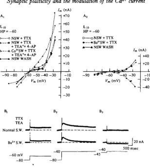
Fig. 7. Steady-state activation of Ca*+ and Cal+-dependent K+ currents. (A) Steady-state current-voltage curves of Lio. In graphs A, and A,, Lio was voltage clamped to a holding potential of —60 mV and 5 s clamp steps were delivered every 30 s to various hyperpolarized and depolarized membrane potentials. The current at the end of each 5 s voltage step was plotted against membrane voltage. In normal sea water and TTX the steady-state I-V curve shows a pronounced region of reduced positive slope in the range of the normal resting potential of Lio ( — 60 to —40 mV). In A[ application of TEA+ and 4-AP have little effect on the steady-state I-V curve since transient K+ channels normally inactivate during the prolonged (5 s) voltage steps. Replacement of extracellular Ca*"1" with 10 mM-Colt ions, however, linearizes the I—V curve by blocking a steady state inward Ca1"1" current. When normal sea water is washed back the normal steady-state I-V curve is restored. When, in A,, Ba'+ ions are substituted for Ca*+ ions, the normally observed area of steady-state reduced positive slope is converted into a region of inward-going rectification. Ba>+ carries the steady-state inward current through the Ca1+ channel, but does not activate an opposing CaI+-dependent current.
130 M. KLEIN, E. SHAPIRO AND E. R. KANDEL
Na* channel
i i
Vesicle
K* channel (open)
Ca2+ channel (closed)
Hyperpolarized
K+ channel (closed)
i
Ca2* channel
(open) Depolarized
Fig. 8. Diagrammatic summary of the effects of resting membrane potential on presynaptic currents and transmitter release. When the neurone is held at a hyperpolarized resting level two currents are altered. One, the steady-state calcium current is reduced and consequently the resting intracellular calcium concentration is reduced. As a result the influx of calcium that occurs with an action potential may not by itself be sufficient to cause maximal transmitter release, and in the limit, to cause any release. Second, the voltage-sensitive potassium currents are activated to a greater degree when an action potential occurs from a hyperpolariied than from a depolarized membrane potential. The resulting spike is decreased in both duration and amplitude and therefore allows a smaller calcium influx to occur. Thus, at depolarized mem-brane potential, increased steady-state calcium currents as well as potassium current inactiva-tion cause a larger amount of calcium to be available for transmitter release, resulting in increased synaptic transmission. Filled symbols represent closed channels and clear symbols represent open channels. Arrows from the K+ channels to the Cal + channels represent the flow of hyperpolarizing current (through the potassium channels) which closes open Ca1+ channels and prevents additional Cal + channels from opening. The filled dots ( # ) represent
the calcium concentration, which is increased at depolarized membrane potentials due to increased number of open calcium channels.
cellular Ca2+. Co2+ blocks this steady-state Ca2+ inward current as well as the Ca2+ -dependent K+ current (Fig. yAv A2).
Ba8+ flows through Ca2+ channels without activating the K+ conductance (see Figs. 6B, C and 7B and Adams et al. 1980). In the presence of Ba2+, a voltage-dependent steady-state inward current flows through the Ca2+ channels at depolarized levels of holding potential (Fig. "]AV 7B); this inward current is not present at hyperpolarized levels (Fig. 7B). To activate this K+ conductance the concentration of intracellular Ca^ in the steady state must be significantly larger at more de-polarized than at more hyperde-polarized membrane potentials.
Synaptic plasticity and the modulation of the Ca
2+current 131
t
sight and duration of the spike. In this way the transient Caa+ current can also be odulated, albeit indirectly. Our findings also suggest that despite a transient Ca2+ current that is not directly affected by changes in holding potential, powerful control over synaptic transmission - ranging from total block to enhanced effectiveness - can be achieved by variationsin the steady-state Ca2+ current (Fig. 8).These results support the idea first proposed by Shimahara & Tauc (1975) that EPSPs and IPSPs have a dual function. In addition to controlling the probability of firing an action potential, synaptic potentials also set the level for transmitter release. The results further suggest a possible mechanism for long-term regulation of synaptic output by metabolic or hormonal actions which affect resting potential (Thomas,
1972; Mayeri, Brownell & Branton, 1979; Mayeri et al. 1979).
Presynaptic inhibition
Presynaptic inhibition has been described in several vertebrate and invertebrate synapses and has been attributed to a depolarization of the synaptic terminals (re-viewed in Burke & Rudomin, 1977; Ryall, 1978). This idea seemed consistent with the release properties described at the squid giant synapse where depolarization reduces transmitter release (Miledi & Slater, 1966). However, as described above, in
Aplysia and in the leech, depolarization enhances rather than depresses transmitter
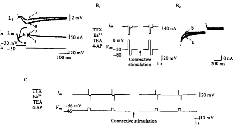
release (Shimahara & Tauc, 1975; Nicholls & Wallace, 1978 a; Shapiro et al. 1980a). At these synapses, a different mechanism must account for presynaptic inhibition. In Aplysia the synapses of cell Lio that are modulated by membrane potential can also be modulated by presynaptic inhibition. Stimulation of one of the fibre pathways to the abdominal ganglion causes a depression of both the monosynaptic excitatory and the inhibitory connexions made by Lio (Fig. 9; Waziri, Kandel & Frazier, 1969; Shapiro et al. 19806). Recently, Byrne (1980) has discovered a group of cells (the L32 cells) which, when stimulated, produce presynaptic inhibition without producing any conductance changes in the postsynaptic follower cells of Lio.
Each of the ways of producing presynaptic inhibition tends to produce an IPSP in the presynaptic neurone, Lio. Thus one change in the presynaptic neurone that clearly contributes to presynaptic inhibition is the hyperpolarization that occurs as a result of the IPSP. This hyperpolarization would reduce the output of transmitter by reducing the steady-state Ca2+ influx and by removing some of the inactivation of the K+ channels. However, presynaptic inhibition outlasts the hyperpolarization and can also be observed in cases in which the cell does not hyperpolarize. To determine whether additional mechanisms are operative we controlled for changes in membrane potential by examining the presynaptic neurone under voltage-clamp conditions.
Presynaptic inhibition can be observed under voltage-clamp conditions
In a solution containing TTX and TEA+, and with the cell held at a membrane potential which inactivates the early outward K+ current, the inward current produced by a depolarizing clamp step is carried by Ca8+ ions, and the residual outward current is mainly due to Ca2+-dependent K+ current and the leakage current. Under these circumstances presynaptic inhibition is correlated with a decrease in transient inward
evoked by the depolarizing command (Fig. 10).
132
A,
M. KLEIN, E. SHAPIRO AND E. R. KANDEL
RB 10mV
5mV
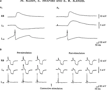
Pre-5 timulation Post-stimulation
RB
L,o A
J
Connective stimulation
J
_ l 2 0 m V 50 ms
10 mV
5mV
[image:16.451.55.409.49.352.2]_ | 2 0 m V 50 ms
Fig. 9. Presynaptic inhibition in Aplytia. (AJ Action potentials in cell L i o elicit, through different branches, EPSPs in some cells (RB) and IPSP in others (L3). (Aj) Cell L3 can be hyperpolarized to reverse the IPSP. (B) Action potentials are produced in cell L i o every 2 s and the excitatory and inhibitory PSPs produced by two of its branches are monitored. The input resistance is monitored by intracellular injection of hyperpolarizing pulses. Stimulation of the connective for 5 s causes a decrease in PSP size not correlated with changes in post-synaptic input resistance. Solution containing 265 mM-Na1+, 60 mM-Ca8+, and 140 mM-Mg1+.
(From Shapiro et al. 19806.)
TTX
TEA Pre-stimulation Post-stimulation
Post
Pre
Connective stimulation
[image:16.451.61.408.454.582.2]Synoptic plasticity and the modulation of the Ca
2+current 133
B, B ,
r m _50
120 mV
1 0 0 m s
°U Connective" j 2 0 m V | 8 nA stimulation ' » 200 ms
1 I J\ I I I I I ,
Ba" ' " ^ L, 1^ L, |20mV TEA
4-AP
At. ' I ' 1. .
[image:17.451.42.417.69.270.2]„ * —110 mV Connective stimulation j s
Fig. 11. Stimulation of presynaptic inhibitory pathway directly reduces current through the Ca'+ channel.
(A) Cell Lio voltage clamped in normal sea water containing TTX and TEA. The depolar-izing command step is from — 50 to — 30 mV. Superimposed currents before (a) and after (6) connective stimulation. Presynaptic inhibition is correlated with reduced CaI+ current and unchanged K.+ current. PSP was reduced by presynaptic inhibition from 22 to 12 mV. The tail-current upon repolarization of Lio to the holding potential is less inward after connective stimulation than prior to connective stimulation. (B^ Lio voltage clamped in high-Ba1+ sea water containing TTX (30 /tM), TEA (25 mM), and 4-AP (10 ITIM). A depolarizing step from — 50 mV to zero mV elicits large inward current. When the cell is repolarized to — 80 mV (the approximate level of EK as determined by the reversal potential of the fast transient outward
current) there is an inward tail current. Stimulation of the presynaptic inhibitory pathway (for 5 s) causes a reduction in the inward net current as well as in the inward tail current upon repolarization to — 80 mV. The PSP was not monitored in this experiment.
(Bt) Same experiment as Bt to show at higher gain and faster sweep speed the tail currents
at EK (— 80 mV) superimposed before (a) and after (6) connective stimulation.
(C) Stimulation of presynaptic inhibitory pathway reduces steady-state inward currents. Cell Lio voltage clamped in high-Ba1+ sea water containing TTX (30 /*M), TEA (25 mM), and 4-AP (1 o mM). A small depolarizing voltage step produces a steady-state inward current. Stimula-tion of presynaptic inhibitory pathway (for 5 s) reduces this inward current. (From Shapiro
et at. 19806.)
be the result of an increase in small residual outward K+ current, or a decrease in the inward Ca2+ current. The observation that the decrease in early inward current during presynaptic inhibition is larger than the increase in late outward and leakage currents (Fig. 10, Fig. 11 A) suggests that the Ca2+ current may be modulated directly. But to distinguish between these alternative possibilities directly we performed two types of experiments: pharmacological block of all the K+ channels, and an examin-ation of the voltage dependence of the presynaptic action.
The inward current decreases even in the presence of K+ channel blockers
M. KLEIN, E. SHAPIRO AND E. R. KANDEL
TTX BaJ+ TEA
4 .A P - 3 0 raV
- 5 0 - 7 0
Y
I 1 s
40 nA
50 mV - 7 0 - 9 0
Jl
Connective stimulation
Fig. 12. The conductance changes caused by stimulation of presynaptic inhibitory pathway are voltage sensitive* Cell Lio is voltage clamped in sea water containing Be.**, TTX (30/*M), TEA (25 miu), and 4-AP (10 rrtM). Under these pharmacologic conditions 30 min intervals were used between runs. (A) From a holding potential of — 50 mV, alternating 20 mV depolarizing and hyperpolarizing voltage steps elicit inward currents. Connective stimulation markedly reduces inward Bai+ current through the Ca1+ channel of the depolarizing step, and only slightly increase leakage current of the hyperpolarizing step. (B) From a holding potential of —70 mV, alternating 20 mV depolarizing and hyperpolarizing voltage steps again elicit in-ward current with hyperpolarizing step but elicit primarily outin-ward leakage currents with depolarizing steps. In this Voltage range depolarizing command pulses do not significantly activate inward currents. Only small leakage currents are produced by both hyperpolarizing and depolarizing steps. Connective stimulation does not affect these currents. (From Shapiro et al.
19806.)
In addition, changes in the steady-state inward current also occurred with connective stimulation (Fig. 11 C). These results imply that the decrease in inward current reflects a decrease in the transient and in the steady-state Ca2+ current during pre-synaptic inhibition.
Presynaptic actions occur only in the voltage range where the calcium current is activated
Synoptic plasticity and the modulation of the Ca
2+current 135
Na* channel
1
CaJt channel I
(open)
Ca2* channel
(closed) "
-Vesicle
K* channel
(open)
0
0 0
Mr
0
0
0 0
0 0
0
0
0
0
- 1 0
0
0
0 0
0
0
0
0
0
Control Preiynaptic inhibition
Fig. 13. Diagram of mechanism of presynaptic inhibition. An unidentified presynaptic inhib-itory transmitter directly reduces both steady-state and transient calcium currents, making less calcium available for transmitter release A specific cell (L32) has recently been found to mediate this presynaptic inhibition (Byrne, 1980). (See legend Fig. 8 for key.)
Ca2+ channel (Fig. 13). A similar mechanism for presynaptic inhibition has been discovered in dissociated dorsal root ganglion cells by Dunlap & Fischbach (1978) and by Mudge, Leeman & Fischbach (1979).
In vertebrate heart muscle Giles & Noble (1976) have found that ACh, acting on mu8carinic receptors in the heart, decreases the Ca2+ current in the heart muscle. This action of ACh is similar to the mechanism of presynaptic inhibition in Aplysia and in dissociated dorsal root ganglion cells. Reuter (1979; Reuter & Scholz, 1977) has suggested that cGMP, or the ratio of cAMP/cGMP in heart muscle, may control the maximum calcium conductance of the muscle. An alternative possibility (Fig. 27) is that the modulatory transmitter closes a Ca2+ channel-receptor complex directly without the mediation of an intracellular second messenger.
In mammals, presynaptic inhibition has been correlated with presynaptic depolar-ization and with increased excitability in afferent fibres. The observed conductance change at some of these synapses follows the chloride Nernst potential (reviewed in Burke & Rudomin, 1977). However, as indicated above, results similar to those reported here have been obtained by Fischbach and his colleagues (Dunlap & Fisch-bach, 1978; Mudge et al. 1979) on chick dorsal root ganglia. Whether the presynaptic depolarization and the increased Cl~ conductance in vertebrates are epiphenomena, that are not directly related to the mechanism of presynaptic inhibition, and are due only to the artificial modes of activation, or whether there are several distinct mech-anisms for presynaptic inhibition, needs now to be determined (for a critical review
136 M. KLEIN, E. SHAPIRO AND E. R. KANDEL
S.N.-Stimulus 1
Stimulus I
|2mV
_)20mV 58 ms
Fig. 14. Homosynaptic depression accompanying habituation in the gill-withdrawal reflex. (A) Diagram of arrangement for recording monosynaptic EPSP from sensory neurone to gill motor neurone in abdominal ganglion of Aplysia. (B) Homosynaptic depression of the monosynaptic sensory-to-motor EPSP. Action potentials evoked in a sensory neurone give rise to an EPSP which undergoes depression when elicited repeatedly every 109 (upper part). Lower part shows partial recovery after rest, and a further decrease in EPSP size with contin-ued stimulation. (From Castellucci & Kandel, 1974.)
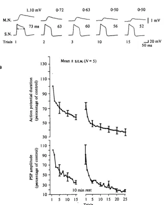
Homosynaptic depression
Homosynaptic depression - a self-induced depression in excitatory transmission at the synapses made by the sensory neurones on their central target cells - is the mechanism for short-term habituation of the siphon and gill-withdrawal reflexes in
Aplysia (Fig. 14; Castellucci et al. 1970). This depression, which is very profound
and rapid at these sensory neurone synapses, is due to a progressive decrease in the amount of transmitter released by each action potential (Castellucci & Kandel,
1974)-The depression is correlated with a decrease in the calcium component of the action potential in TEA
Synaptic plasticity and the modulation of tlie Ca
2+current 137
A Sea water+ T.E.A.
M.N.
S.N. Trials 1
l.lOmV 0-72
73 ms 63
0-63
60
0-50 0-50
52 I 1 mV
10 15 _ l 2 0 m V 50 ms
130-g _ 110 H
1 ! 90^
O ^c
I 2
I 2
•3 • V 70 H
50
3 0
-110
£ o o, o 70
-f
501 3 0
-10
M e a n t S.E.M.(A'=5)
10 min rest
[image:21.451.62.384.85.485.2]1 5 10 15 1 5 10 15 20 25 Trials
Fig. is- Change in Ca1+ current during homo8ynaptic depression. (A) Correlation between sensory neurone action potential in sea water containing ioomM-TEAand monosynaptic EPSP with repeated stimulation. Action potentials fired at O'i HZ. (B) Average action potential duration and EPSP amplitude (based on 5 preparations) during a first habituation run, after a 10 min rest, and during a second run. Spikes evoked at o-i Hz in sea water containing TEA.
138 M. KLEIN, E. SHAPIRO AND E. R. KANDEL
be abolished with cobalt or nickel ions (Klein & Kandel, 1978), which block the calciu channels of other neurones (Adams et al. 1980).
The duration of the action potential in TEA is a sensitive measure of the calcium current because it is determined by the balance between the inward calcium current and the outward potassium and leakage currents. As long as the calcium current is at least equal to the outward currents (the leakage and the K+ currents) the plateau is maintained; when the outward currents outweigh the inward current the membrane repolarizes. The total calcium influx during the plateau phase can therefore be regulated both by intrinsic changes in the calcium channels as well as by changes in potassium or leakage currents. Hence, changes in the duration of the TEA spike are a good indication of changes in total Ca2+ influx, but they do not indicate whether the changes result from a direct action on the calcium channel or whether the influx is affected indirectly by a potassium conductance not blocked by TEA. In addition, some part of the plateau also represents firing of other parts of the neurone outside the cell body. Calcium spikes in the axon and terminal regions can contribute to the duration of the TEA action potential recorded in the cell body, except, of course, when the cell body is isolated from its processes by tying it off, for example.
We next examined transmitter release from the terminals of the sensory neurones, as measured by the size of the synaptic potential in postsynaptic neurones, and the simultaneous change in calcium current, as measured by changes in the duration of the action potential (Fig. 15). Repeated stimulation of the sensory neurone at rates that produce habituation led to a progressive decrease in the PSP (with kinetics similar to what is observed in normal sea water) together with a reduction in the duration of the TEA action potential, indicating a progressive decrease in the calcium current of the action potential. Spontaneous recovery of synaptic transmission with rest was accom-panied by an increase in the calcium current.
Depression parallels the decrease in the Cai+ current
To determine whether the decrease in Ca2+ current was due to a direct action on the Caa+ current or an action on a K+ current, we voltage clamped the cell body of the sensory neurone. In normal sea water, depolarizing commands elicit EPSPs in the follower cells and an inward current in the sensory neurone which is due largely to Na+ and a lesser degree to Ca2+, followed by an outward current (Fig. 16 A). With repeated depolarizing commands repeated at every 10 sec the EPSPs decrease in amplitude but there is no change in the presynaptic current. This is consistent with the lack of a change in the action potential in normal sea water in the absence of >Ja+ or K+ channel-blocking agents.
Synaptic plasticity and the modulation of the Ca
2+current 139
Sea water
3mV
10 nA
Trials 1 10 10 ms
450 HIM TEA 50 nw Na+ 30 IM TTX
M.N.
S.N.
,+20 mV - 4 0 Trials 1
5mV
J 2 n A 50 ms
10 15
450 mM TEA 11 mM Ba1*
10mMl4-AP OCa5*
ONa* Isolated
cell body + 10mV -40
[image:23.451.45.393.59.406.2]Trials 1 15
Fig. 16. Homosynnptic depression and decline in calciurn current under voyage clamp. (A) In normal sea water the inward current is due to sodium and calcium. The presynaptic voltage clamp pulses elicit EPSPs that undergo depression with repetition of the depolarizing command. The decrease in EPSP is not accompanied by any change in either the outward K+ or the inward Na+ current. (B) With the sodium current blocked with T T X and a large
part of the potassium currents blocked with TEA presynaptic voltage-clamp pulses elicit EPSPs that undergo depression with repetition of the depolarizing command. The decrease in EPSP is paralleled by a decrease in the inward current. (C) In the absence of Na+ ions with
virtually all K+ currents blocked (by bathing in a high concentration of 4-AP in addition to
TEA and Ba14) and with voltage-clamp control maximized by ligating the sensory neurone cell body, inward current decreases with o-i Hz stimulation, indicating that current moving through the calcium channels undergoes a progressive decline independent of changes in Na+ or K+ currents.
neurone, including the fine terminal processes of the neurone where transmitter release occurs. As a result, the interpretation of the decline in inward current is not unambiguous. It is possible, for example, that the threshold for firing calcium spikes in fine processes increases during habituation, resulting in transmitter release from fewer terminals. Alternatively (or in addition), the decline in calcium current could occur in a graded manner throughout all parts of the neurone, although perhaps not equally everywhere, so that progressively less transmitter is released from each
ter-•
140
A 10
a.
M. KLEIN, E. SHAPIRO AND E. R. KANDEL
B
10
10
8
6
4
2
n
-m ••
1
•
•
i i i
1 5
Fig. 17. Correlation of the amplitude of the monosynaptic EPSP produced in the follower cell by the sensory neurone and presynaptic inward current. (A) Two consecutive habituation training sessions were separated by a 5 min period of rest. The sensory neurones were voltage clamped in artificial sea water containing Na+ and K+ current blockers, and EPSPs were elicited by 50 ms depolarizing steps every 10 s. (B) The amplitude of the EPSP in the follower cell is plotted as a function of the peak inward CaI+ current.
the EPSP. The parallel decline of the PSP and of the inward current therefore appears to result from an intrinsic change in the calcium current.
To examine the inward current in a situation where the cell body is optimally space-clamped so that uncontrolled responses are eliminated we isolated the cell body of the sensory neurone by tying it off. We next blocked all the K+ currents by substituting Ba2+ for Ca2+ and using 4-AP in addition to high concentrations of TEA. Under these circumstances we still found a progressive decrease in the inward currents with repeated depolarizing commands (Fig. 16C).
Synoptic plasticity and the modulation of the Ca
2+current 141
Na+ channel Ca2* channel I
(open)
CaJ* channel
(closed) —
Vesicle
K+ channel
(open)
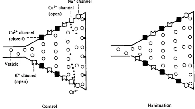
[image:25.451.65.382.80.257.2]Control Habitation
Fig. 18. A schematic model of habituation. With repeated activation, calcium channels undergo inactivation of calcium inflow, either as a direct effect of depolarization, or as a result of some other process. Arrows from potassium to calcium channels represent the flow of hyperpolariz-ing current (through the potassium channels) which repolarizes the action potential and thereby closes open calcium channels and prevents new ones from opening (see legend of Fig. 8 for key).
decreases with repeated depolarization, whereas the depression of the PSP is unaffected
(see, for example, Fig. 22 B, left half of figure).
The term ' inactivation' has been applied to a decrease in an ionic current elicited at a constant membrane voltage that is not due to an increase in an opposing current or to a change in driving force. We have provided evidence that the decrease in inward current is not the result of the activation of a residual K+ current (Fig. 16 C). It is also unlikely that the reduction in inward current is due to a change in driving force (resulting from an accumulation of divalent cations in the cell or near the inner surface of the membrane) since we have found that while the transient inward current de-creases with repeated depolarizing command pulses of 60 to 70 mV (designed to simulate the action potential), the steady-state current through the Ca8+ channel (elicited with small voltage-clamp steps) does not decrease. A change in driving force should affect both transient and steady-state components of the current. Thus, the homosynaptic depression that underlies habituation is correlated with a prolonged inactivation in the transient inward current resulting from its repeated activation (Fig. 18).
142 M. KLEIN, E. SHAPIRO AND E. R. KANDEL
L. connective
Siphon N.
Depression 0 min lOmin 20 min 30 min 50 min
M.N.
S.N.
i(\ - ft ft— A ft—
| 2 m VS.N.
Facilitation 10 s SOs 10 min 30 min 50 min
M.N.
S.N.
J 2 0 m V 50 ms
| 5 m V
5mV
A_
20 mV2mV50 ms
C,
140%
Connective 100% I
1
148%
5-HT 100% Jf
Synaptic plasticity and the modulation of the Ca
2+current 143
the charge carrier. In addition, the time course of recovery from inactivation is nuch slower in the sensory neurones than in cells R2 and R15. It would therefore be of interest to explore the specific mechanisms that underlie the prolonged Ca2* current inactivation in the sensory neurones.
Presynaptic facilitation
As is often the case with behavioural habituation, reflexes that illustrate this form of learning also show an opposite learning process: sensitization. Presynaptic facili-tation was first encountered while trying to develop a cellular analogue for reflex sensiti-zation, a simple form of learning which involves the enhancement of a reflex response by a strong stimulus (Kandel & Tauc, 19650,6). This process was subsequently shown to be actually utilized in behavioural sensitization of the gill-withdrawal reflex (Kupfermann et al. 1970).
As we have seen, repeated sensory stimulation at rates that produce habituation in the intact animal produces a depression in the monosynaptic excitatory connexions between sensory neurones and their followers due to a decrease in the number of transmitter quanta released per impulse (Castellucci & Kandel, 1974). On the other hand, stimulation of a different pathway for a few seconds causes an increase in transmitter release from the sensory neurones (Castellucci & Kandel, 1976 and Fig. 19). This presynaptic facilitation is simulated by cyclic AMP and by serotonin, but not by a number of other candidate transmitter substances that were examined. A sensitizing stimulus produces two actions on the sensory neurones: (1) a slow EPSP thatlastsupto half an hour (Klein & Kandel, 1978), and (2) a similarly long facilitation of synaptic transmission at the sensory-to-motor synapses (Fig. 19; Castellucci et al. 1970). To determine what was responsible for the increase in transmitter release, we examined the slow EPSP and other changes in the membrane properties of the sensory neurone cell body that accompany presynaptic facilitation.
The slow EPSP is due to a decrease in the conductance to K+
We first examined the changes in the membrane conductance produced in the presynaptic neurone by the slow EPSP. Using electrotonic potentials produced by
Fig. 19. Synaptic facilitation at the synapse between mechanoreceptor neurones and motor neurones. (A) Ventral aspect of the abdominal ganglion of Aplysia, illustrating simultaneous recording from gill motor neurone L7 and a mechanoreceptor sensory neurone (SN). Stimula-tion of the left connective (LC), which carries informaStimula-tion from the head to the abdominal ganglion, is used as the facilitating stimulus. (B) Depression and subsequent facilitation of the monosynaptic EPSP after a strong stimulus. Top two series of traces illustrate selected records of depression of the EPSP in the motor cell (MN) during a series of 50 min of consecutive intracellular stimuli to the sensory neurone (SN) (every 10 s) prior to the presentation of the facilitating stimulus. Middle two traces illustrate the effects of the facilitating stimulus (a train of shocks to the LC at 6 Hz for 10 9). Arrows indicate the last EPSP before the facilitating stimulus and the first EPSP after the stimulus. Bottom series of two traces illustrate the gradual decline of the facilitation over the subsequent 50 min, during which the sensory neurone was again stimulated once every 10 s. The middle two traces are at lower gain and at a slower time scale. (From Castellucci & Kandel, 1976.) (C) Effects of connective stimulation and 5-HT on sensory neurone membrane potential and resistance. (CJ Connective stimulation caused a depolarization and sin increase (40%) in membrane resistance as measured with electrotonic potentials elicited by constant-current pulses applied through the second barrel of a double-barrelled microelectrode. (Ct) Incubation with 5-HT also produces a long-lasting
144 M. KLEIN, E. SHAPIRO AND E. R. KANDEL
constant current pulses in the sensory neurones, we found that the slow EPSP was accompanied by an apparent decrease in the conductance of the membrane and that this decrease was simulated by serotonin, the putative facilitating transmitter, by cyclic AMP, and by phosphodiesterase inhibitors (Klein & Kandel, 1978). This was confirmed with voltage-clamp experiments. Stimulating the facilitatory pathway or incubating the ganglion or the isolated cell body with serotonin under voltage clamp caused an inward shift in the holding current and a decrease in the ' leakage' currents elicited by small step hyperpolarizations. Moverover, the slow EPSP and the inward shift in the holding current produced by connective stimultion or by serotonin were blocked by K+ channel-blocking agents (compare Fig. 22 Ct and Ca), and by replacing intracellular K+ in the sensory neurones with Cs+, an impermeant cation. These results imply that this EPSP is due to a decrease in K+ conductance. The EPSP is voltage dependent. Current-voltage curves are highly non-linear. At levels of membrane potentials more depolarized than — 50 mV the non-linearity is reduced by connective stimulation and serotonin but at more hyperpolarized potentials than
— 60 mV synaptic stimulation or serotonin gave rise to little or no synaptic current. By contrast, when the external K+ concentration was doubled, the current reversed at about — 50 mV. The association of a decrease in a voltage-sensitive K+ con-ductance with presynaptic facilitation suggested that the facilitation might occur through the reduction of a presynaptic K+ current that is activated by the action potential, thereby leading to an increase in the duration of the action potential and increased transmitter release. We therefore examined the presynaptic action potential and the currents activated by them under a variety of conditions to see whether or not a reduction in the K+ current of the presynaptic action potential could account for presynaptic facilitation.
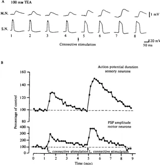
Presynaptic facilitation produces an increase in the calcium current of the action potential When we bathed the abdominal ganglion in TEA and examined action potentials in the sensory neurones, we found that they were prolonged by stimulating the connective, indicating an increase in the Ca2+ current. As with homosynaptic depression, the changes in the duration of the action potential as a result of presynaptic facilitation paralleled the changes in transmitter release (Fig. 20). A similar prolongation of the action potential was also produced by serotonin, by cAMP, and by phosphodiesterase inhibitors (Klein & Kandel, 1978).
Calcium current prolongation is due to a depression of the K+ current
The increase in Ca2+ current could be produced by a direct action on the Caa+ channel or by an indirect action by means of a reduction in an opposing K+ current. To distinguish between direct and indirect actions on the Caa+ current, we voltage clamped the cell body of the sensory neurone and analysed the changes in ionic currents produced by connective stimulation and by serotonin, the presumed facili-tating transmitter.
Synaptic plasticity and the modulation of the Ca
i+current 145
A 100 mM TEA
M.N.
S.N.
Connective stimulation
1 mV
_ J 2 0 m V 50 ms
I6O-1
140-S
120-8
"o
100-80-1
I
£ 400-1
300-
200-
1000
-Action potential duration sensory neurone
PSP amplitude motor neurone
L connective stimulation!,, connective stimulation 4 5 6
Time (min)
[image:29.451.64.394.82.421.2]8
Fig. 20. Increase in Ca1+ current that parallels presynaptic facilitation. (A) Broadening of the action potential in TEA accompanying presynaptic facilitation of monosynaptic sensory-to-motor EPSP. (B) Graph of EPSP amplitude and presynaptic spike duration during an experi-ment in which the facilitating pathway was first stimulated weakly, and then more strongly. In both A and B spikes were evoked at o-i Hz in ioo mM-TEA sea water. (From Klein and Kandel, 1978.)
of drugs the finding made in TEA solution that connective stimulation can alter the configuration of the currents contributing to the action potential. To analyse this change in the presynaptic current, we isolated the cell body to maximize voltage-clamp control, using serotonin to simulate the presynaptic facilitation. In sea water con-taining no drugs, serotonin caused a decrease in transient outward currents elicited with depolarizing steps, as well as the decrease in resting conductance (Fig. 22 B, Cx). However, when potassium currents were blocked with TEA and 4-AP, serotonin had no effect on any membrane conductance (Fig. 22 A, C2). The effects of serotonin were blocked with either Ca2+ or Ba2+ as the divalent cation that was carrying current through the Ca2+ channels.
146 M. KLEIN, E. SHAPIRO AND E. R. KANDEL
Post
Trials 1 I
Connective stimulation , Control (1)
-Depressed (15) Facilitated (20)
25 31
5 msec
10 nA
5 msec
Fig. 21. Decrease in outward current parallels presynaptic facilitation. (A) In normal sea water depolarizing commands in the sensory neurones produce inward and outward currents in the sensory neurone and EPSPs in the motor cells. The cell was stimulated at one per 10 s to simulate habituation and the repeated commands led to a depression of the EPSP. Connec-tive stimulation produced a decrease in the outward current and a parallel facilitation of the EPSP. (B) Three traces from Part A arc superimposed to illustrate the lack of significant change in the outward currents with the repeated depolarizing commands designed to simulate habituation (traces 1 and 15) and the depression of the outward current following stimulation of the connective designed to simulate sensitization (trace 20).
current in a different way. We substituted the non-permeant cation Cs+ for the intra-cellular K+ using the antibiotic nystatin, which makes the membrane permeable to monovalent cations (Russell, Eaton & Brodwick, 1977). We then washed out the nystatin and recorded from the cell in K+-free solution with electrodes filled with CsCl. Under these circumstances, serotonin produced no modulation of the current. By contrast, when a small amount of K+ was reintroduced into the cell with a micro-electrode filled with KC1, the net current became outward, and now was again affected by serotonin (Fig. 23). These experiments imply that serotonin, and presumably presynaptic facilitation, act on K+ currents, and have no significant direct action on the calcium channel.
Synaptic plasticity and the modulation of the Ca
2+current 147
450 mw TEA lm
11 mM BaJ*
10 mM 4AP 0Ca5+ ONa*
Vm + 10 mV
- 4 0 Trials 1
"Irlri/Irtr trirtriftr
5 10 12 15 16 20 25 29
B
Normal ASW +15 mV
Trials 1 2 5 10 12 15 20 23 25 30
Serotonin
C,
Normal ASW - 5 0 mV
- 7 0
nA450mMTEA 100ms lOrnu 4 AP
11 m« 0 Na*
v
m- S O r n V . - 7 0
_|0-5nA 100 ms
Control Serotonin Control Serotonin Fig. 22. Changes in presynaptic currents of the cell body of sensory neurones isolated by ligation, with stimuli that produce habituation and sensitizntion. Parts A and B are from the same cell examined first with all the K+ channels blocked and then in normal sea water. (A) With all K+ currents blocked, a step depolarization every io s (bottom records) still cause gradual inactivation of inward current (top records); however, 5-HT produces no effect. (B) When the same cell as in part A was placed in normal sea water after washing out the K+ channel blocking agents a step depolarization every 10 s (bottom records) again causes gradual inactivation of inward current (top records). But now 5-HT again produces an increase in the transient inward current and a depression of the outward current. In addition the holding current becomes more inward and the leakage current is reduced. (C) Holding and leakage current (produced by a small hyperpolarizing command) in normal sea water (C,) and in K+ channel-blocking solution (Ct). Ci. In normal sea water, 5-HT causes the holding current to
become more inward and the leakage to decrease (C,). In sea water containing K+ blockers both effects of serotonin are abolished.
Presynaptic facilitation produces an increase in duration of the action potential
148 M. KLEIN, E. SHAPIRO AND E. R. KANDEL
Nystatin-Na+,
K+-free
5 mu M
+ 10 mV
- 4 5
nA
+ 10
- 4 5
_ | 2 5 m V 25 ms
1
Nystatin - Nat K*-free Nystatin-Na* K*-free KC1 electrode Control 5-HT
5-HT, Control
,,M
5 msJO-5 nA 10ms_J 1 nA+ 15 mV
-45
+15
-45
Fig. 23. Effect of serotonin on calcium current after exchange of intracellular K+ for Cs+.
The ganglion was first bathed in a solution containing the antibiotic nystatin, which greatly increases membrane permeability for monovalent cations. K+ and Na+ ions were washed out and replaced with Cs+ ions. After ion substitution, nystatin was washed out, and a test
solution containing only Ca*+, Mg*+, Cl~ and TRIS ions was used. Cells were impaled with 3M-CsCl electrodes. (A) Aj. The inward current after nystatin ion exchange is due to Ca1+ and there are no significant outward currents. Calcium current after Cs+ substitution. (A,.)
Synoptic plasticity and the modulation of the Ca
2+current 149
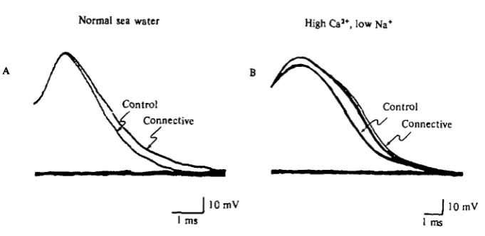
Normal sea water High Ca5*, low Na+
Control \ \ C o n t r o 1
[image:33.451.56.397.63.229.2]Connective W ^ Connective
|lOmV |lOmV 1 ms 1 ms
Fig. 24. Presynaptic facilitation produces action potential broadening in solutions containing no K+ blocking agents. (A) Normal sea water. (B) Sea water containing 0 5 x sodium,
5-5 x calcium, 2 x magnesium.
Prolongation of the depolarizing command enhances transmitter release
To strengthen the conclusion that increased transmitter release during sensitization is caused by broadening of the action potential, we next increased the duration of the presynaptic depolarization under voltage clamp in an attempt to simulate the changes that occur in the action potential. We clamped a sensory neurone in the presence of Na+ and K+ channel-blocking agents and elicited an EPSP in a follower cell with a depolarizing command step. When we increased the duration of a 20 msec depolariz-ing step by 25%, the EPSP doubled in amplitude (Fig. 25). This occurs because the Ca2+ current has a very long rise time (approximately 10-30 msec) compared to the duration of the step, and, in this cell, is still increasing at the end of the 20 msec command. Lengthening the command by 5 msec increases appreciably the calcium influx (measured by the area under the current). If the depolarization were as short as an action potential (approximately 2 msec at half amplitude), an even smaller percentage increase in duration of the spike might increase the EPSP significantly. The observed changes in action potential duration could therefore cause the increase in transmitter release that underlies sensitization.
Presynaptic facilitation is accompanied by a change in the shape of the EPSP
If increased release were a result of broadening of the presynaptic action potential, the Ca2+ current would be activated for a longer time, and we might expect to see a corresponding change in the configuration of the EPSP. When we superimposed the two EPSPs of Fig. 25 produced by command pulses of different duration, adjusting for the amplitude difference, we found, not surprisingly, that the PSP elicited with the 25 msec depolarization reached its peak later than the PSP elicited with a 20 msec step (Fig. 25 C). Similarly, the PSPs produced by action potentials after a sensitizing stimulus show longer rise times compared to the PSPs elicited prior to sensitization. These changes in shape are qualitatively similar to those produced by increasing the duration of a voltage-clamp command (Fig. 26).
M. KLEIN, E. SHAPIRO AND E. R. KANDEL
450 mu TEA 50 mil Na+
3 0 MM TTX Post
Pre
20 ms 25 ms
1 mV
| 2 n A
J 20 mV 25 ms
25 ms
25 ms
Fig. 25. Increasing the duration of the presynaptic command pulse increases the amplitude of the EPSP. (A) A 20 ms voltage-clamp pulse gave rise to an EPSP (top) approximately 1 mV in amplitude. (B) When the step was prolonged to 25 ms, the EPSP doubled in size. Inset shows superimposition of the voltage records. (C) Superimposition of the EPSPs in Parts A and B to illustiate the change in shape of the facilitated EPSPs. The EPSPs were scaled so as to match their peak amplitudes.
and after facilitation. The changes in PSP amplitude produced by habituation and spontaneous recovery from habituation do not affect the shape of the synaptic potential (Fig. 26). Similarly, changes in PSP amplitude produced by altering the calcium and magnesium concentrations do not produce shape changes (Castellucci & Kandel, 1974). On the other hand, when the PSP is increased by adding TEA to the bathing solution, blocking K+ current, the increase in PSP amplitude is accompanied by a prolongation of its rise time, similar to that seen in presynaptic facilitation (Castellucci & Kandel, 1974). These findings strengthen our earlier conclusion that habituation is caused directly by an intrinsic decline in calcium current rather than increased potas-sium current, and that habituation and sensitization reflect different modes of calcium current modulation.
Synoptic plasticity and the modulation of the Ca
z+current 151
Normal ASW
Initial habituation Steady state habituation Facilitation
h
PSP
Average
[image:35.451.40.413.95.333.2]10 mi
Fig. 26. Comparison of EPSP shapes after habituation and sensitization. Monosynaptic EPSPs were evoked with sensory neurone action potentials fired once every 10 s in normal sea water. Blocks of five EPSPs at the beginning and end of the habituation run and after connective stim-ulation (uppermost traces) were averaged using a signal averager (middle traces). The averages were scaled so that their peak amplitudes were the same and were then superimposed in pairs (bottom traces). The traces at the bottom left show superimposition of the averages taken from the beginning and end of the habituation run. Note that despite the fact that the EPSP has decremented considerably during the run, its shape has remained constant. The traces on the bottom right show the superimposition of the averages of the habituated EPSP and the facilit