REVIEW
PKM2, function and expression
and regulation
Ze Zhang
1, Xinyue Deng
2, Yuanda Liu
3, Yahui Liu
1*, Liankun Sun
2*and Fangfang Chen
4*Abstract
Pyruvate kinase (PK), as one of the key enzymes for glycolysis, can encode four different subtypes from two groups of genes, although the M2 subtype PKM2 is expressed mainly during embryonic development in normal humans, and is closely related to tissue repair and regeneration, with the deepening of research, the role of PKM2 in tumor tissue has received increasing attention. PKM2 can be aggregated into tetrameric and dimeric forms, PKM2 in the dimer state can enter the nuclear to regulate gene expression, the transformation between them can play an important role in tumor cell energy supply, epithelial–mesenchymal transition (EMT), invasion and metastasis and cell proliferation. We will use the switching effect of PKM2 in glucose metabolism as the entry point to expand and enrich the Warburg effect. In addition, PKM2 can also regulate each other with various proteins by phosphorylation, acetylation and other modifications, mediate the different intracellular localization of PKM2 and then exert specific biological functions. In this paper, we will illustrate each of these points.
Keywords: Cancer metabolism, Glycolysis, Pyruvate kinase, Warburg effect
© The Author(s) 2019. This article is distributed under the terms of the Creative Commons Attribution 4.0 International License (http://creat iveco mmons .org/licen ses/by/4.0/), which permits unrestricted use, distribution, and reproduction in any medium, provided you give appropriate credit to the original author(s) and the source, provide a link to the Creative Commons license, and indicate if changes were made. The Creative Commons Public Domain Dedication waiver (http://creat iveco mmons .org/ publi cdoma in/zero/1.0/) applies to the data made available in this article, unless otherwise stated.
Introduction
At the beginning of the twentieth century, German sci-entist Warburg discovered that in tumor tissues, even if oxygen is sufficient, malignant tumor cells still undergo active glucose glycolysis, the metabolic characteristic of this aerobic glycolysis is called Warburg effect [1]. And it is characterized by high glucose uptake rate, active gly-colysis, and decrease of local microenvironment pH [2]. Pyruvate kinase (PK), as one of the key enzymes of gly-colysis, acts on its substrate phosphoenolpyruvate (PEP) to form pyruvate [3], and pyruvate kinase (PK) has four different subtypes: L, R, M1, M2 [4]. PKL isoforms are mainly found in liver, kidney and red blood cells; while PKR is mainly expressed in red blood cells, biological activity is not clear; PKM1 is distributed in myocardium, skeletal muscle and brain tissue; PKM2 is distributed in
tissues such as brain and liver [5]. Although the PKM2 tetramer and dimer are composed of the same mono-mer [6], the biological effects between the tetramer and the dimer are significantly different [7]. The tetramer mainly plays the role of pyruvate kinase and regulates the glycolysis and the dimer PKM2 in the context of glucose metabolism can be used as a switch for energy metabolism and material synthesis [8], routing glucose metabolism to pyruvate into the tricarboxylic acid cycle, converting to the pentose phosphate pathway, the uronic acid pathway, and the polyol pathway. In turn, a carbon source and a oxidation–reduction (REDOX) equivalent are provided for quinochrome ribose anabolism and non-essential amino acid anabolism [9, 10]. If in the context of non-glucose metabolism, after the tetramer is converted into a dimer, PKM2 can exist in a variety of different intracellular localizations, enter the nuclear to regulate gene expression, and attaches to the mitochondrial outer membrane to maintain mitochondrial function and localizes to the endoplasmic reticulum to inhibit endo-plasmic reticulum stress [11]. Once again, PKM2 can also be modified with phosphorylation, acetylation and other proteins to regulate protein activity and intracellu-lar localization. PKM2 can increase or even replace the
Open Access
*Correspondence: [email protected]; [email protected]; [email protected]
1 Department of General Surgery, The First Hospital of Jilin University, Changchun 130021, China
2 Department of Pathophysiology, College of Basic Medical Sciences, Jilin University, Changchun 130021, China
original PK form regardless of the tissue-derived cells. Therefore, some researchers refer to PKM2 as tumor-specific PK [12].
PKM2 dimer, tetramer and glucose metabolism Glucose is the main energy supply substance in nor-mal tissues, under the condition of sufficient oxygen, glucose undergoes biological processes such as gly-colysis, tricarboxylic acid cycle (TCA) and oxidative phosphorylation (OXPHOS) to completely decompose glucose into carbon dioxide and water, and when con-sumes oxygen, the cell itself is supplied with a large amount of ATP at the same time [13]. Warburg found that abnormal glucose metabolism is an important feature of tumor cells, that is, tumor cells under the conditions of oxygen enrichment, but with a less effi-cient aerobic glycolysis, and then proposed the famous “Warburg effect”, the tumor supplies energy through a low-efficiency ATP production process that uses glu-cose uptake for aerobic glycolysis [14]. Although the Warburg effect has been practiced for nearly a century, with the deepening of research on glucose metabolism in tumor cells, it has been found that although there is indeed high consumption of glucose in tumor tissues, there is a proportional difference between glucose con-sumption and ATP supply, the production of ATP is more than the corresponding amount of ATP produced by glucose aerobic glycolysis [15], therefore, after years of academic debate and continuous improvement of the second stage of Warburg effect, the speculation that most scholars can accept at this stage is that tumor cells divide glucose metabolism into three separate parts of glycolysis, tricarboxylic acid cycle (TCA), and oxida-tive phosphorylation (OXPHOS) [16], Dr. Warburg only explained the glycolysis part of glucose metabo-lism in tumor cells, namely aerobic glycolysis, which is produced by the effect is currently thought to be the interception of glucose metabolism rather than the shunt [17], the purpose of which is to switch glucose metabolites pathway of entering the tricarboxylic acid cycle, oxidizing the respiratory chain for complete oxi-dative decomposition into the pentose phosphate path-way, aldose acid pathpath-way, polyol pathway etc. (Fig. 1
the section of glucose metabolism) for the synthesis of five-carbon ribose and non-essential amino acids [18], after that, provide the biomass needed for prolif-eration to the tumor cells, although a small amount of glucose can still follow the original Warburg effect to produce pyruvate by glycolysis, and the pyruvate shut-tles to form Ac-CoA into the tricarboxylic acid cycle, which means both aerobic fermentation and the aero-bic oxidation are parallel [19], but subsequent studies have found that a large amount of acetyl-CoA (Ac-CoA)
entering the tricarboxylic acid cycle (TCA) is more likely to come from fatty acid oxidation (FAO), amino acid replenishment and gluconeogenesis pathway etc. [20], and the main purpose of the tricarboxylic acid cycle (TCA) is no longer supplies H+ and REDOX
equivalents to the subsequent electron transport chain, but provides tumor cells such as Glutamine, Proline, Ornithine, Lysine, Methionine and other non-essential amino acids which tumor cells biosynthesis required for (Fig. 1 the section of amino acid complement) [21,
membrane potential of the mitochondria and maintain the operation of the electron transport chain [21, 31]. The integrated metabolic mode of glucose, amino acid and fatty acid of the tumor cells protects the mitochon-drial membrane function, ensures a large amount of ATP supply to the tumor cells [32], and it also provides a material basis for the proliferation of tumor cells [33,
34]. Therefore, when PKM2 is newly knocked out from the tumor and PKM1 is expressed, the mitochondrial respiration of the cancer cells is converted from aerobic glycolysis to mitochondrial respiration, and the tumor cell proliferation ability, invasion and metastasis ability are all decreased [35]. This conjecture is also referred to by some scholars as the “post–post-Warburg effect”, or maybe we can call it as “ZZ effect”, and this energy
supply model is called “reprogramming of tumor cell energy metabolism (EMR)” [36, 37]. PKM2 combined with other key enzymes in glucose metabolism, such as glycosyl kinase (GK), pyruvate kinase (PK), pyruvate dehydrogenase kinase (PDK), lactate dehydrogenase (LDH), glucose transporters (GLUT), etc. [38]. These key enzymes work together to regulate tumor energy metabolism, and the effect is not the mode of the bar-rel short plate, that is, simply inhibiting the activity of an enzyme, although it can inhibit the metabolism of tumor ability in a short time course, it is quickly regulated by other key enzymes after a while and per-form compensate [39]. The effect of inhibiting a single enzyme is called to anti-Matthew Effect would more closely. That is, the weakened enzyme has its activity
Fig. 1 PKM2: Junction of Metabolic Networks and Signal Cascades. POST-Warburg effect, in glucose metabolism, tumor cells divide glucose metabolism into three separate parts of glycolysis, tricarboxylic acid cycle and oxidative phosphorylation (OXPHOS). The effect of PKM2 is currently considered to be the interception of glucose metabolism and the metabolic pathway is transferred to the pentose phosphate pathway (PPP), the uronic acid pathway (UAP), the polyol pathway (PYP), etc. for the synthesis of the subsequent five-carbon ribose and non-essential amino acids. The TCA circle is backed up by fatty acid metabolism and amino acid metabolism, and its main purpose is to provide raw materials for the synthesis
of non-essential amino acids, and the secondary purpose is to supply REDOX equivalents [43]. Although the classic glutathione replenishment
pathway is well known. But there are more similar pathways in tumor cells which marked with blue text in the picture. PKM2 can be replenished by many amino acids such as alanine (Ala), cysteine (Cys), glycine (Gly), threonine (Thr), tryptophan (Try), etc. Although Ac-CoA mainly relies on fatty acid metabolism for supply, it can also be replenished by some amino acids such as leucine, isoleucine, tryptophan etc. The tricarboxylic acid cycle (TCA) serves as the focal point for the metabolism of the three major metabolic processes, the amino acid is also the most abundant in its form of replenishment. For example, a variety of amino acids such as aspartic acid, arginine, glutamic acid, glutamine, histidine, isoleucine, methionine, phenylalanine, proline, tyrosine, threonine, valine etc. can complement the eight intermediate metabolites in the tricarboxylic acid cycle (TCA). In the three major metabolic processes, the REDOX equivalents produced will converge in the mitochondria and eventually promote the oxidative phosphorylation (OXPHOS) of the electron transport chain, while protecting the mitochondrial function of the tumor cells, also providing the cells
compensation enhanced or compensated by the biolog-ical action of other enzymes, and the relatively strong enzyme weakens its biological function, thereby achiev-ing a dynamic equilibrium relationship with the weak-ened enzyme [40–42] (Additional file 1: Table S1).
POST-Warburg effect, in glucose metabolism, tumor cells divide glucose metabolism into three separate parts of glycolysis, tricarboxylic acid cycle and oxidative phos-phorylation (OXPHOS). The effect of PKM2 is currently considered to be the interception of glucose metabolism and the metabolic pathway is transferred to the pen-tose phosphate pathway (PPP), the uronic acid pathway (UAP), the polyol pathway (PYP), etc. for the synthesis of the subsequent five-carbon ribose and non-essential amino acids. The TCA circle is backed up by fatty acid metabolism and amino acid metabolism, and its main purpose is to provide raw materials for the synthesis of non-essential amino acids, and the secondary purpose is to supply REDOX equivalents [43]. Although the classic glutathione replenishment pathway is well known. But there are more similar pathways in tumor cells which marked with blue text in the picture. PKM2 can be replenished by many amino acids such as alanine (Ala), cysteine (Cys), glycine (Gly), threonine (Thr), tryptophan (Try), etc. Although Ac-CoA mainly relies on fatty acid metabolism for supply, it can also be replenished by some amino acids such as leucine, isoleucine, tryptophan etc. The tricarboxylic acid cycle (TCA) serves as the focal point for the metabolism of the three major metabolic processes, the amino acid is also the most abundant in its form of replenishment. For example, a variety of amino acids such as aspartic acid, arginine, glutamic acid, glu-tamine, histidine, isoleucine, methionine, phenylalanine, proline, tyrosine, threonine, valine etc. can complement the eight intermediate metabolites in the tricarboxylic acid cycle (TCA). In the three major metabolic processes, the REDOX equivalents produced will converge in the mitochondria and eventually promote the oxidative phosphorylation (OXPHOS) of the electron transport chain, while protecting the mitochondrial function of the tumor cells, also providing the cells with a large amount of adenosine triphosphate (ATP) required for survival as well [44]. REDOX equivalents marked with purple text in the picture (Additional file 2: Fig. S1).
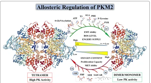
Although PKM1, PKM2, PKL, and PKR all have a tetra-meric form and pyruvate kinase activity, only PKM2 has both a dimeric form and a tetrameric form. In Fig. 2, we will show the characteristics of PKM2 in different states and its unique allosteric adjustment mode. When PKM2 is in a tetrameric state, it has a higher affinity with its substrate phosphoenolpyruvate (PEP), and a higher PK enzymatic activity, to catalyze the production of pyru-vate by phosphoenolpyrupyru-vate (PEP). PKM2 also has a
to an increase in upstream metabolites of the pyruvate kinase reaction in glycolysis, and is used for the synthesis of nucleotides, NADPH and phospholipids to ensure cell proliferation. Similarly, when tumor cells are exposed to stress or anti-chemotherapeutic drugs, such as increased expression of hypoxia inducible factor-1α (HIF-1α) in cells during hypoxia, both the transcriptional activity of PKM2 and the proportion of tetrameric PKM2 can be increased, similarly when cisplatin etc. using ROS as a killing chemotherapeutic agent that acts on tumor cells is used [52], PKM2 reforms into a tetrameric form, open the tricarboxylic acid cycle (TCA) and the electron trans-port chain to consume excess reactive oxygen species in the cell and protect the mitochondria from drug attack. In summary, amino acid, fatty acid, glucose intermediate and bypass metabolites can regulate PKM2 enzyme com-position and enzyme activity [53] (Additional file 3: Fig. S2).
The transition between PKM2 dimers and tetramers is allosterically regulated by endogenous and exogenous activators and inhibitors. PKM2 has PK enzyme activ-ity only when it serves as a tetramer. PKM2 is activated
by the glycolytic intermediate products named fructose 1,6-bisphosphate (FBP). It can also be activated by the allosteric effects of serine and succinylaminoimidazole-carboxamide ribose-50 phosphate SDH succinate dehy-drogenase (SAICAR) [54, 55]. The PK enzymatic activity of PKM2 can be inhibited by many endogenous inhibi-tors and cellular signaling events including 0-GlcNAcyla-tion, pyruvate (PYR), P-tyrosine (P-TYR), phenylalanine (PHE), alanine (ALA), adenosine triphosphate (ATP), and thyroid hormone T3 [56–58]. In addition, due to the number of related molecules involved in PKM2’s post-translational modification (PTM), I will not list them in Fig. 2, but in the form of Tables 2 and 3 in the fourth part of this article “Interaction of PKM2 with other proteins” (Additional file 4: Fig. S3).
It is now accepted that the PKM2 in the tetrameric state has an allosteric regulatory domain within its spa-tial structure, forming a pattern similar to the seesaw, when some allosteric regulators are inserted into the spatial structure involved in PKM2 allosteric regula-tion. After the domain (Fig. 3a), the tetramer PKM2 can be transferred from a compact state (R-state) to a loose
Fig. 2 Relationship between PKM2 enzyme activity and spatial conformation. The transition between PKM2 dimers and tetramers is allosterically regulated by endogenous and exogenous activators and inhibitors. PKM2 has PK enzyme activity only when it serves as a tetramer. PKM2 is activated by the glycolytic intermediate products named fructose 1,6-bisphosphate (FBP). It can also be activated by the allosteric effects of serine
and succinylaminoimidazolecarboxamide ribose-50 phosphate SDH succinate dehydrogenase (SAICAR) [54, 55]. The PK enzymatic activity of
PKM2 can be inhibited by many endogenous inhibitors and cellular signaling events including 0-GlcNAcylation, pyruvate (PYR), P-tyrosine (P-TYR),
phenylalanine (PHE), alanine (ALA), adenosine triphosphate (ATP), and thyroid hormone T3 [56–58]. In addition, due to the number of related
molecules involved in PKM2’s post-translational modification (PTM), I will not list them in Fig. 2, but in the form of Tables 2 and 3 in the fourth part of
state (T-state) and finally disassembled into a dimeric form [59]. When these allosteric regulators bind to PKM2, they will change the spatial conformation of PKM2, and affect the electrostatic force inside the mol-ecule, and then affect the transition state of PKM2. The allosteric form a stable and compact PKM2 R-state to form a tetramer and perform PK enzyme activity. After allosteric adjustment the PKM2 forms a loose and unsta-ble T-state, and eventually breaks the linked fragment in the tetramer to form a PKM2 dimer form with lower PK enzymatic activity. When PKM2 is allosteric to form a dimer, it will expose the active region inside the mol-ecule, although the PK enzyme activity is low, it has pro-tein activation activity [60]. In Fig. 3b we specifically list the each participating allosteric regulates the binding site of the small molecule and the binding site of the activa-tor in the PKM2 protein spatial structure [61]. In Fig. 3c we simply describe the seesaw structure of PKM2 and specifically identify the specific spatial domains that par-ticipate in the seesaw pattern: α-9 and 10 and 11 and 13 and 14 and 15 and 18 and β-20 which marked with blue text in Fig. 3c. The residues at the active site which was mentioned in Fig. 3a are highlighted by red box in Fig. 3c. Residue Arg342 and Residue Lys342, which is responsible for active site “RGD” stabilization is colored in red. There is one point to be noted here: whether PKM2 has protein kinase activity or not, there is a negative attitude, how-ever in some of the researchers’ experiments described later, phosphorylation was indeed found [62].
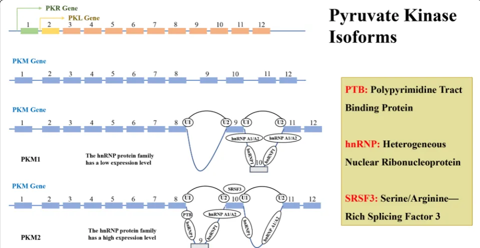
The splicing of PMK2
There are four PK subtypes encoded by two genes in mammals which was shown in Fig. 4. The PK enzyme is encoded by two genes, PKLR and PKM, which PKL gene encodes PKL, and is expressed in the liver and pancreas, intestine, and kidney, while PKR is mostly expressed in red blood cells [63]. PKM1 and PKM2 encoded by PKM
gene have the same length of gene coding and alterna-tive mutually exclusive exons, could encode 56 amino acid residues, and the regions differ in the splicing dif-ference at the 22nd position [64, 65]. The PKM subtypes perform the same catalytic function. However, in view of the fact that PKM1 is a tetrameric enzyme with sus-tained activity, there is a difference of 22 amino acids when compared with PKM2, and the mRNA generated by transcription of PKM under the action of cleavage fac-tor, can form PKM1 containing exon 9 or PKM2 contain-ing of exon 10 [66]. In Fig. 5 and Table 1, we could see the splicing factors of PKM gene include: hnRNPL (PTB), hnRNPAI, hnRNPA2 three heterogeneous riboproteins, which release exon 10 by binding exon 9, and promote PKM2 expression while inhibit PKM1 expression, NEK2 (never in mitosis (NIMA)-related kinase 2) can promote the release of exon 10 by binding to hnRNPAI/A2, fur-ther increase the expression of PKM2 [67–69]. Intensive studies have shown that under the regulation of C-MYC, three hnRNPs (heterogeneous ribonucleoproteins): hnRNPL (PTB), hnRNPA1, hnRNPA2, bind to the intron sequence between exon9 and exon10, inhibits the cleav-age of exon9, while serine/arginine-rich protein-specific kinase (SRSF-3) combines with the exon10 sequence to facilitate exon10 cleavage, thus completing the conver-sion of PKM1 to PKM2 [70, 71]. This difference is located in the spatial groove of PKM2 combined with fructose-1,6-diphosphate (FBP), allowing FBP to bind to PKM2 to deform the latter into an active tetramer. HIF-1α induces the PI3 K-AKT-mTOR signaling pathway and also regu-lates PKM2 by down-regulating C-MYC expression and up-regulating hnRNPs [72, 73]. NF-κB can mediate tran-scriptional upregulation of the PKM gene [74]. When epidermal growth factor (EGF) acts and activates its receptor (EGFR), EGFR can also induces NF-κB activa-tion following inflammatory and cytokine stimulaactiva-tion, in which polyubiquitination of IKK and phosphorylation
Fig. 3 The Specific Site of PKM2 Allosteric Regulation and The Amino Acid Sequence of PKM2. It is now accepted that the PKM2 in the tetrameric state has an allosteric regulatory domain within its spatial structure, forming a pattern similar to the seesaw, when some allosteric regulators are
inserted into the spatial structure involved in PKM2 allosteric regulation. After the domain (a), the tetramer PKM2 can be transferred from a compact
state (R-state) to a loose state (T-state) and finally disassembled into a dimeric form [59]. When these allosteric regulators bind to PKM2, they will
change the spatial conformation of PKM2, and affect the electrostatic force inside the molecule, and then affect the transition state of PKM2. The allosteric form a stable and compact PKM2 R-state to form a tetramer and perform PK enzyme activity. After allosteric adjustment the PKM2 forms a loose and unstable T-state, and eventually breaks the linked fragment in the tetramer to form a PKM2 dimer form with lower PK enzymatic activity. When PKM2 is allosteric to form a dimer, it will expose the active region inside the molecule, although the PK enzyme activity is low, it has protein
activation activity [60]. In b we specifically list the each participating allosteric regulates the binding site of the small molecule and the binding site
of the activator in the PKM2 protein spatial structure [61]. In Fig. 3c we simply describe the seesaw structure of PKM2 and specifically identify the
specific spatial domains that participate in the seesaw pattern: α-9 and 10 and 11 and 13 and 14 and 15 and 18 and β-20 which marked with blue
text in c. The residues at the active site which was mentioned in a are highlighted by red box in c. Residue Arg342 and Residue Lys342, which is
responsible for active site “RGD” stabilization is colored in red. There is one point to be noted here: whether PKM2 has protein kinase activity or not,
there is a negative attitude, however in some of the researchers’ experiments described later, phosphorylation was indeed found [62]
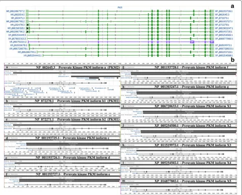
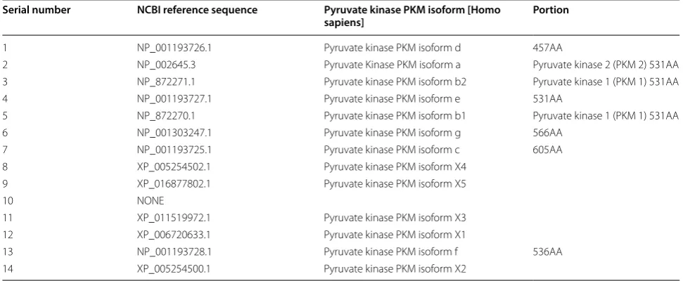
of TAK1 plays a crucial role this process. Activation of EGFR mediates PLCγ1-dependent PKCε activa-tion, resulting in PKCε monoubiquitination of Lys321 by RINCK1 ubiquitin ligase. Monoubiquitinated PKCε interacts with the NEMO zinc finger domain and recruits the cytoplasmic IKK complex to the plasma membrane, where PKCε phosphorylates IKKβ and activates IKKβ at Ser177 [75]. Activated RelA interacts with hypoxia-inducible factor 1 alpha (HIF1α), ultimately binds RelA to the PKM gene promoter and activates PKM transcrip-tion [76]. In turn, PTB is also up-regulated by EGFR acti-vation, and the PKM pre-mRNA is spliced into PKM2 mRNA to up-regulate PKM2 expression. These results indicate that both PKM and precursor mRNA are pro-duced by transcription of the PKM gene, but the tran-sition of PKM1 to PKM2 expression is a coordinated regulation of PTB-dependent splicing [77]. A total of 14 transcripts and 12 protein subtypes of the PKM gene are recorded in the NCBI and UCSC databases, see Table 1
for details. For broader regions represent the exons and the narrower regions represent introns. The dark regions represent the sequence between the translation initiation codon and the stop codon, and the light regions repre-sent the 57 UTR and 37 UTR regions [78] (AA: amino acid residues).
In addition to the different cleavage patterns, more and more studies have shown that the expression of PKM2 is also closely related to MicroRNAs (miR/miRNA), Long coding RNAs (LncR/LncRNA), etc., in non-coding RNA families, among which miRNAs are a class of short-chain non-coding RNAs, and are bound to the seed region at the ‘-UTR end of mRNAs and conduct functions, therefore affect protein synthesis and folding [79]. Some scholars have found that there are two bind-ing sites at the 3′ UTR end of miRNA-326 and PKM2, and miRNA-326 can inhibit the expression of PKM2 in glioma cells [80]. In intestinal cancer cells, miRNA-let-7a inhibits the proliferation, invasion and migration of intestinal cancer by down-regulating the expression of PKM2 [81–83]. Some microRNAs have organ specifica-tion, such as miRNA-122 [84], miRNA-124 [85], miRNA-133-3p [86], miRNA-137 [84], miRNA-20 [68] [4, 87] and miRNA-3662 [88] etc., which regulate the expression of PKM subunits by directly targeting polypyrimidine bundle binding protein 1 (PTBP1), while polypyrimi-dine bundle binding protein 1 (PTBP1) is a splice that regulates PKM2 dominant expression. Similar to: miRNA-29b [89], miRNA-99a [90], miRNA-133b [91], miRNA-145 [92], miRNA-148a [93], miRNA-152 [93,
94], miRNA-290 [95], miRNA-326 [96], miRNA-338-3P
[97], miRNA-340 [98], miRNA-369 [99], miRNA-371 [95], miR-379 [100], miRNA-675 [101], miRNA-4417 etc. [102]. While these miRNAs are targeted to the splic-ing factors PTBP1, hnRNPA1, and hnRNPA2, [103] the expression of PKM mRNA is shifted from PKM1 to PKM2, and the expression of PKM2 is increased [104]. In addition to the important total use of miRNAs in epigenetics, there is also a long-chain non-coding RNA called LncRNA in cells. Although these RNAs are dif-ficult to directly regulate the mRNA of PKM2, they can regulate miRNAs binding of PKM2, in turn to regulate the expression of PKM2. For example, miRNA-675 can form a ceRNA model with LncRNA-H19 [105], which in turn affects PKM2 expression. The same regulation
exists between LncRNA-MEG3 and miRNA-122 [106], LncRNA-MIF and miRNA-586 [107], LncRNA-CASC2c and miRNA-101 [97]. The double-mutant P53 can regu-late LncRNA CUDR and down-reguregu-late PKM2 to inhibit tumor growth [108]. In addition to the regulation of tumor suppressor genes, these long-chain non-coding RNAs can also influence intracellular signaling path-ways, for example, LncRNA-Ftx [109], LncRNA-SchLAH [110], LncRNA-ROR [111], LncRNA-DACOR1 [112] can mediate PTEN signaling pathway in cells. The Pi3k/AKT/ mTOR signaling pathway has an effect that produces dif-ferent biological effects [72, 113].
The genes encoding pyruvate kinase can be divided into two types: PKLR and PKM. PKLR binds to the
Fig. 5 Expression Patterns of The Transcript and The Protein Subtypes of PKM. a That there are 14 different subtype sequences in the PKM gene transcript, and there are some differences between them, among which the PKM1 (No. 3 and No. 5) and PKM2 (No. 2) subtypes are more compared,
b the coding sequence NO. 12 expresses PKM1 and the coding sequence expresses NO. 13 PKM2. There are only 23 amino acid residues between
coding gene through a tissue-specific promoter, encoding two subtypes of PKL and PKR (green for PKR and yellow for PKL). PKM encodes PKM1 and PKM2 subtypes by alternative splicing of mutually exclusive exon 9 and 10, and a high expression level of PTB, hnRNP 1 and hnRNP A1/A2 are required for during the cleavage process of exon 9 of PKM2, while the cleavage process of exon 10 of PKM1 is not required. Transcription factor SRSF3 also plays an important role in the conversion of PKM2 and PKM1. Each pyruvate kinase subtype has a different tis-sue expression pattern (Additional file 5: Fig. S4).
Figure 5a shows that there are 14 different subtype sequences in the PKM gene transcript, and there are some differences between them, among which the PKM1 (No. 3 and No. 5) and PKM2 (No. 2) subtypes are more compared, Fig. 5b shows the coding sequence NO. 12 expresses PKM1 and the coding sequence expresses NO. 13 PKM2. There are only 23 amino acid residues between the two protein sequences. And there are few studies on other PKM protein subtypes, and the functions are not sure yet.
Non‑glycolysis enzyme function of PKM2
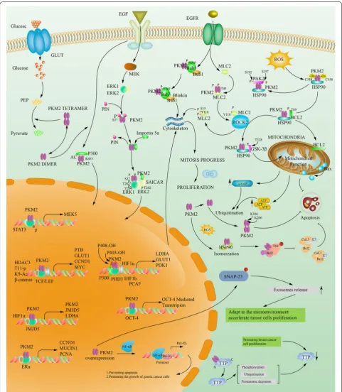
By learning of that, it could be found PKM2 not only plays an important role in cytosolic glucose metabolism, but also can transfer from the cytoplasm to the nucleus rely on its c-terminal nuclear localization signal in inter-leukin-3, growth hormone inhibitor analogue TIT-232, peroxide, epidermal growth factor (EGFR), ultraviolet radiation and other factors, and in the form of dimers to play a role in protein kinase activity in the nucleus of a variety of transcription factors and thus affect a variety of signaling pathways to promote tumor development
[114, 115]. Recent studies have found that PKM2-spe-cific exon 10 can recruit extracellular signal-regulated kinase 2 (ERK2) and bind to the Iso429/Leu431 region of PKM2, mediate Ser37 site phosphorylation on PKM2, and recruit PIN1 to form PRKM2/PIN1 complex, if the Ser37 site is mutated to other amino acids, although PKM2 can still form tetramers in the cytoplasm, PKM2 cannot enter the nucleus [116], which suggests that the complex is an important transporter that mediates PKM2 entry into the nucleus, all these results suggest that PKM2 can play a role in regulating transcription and post-translational modification, and these effects depend on the interaction between PKM2 and ERK1/2, PIN1 and Importin 5α [117]. Moreover, if PKM2 exists in the form of a tetramer, the Arg399 site of nuclear localization sequence (NLS) in one side of the PKM2 monomer in its symmetrical structure can form a stable charge–charge interaction with Glu418 and Glu396 of the opposite mirror PKM2 monomer (the other two is located in the tetramer PKM2), which also maintains the spatial confor-mation of the PKM2 tetramer to some extent. Only when the spatial structure of PKM2 changes, such as PKM2 in the dimeric form, fully exposes the NLS of PKM2 which buried in the 3-dimensional space, through interacting with Importin 5α and NLS to change the electrostatic attraction inside the PKM2 protein molecule after bind-ing the PIN1 molecule, in turn, the dimeric structure of PKM2 is maintained [118]. PKM2 also transactivates SLC2A1, LDHA, PDK1, HK1, and VEGFA gene expres-sion via hypoxia inducible factor-1α (HIF-1α) transcrip-tion factor [56, 119]. In addition, PKM2 up-regulates gene expression through hypoxia inducible factor-1α (HIF-1α), β-catenin (β-cat), insulin, signal transducers Table 1 Subtype classification of pyruvate kinase in mammals
Serial number NCBI reference sequence Pyruvate kinase PKM isoform [Homo
sapiens] Portion
1 NP_001193726.1 Pyruvate kinase PKM isoform d 457AA
2 NP_002645.3 Pyruvate Kinase PKM isoform a Pyruvate kinase 2 (PKM 2) 531AA
3 NP_872271.1 Pyruvate kinase PKM isoform b2 Pyruvate kinase 1 (PKM 1) 531AA
4 NP_001193727.1 Pyruvate kinase PKM isoform e 531AA
5 NP_872270.1 Pyruvate kinase PKM isoform b1 Pyruvate kinase 1 (PKM 1) 531AA
6 NP_001303247.1 Pyruvate kinase PKM isoform g 566AA
7 NP_001193725.1 Pyruvate kinase PKM isoform c 605AA
8 XP_005254502.1 Pyruvate kinase PKM isoform X4
9 XP_016877802.1 Pyruvate kinase PKM isoform X5
10 NONE
11 XP_011519972.1 Pyruvate kinase PKM isoform X3
12 XP_006720633.1 Pyruvate kinase PKM isoform X1
13 NP_001193728.1 Pyruvate kinase PKM isoform f 536AA
and activators of transcription 3 (STAT3) and other tran-scription factors to promote cell growth and prolifera-tion [74, 120, 121]. PKM2 can also act as a protein kinase, which forms a complex with β-catenin and binds to the CCND1 promoter region to phosphorylate histone H3, and phosphorylated histone H3 is separated from histone deacetylase 3, which will make the 9th tyrosine of histone H3 acetylation [122], then promotes the expression of an important cytokine of cell proliferation namely CycinD1, and then regulates the cell cycle. PKM2 can also affect the target gene C-MYC by affecting β-catenin, and C-MYC can also be used to promote PKM2 gene tran-scription and hnRNA cleavage in turn [123]. In addition to regulating the expression of CycinD1, which affects the transformation of G1-S stage, PKM2 also regulates the filamentous manner, which regulates the tyrosine at position 207 of the mitotic protein Bub3, promotes the formation of the Bub3-Bubl complex, and the Bub3-Bubl complex further act on the exocentric protein Blinkin to promote its binding to the ligand Bub and thus precisely regulates chromosome segregation and cell proliferation [124]. Studies have shown that under various conditions, the MER/ERK pathway is activated, phosphorylating the serine at position 37 on PKM2, which could help PKM2 enter the nucleus [125]. C-src can also be activated by EGFR activation, and C-src phosphorylates tyrosine at position 333 on β-catenin, so that PKM2 can bind to β-catenin/TCF/LEF after entering nucleation, thereby activates the expression of β-catenin downstream gene, such as CCND1 and MYC promote cell proliferation, but also activate PTB, LDHA, GLUT1. PTB can again acti-vate the transcription of PKM2 to form a chain reaction [76]. LDHA and GLUT1 can regulate glucose uptake and tumor cell glycolysis, thereby regulate glucose metabo-lism. It was confirmed by research that PKM2 in the nucleus can phosphorylate tyrosine at 705th position of signal transduction factor and transcriptional activator 3, further promoting: the transcription of mitogen acti-vated protein kinase kinase 5 (MAPKK5/MEK5), and MEK5 promotes the growth of tumor cells [126]. Li et al’s study confirmed that PKM2 is resistant to gemcitabine in patients with intestinal cancer by phosphorylating tran-scriptional activator factor 3. It has also been reported in the literature that PKM2 can make the serine at posi-tion 202/203 of protein kinase B substrate l (AKT1S1), promote its binding to 14-3-3 to activate mammalian rapamycin target protein sensitive complex 1 (mTORC l) signaling pathway to promote tumor growth [127]. PKM2 can directly phosphorylate extracellular signal-regulated kinase 1/2 (ERK l/2) to promote tumor growth [55]. Stud-ies have shown that PKM2 can directly bind to the active domain of hypoxia inducible factor-1α (HIF-1α) to pro-mote the activation of downstream vascular endothelial
growth factor (VEGF) expression [128]. NAD-dependent deacetylase sirtuin6 (SIRT 6) could deacetylate the 433th lysine of PKM2 in the nucleus to cause it to transfer out of the nucleus and inhibit tumor growth. In summary, PKM2 in the nucleus is associated with cell proliferation [129]. Tables 2, 3 and Fig. 6 summarizes the specific sites, source literature, and biological effects of the various roles mentioned above.
Interaction of PKM2 with other proteins
Phosphorylation
1. The phosphorylated PKM2 can protect mitochon-drial from oxidative stress. At the beginning of the period, ROS can oxidize the Cys358 site of PKM2 when tumor cells are exposed to oxidative stress damage. PKM2 acts as a phosphorylase to phospho-rylate its substrate [130]. PKM2 can phosphorylate Bcl2 threonine (Thr69), which prevents the bind-ing of Cul3-based E3 ligase to Bcl2 and subsequent inhibits degradation of Bcl2. Then heat shock protein 90 (HSP90) acts as a bridge, stabilizes and transferres the PKM2-BCL2 complex to Mitochondrial outer membrane, which regulates the ratio of Bax/Bcl2. Liang, Cao et al. believe that mitochondrial function still exists in the background of Warburg effect, and the maintenance of mitochondrial function in this case depends on the binding of PKM2 and Bcl2 on the outer membrane of mitochondria, inhibiting the release of ROS and oxidative stress-induced apopto-sis [131].
2. The phosphorylated PKM2 can increase its nuclear localization and promote cell proliferation [55].
1. PKM2 phosphorylates PAK2 directly on Ser20, Ser141 (phosphorylated but weakly acting) and Ser192/197. PKM2-mediated phosphorylation of Ser192/197 is essential for maintaining PAK2 activity in PDAC cells, promotes HSP90 associa-tion with PKM2-PAK2 complex, which prevents the degradation of ubiquitin and protease of PAK2, and ultimately mediates tumor cell inva-sion, metastasis and cell proliferation [132]. 2. After binding to SAICAR, PKM2 phosphorylates
the Thr202, Tyr204 and ERK2 Thr202 of ERK1, which in turn phosphorylates the Ser37 of PKM2. This cascaded phosphorylation pattern can form an activation loop and activate the ERK/MAPK signal pathway [55].
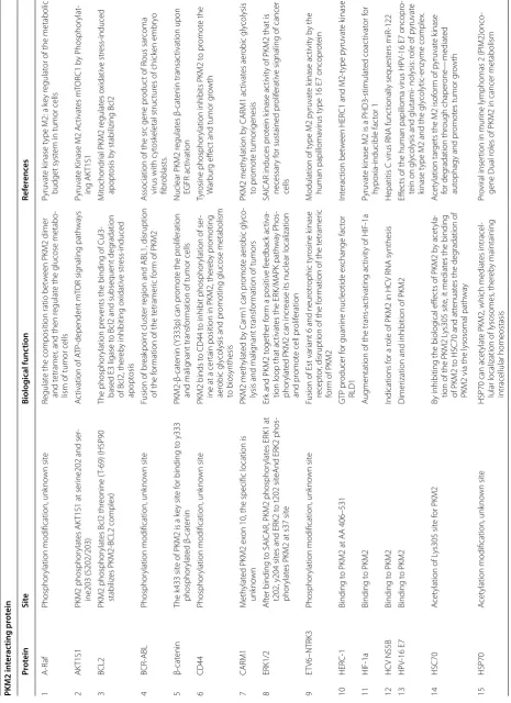
PKM2-β-Table 2 PKM2 in ter ac ting pr ot
ein & I
nt
er
ac
tion sit
e & Biolo
gic
al F
unc
tion & Ref
er enc es PKM2 in ter ac ting pr ot ein Pr ot ein Sit e Biolog ical func tion Ref er enc es 1 A-R af Phosphor
ylation modification, unk
no
wn sit
e
Regulat
e the composition ratio bet
w
een PKM2 dimer
and t
etramer
, and then r
egulat
e the glucose metabo
-lism of tumor cells
Pyruvat
e k
inase t
ype M2: a k
ey r
egulat
or of the metabolic
budget syst
em in tumor cells
2 AK T1S1 PKM2 phosphor ylat es AK
T1S1 at ser
ine202 and ser
-ine203 (S202/203)
A
ctivation of A
TP -dependent m TOR sig naling path wa ys Pyruvat e K
inase M2 A
ctivat es m TOR C1 b y P hosphor ylat -ing AK T1S1 3 BCL2 PKM2 phosphor ylat
es Bcl2 thr
eonine (
T-69) (HSP90
stabiliz
es PKM2-BCL2 complex)
The phosphor
ylation pr
ev
ents the binding of C
ul3-based E3 ligase t
o Bcl2 and subsequent deg
radation
of Bcl2, ther
eb
y inhibiting o
xidativ e str ess-induced apopt osis M itochondr
ial PKM2 r
egulat es o xidativ e str ess-induced apopt osis b
y stabilizing Bcl2
4
BCR-ABL
Phosphor
ylation modification, unk
no
wn sit
e
Fusion of br
eak
point clust
er r
eg
ion and ABL1, disruption
of the f
or
mation of the t
etramer
ic f
or
m of PKM2
A
ssociation of the sr
c gene pr
oduc
t of R
ous sar
coma
virus with c
yt
osk
eletal struc
tur
es of chick
en embr yo fibr oblasts . 5 β-cat enin
The k433 sit
e of PKM2 is a k
ey sit
e f
or binding t
o y333 phosphor ylat ed β-cat enin PKM2-β-cat enin (
Y333p) can pr
omot
e the pr
olif
eration
and malig
nant transf
or
mation of tumor cells
Nuclear PKM2 r
egulat es β-cat enin transac tivation upon EGFR ac tivation 6 CD44 Phosphor
ylation modification, unk
no
wn sit
e
PKM2 binds t
o CD44 t
o inhibit phosphor
ylation of ser
-ine at a cer
tain position in PKM2, ther
eb
y pr
omoting
aer
obic gly
colysis and pr
omoting glucose metabolism
to biosynthesis
Tyr
osine phosphor
ylation inhibits PKM2 t
o pr omot e the W ar bur g eff ec
t and tumor g
ro wth 7 CARM1 M eth ylat
ed PKM2 ex
on 10, the specific location is
unk no wn PKM2 meth ylat ed b y C ar
m1 can pr
omot
e aer
obic gly
co
-lysis and malig
nant transf
or
mation of tumors
PKM2 meth
ylation b
y CARM1 ac
tivat es aer obic gly colysis to pr omot e tumor igenesis 8 ERK1/2 A ft
er binding t
o SAICAR, PKM2 phosphor
ylat
es ERK1 at
t202, y204 sit
es and ERK2 t
o t202 sit
eAnd ERK2 phos
-phor
ylat
es PKM2 at s37 sit
e
Er
k and PKM2 t
ogether f
or
m a positiv
e f
eedback ac
tiva
-tion loop that ac
tivat
es the ERK/M
APK path wa y P hos -phor ylat
ed PKM2 can incr
ease its nuclear localization
and pr
omot
e cell pr
olif
eration
SAICAR induces pr
ot
ein k
inase ac
tivit
y of PKM2 that is
necessar
y f
or sustained pr
olif
erativ
e sig
naling of cancer
cells
9
ET
V6–NTRK3
Phosphor
ylation modification, unk
no
wn sit
e
Fusion of Est var
iant 6 and neur
otr ophic t yr osine k inase recept or
, disruption of the f
or
mation of the t
etramer
ic
for
m of PKM2
M
odulation of t
ype M2 p
yruvat e k inase ac tivit y b y the human papilloma virus t
ype 16 E7 oncopr
ot ein 10 HER C-1 Binding t
o PKM2 at AA 406–531
GTP pr
oducer f
or guanine nucleotide ex
change fac tor RLD1 Int erac tion bet w een HER
C1 and M2-t
ype p yruvat e k inase 11 HIF-1a Binding t o PKM2 A ug
mentation of the trans-ac
tivating ac
tivit
y of HIF-1a
Pyruvat
e k
inase M2 is a PHD3-stimulat
ed coac tivat or f or hypo xia-inducible fac tor 1 12 HC V NS5B Binding t o PKM2 Indications f
or a r
ole of PKM2 in HC
V RNA synthesis
Hepatitis C virus RNA func
tionally sequest ers miR-122 13 HP V-16 E7 Binding t o PKM2 Dimer
ization and inhibition of PKM2
Eff
ec
ts of the human papilloma virus HP
V-16 E7 oncopr
o-tein on gly
colysis and glutami- nolysis: r
ole of p
yruvat
e
kinase t
ype M2 and the gly
colytic-enz yme complex. 14 HSC70 A cet
ylation of L
ys305 sit
e f
or PKM2
By inhibiting the biolog
ical eff
ec
ts of PKM2 b
y acet
yla
-tion of the PKM2 L
ys305 sit
e, it mediat
es the binding
of PKM2 t
o HSC70 and att
enuat
es the deg
radation of
PKM2 via the lysosomal path
wa
y
A
cet
ylation tar
gets the M2 isof
or
m of p
yruvat e k inase for deg radation thr ough chaper one —mediat ed aut
ophagy and pr
omot
es tumor g
ro wth 15 HSP70 A cet
ylation modification, unk
no
wn sit
e
HSP70 can acet
ylat
e PKM2, which mediat
es intracel
-lular localization of lysosomes
, ther eb y maintaining intracellular homeostasis Pr oviral inser
tion in mur
ine lymphomas 2 (PIM2)onco
-gene Dual r
Table 2 (c on tinued) PKM2 in ter ac ting pr ot ein Pr ot ein Sit e Biolog ical func tion Ref er enc es 16 H3
PKM2 can phosphor
ylat e hist one H3 T11 Phosphor ylat ed hist
ones can pr
omot
e the G1-S phase
transition of tumor cells
, phosphor
ylation of stat3 can
pr
omot
e the pr
oduc
tion of c
yclinD
, phosphor
ylation
of ML
C2 can enhance the ac
tivit
y of M
APK path
wa
y,
and phosphor
ylat
ed Bub3 can enhance the ac
tivit y of EGFR path wa y Pyruvat e k
inase M2 at a glance
17
ST
AT3
PKM2 can phosphor
ylat e stat3 Y705 18 ML C2
PKM2 can phosphor
ylat e ML C2 Y118 19 Bub3
PKM2 can phosphor
ylat e Bub3 Y27 sit e 20 FL T3 Phosphor
ylation modification, unk
no wn sit e Fms-r elat ed t yr osine k inase , int er
nal tandem duplica
-tion (ITD) mutant, disrup-tion of the f
or
mation of the
tetramer
ic f
or
m of PKM2
A
ssociation of the sr
c gene pr
oduc
t of R
ous sar
coma
virus with c
yt
osk
eletal struc
tur
es of chick
en embr yo fibr oblasts . 21 FGFR1
FGFR1 can phosphor
ylat
es PKM2 atT
yr 83, Tyr105, Tyr148, T yr175, T yr370, T yr390 sit e
Inhibits the biolog
ical ac
tivit
y of PKM2 which coule
regulat
e the glucose metabolism in tumor cells
TRIM35 I
nt
erac
ts with p
yruvat
e k
inase isof
or
m M2 t
o suppr ess the W ar bur g eff ec
t and tumor
igenicit y in hepat ocellular car cinoma 22 JAK2 Phosphor
ylation modification, unk
no
wn sit
e
Disruption of the f
or
mation of the t
etramer ic f or m of PKM2 A
ssociation of the sr
c gene pr
oduc
t of R
ous sar
coma
virus with c
yt
osk
eletal struc
tur
es of chick
en embr yo fibr oblasts . 23 JNK1 JNK1 phosphor ylat
es PKM2 at
Thr365 sit
e
Inhibits the biolog
ical ac
tivit
y of PKM2 which coule
regulat
e the glucose metabolism in tumor cells
, and
the nuclear transf
er of PKM2 is inhibit
ed
, which in tur
n
inhibits the biolog
ical r
ole of PKM2 in the nucleus
PARP14 pr omot es the W ar bur g eff ec
t in hepat
ocellular
car
cinoma b
y inhibiting
JNK1-dependent PKM2 phos
-phor
ylation and ac
tivation 24 GSK -3β GSK -3β phosphor
ylation of PKM2
Thr
-328 (HSP90 stable
PKM2-GSK3β complex)
Thr
-328 phosphor
ylation is essential f
or maintaining
PKM2 stabilit
y and its biolog
ical func
tion in r
egulating gly colysis , mit ochondr ial r espiration, pr olif eration and apopt osis HSP90 pr omot
es cell gly
colysis
, pr
olif
eration and inhibits
apopt
osis b
y r
egulating PKM2 abundance via
Thr
-328
phosphor
ylation in hepat
ocellular car
cinoma
25
P53
Phosphor
ylation modification, unk
no
wn sit
e
PKM2-P53 can pr
omot
e the pr
olif
eration and malig
nant
transf
or
mation of tumor cells
Dual r
oles of PKM2 in cancer metabolism
26
p300
PKM2 could be acet
ylat
ed b
y p300 at K433 sit
e
PKM2 K433 acet
ylation con
ver
ts cell pr
olif
eration and
cyt
oplasmic metabolic k
inase t
o nuclear pr
ot ein kinase ac tivit y M
itogenic and oncogenic stimulation of K433 acet
yla
-tion pr
omot
es PKM2 pr
ot
ein k
inase ac
tivit
y and nuclear
localization 27 PAK2 PKM2 dir ec tly phosphor ylat es P AK2 (HSP90-stabiliz ed PKM2-P
AK2 complex) on S
er20, S
er141 (phosphor
yl
-at
ed but w
eak
ly ac
ting) and ser192/197 (ac
tion sit
e)
Phosphor
ylation of ser
ine 192/197 mediat
ed b
y pk
m2
in PD
A
C cells is cr
itical f
or maintaining P
AK2 le
vels
phosphor
ylation of ser192/197 pr
omot
es the associa
-tion of HSP90 with P
AK2, ther eb y pr ev enting ubiquitin and pr ot ease deg
radation of P
AK2
Pyruvat
e k
inase M2 pr
omot es pancr eatic duc tal adeno -car cinoma in
vasion and metastasis thr
ough phospho
-rylation and stabilization of P
AK2 pr ot ein 28 PANK -4 Binding t o PKM2 Reduc
tion of the ac
tivit
y of the t
etramer
ic f
or
m
Dual r
oles of PKM2 in cancer metabolism
29 Par kin Par kin pr omot
es ubiquitination of L
ys186 and L
ys206
sit
es in PKM2
Par
kin inhibits the biolog
ical ac
tivit
y of PKM2 and r
egu
-lat
es glucose metabolism b
y pr
omoting ubiquitina
-tion of L
ys186 and L
ys206 sit
es of PKM2
Par
kin r
egulat
es the ac
tivit
y of p
yruvat e k inase M2 30 PCAF A cet ylat
es modification, unk
no
wn sit
e
Reduc
tion of the ac
tivit
y of the t
etramer
ic f
or
m
Dual r
oles of PKM2 in cancer metabolism
31
PHD3
PDH3 can h
ydr
ox
ylat
e the P
ro403 and P
ro408 sit
es of
PKM2
PKM2 binds t
o PDH3, and modified PKM2 is mor
e
susceptible t
o HIF-1α binding and f
or
ms an ac
tivation
loop that pr
omot
es anaer
obic gly
colysis and meta
-bolic r
ecombination
Pyruvat
e k
inase M2 is a PHD3—stimulat
Table 2 (c on tinued) PKM2 in ter ac ting pr ot ein Pr ot ein Sit e Biolog ical func tion Ref er enc es 32 PK C ♁ Phosphor
ylation modification, unk
no
wn sit
e
H
ypothesis: r
egulation of stabilit
y or deg
radation of
M2-PK
Dual r
oles of PKM2 in cancer metabolism
33
PIAS3
Binding t
o PKM2 at AA 1–348
Sumo
ylation of PKM2 and nuclear translocation of PKM2
The SUMO
-E3 ligase PIAS3 tar
gets p yruvat e k inase M2 34 PM L Binding t o PKM2 Reduc
tion of the ac
tivit
y of the t
etramer
ic f
or
m
M
odulation of M2-t
ype p yruvat e k inase ac tivit y b y the cyt oplasmic P
ML tumor suppr
essor pr
ot
ein
35
PIM2
PIM2 can phosphor
ylat
e PKM2 at
Thr454 sit
e
PIM2 phosphor
ylat
es the PKM2
Thr454 sit e, mediat es PKM2-dependent anaer obic gly colysis
, and maintains
mit
ochondr
ial func
tion in tumor cells
Pr
oviral inser
tion in mur
ine lymphomas 2 (PIM2)onco
-gene phosphor ylat es p yruvat e k
inase M2 (PKM2) and
pr
omot
es gly
colysisin cancer cells
36 PTP1B PTP1B phosphor ylat es the Tyr105 and Tyr148 sit e of PKM2
PTP1B inhibits the biolog
ical ac
tivit
y of PKM2 b
y phos -phor ylating the Tyr105 and Tyr148 sit
e of PKM2
Pr
ot
ein t
yr
osine phosphatase 1B r
egulat
es p
yruvat
e
kinase M2 t
yr osine phosphor ylation 37 PRM T4 PRM T4 meth ylat
es specifically the dimer
ic f
or
m of PKM2
at Ar
g445/447/455 r
esidues in the C domain
Allost
er
ic ac
tivat
ors inhibit PKM2 t
etramer
ization f
or
m
thought PKM2 meth
ylation
Posttranslational modifications of p
yruvat
e k
inase M2:
tw
eaks that benefit cancer
38
MG
MG can gly
cosylat
e the Ar
g399 sit
e of PKM2
MG can gly
cosylat
e the Ar
g399 sit
e of PKM2, the r
esult
of which can change the spatial configuration of PKM2
M
olecular association of glucose
-6-phosphat
e isomerase
and p
yruvat
e k
inase M2 with gly
ceraldeh yde -3-phos -phat e deh y-dr
ogenase in cancer cell
39
O
ct No
. 4
Binding t
o PKM2 at AA 307–531
A
ug
mentation of the trans-ac
tivating ac
tivit
y of O
ct 4
Pyruvat
e k
inase iso
zyme t
ype M2 (PKM2) int
erac
ts and
cooperat
es with O
ct
-4 in r
egulating transcr
iption.
40
Opa
Binding t
o PKM2 at AA 367–531
Out
er membrane pr
ot
eins in
volv
ed in gonococcal
adhesion t
o and in
vasion of human epithelial cells
cr
eation of a micr
oen
vir
onment of high p
yruvat
e
concentration
Posttranslational modifications of p
yruvat
e k
inase M2:
tw
eaks that benefit cancer
41
O_GlcNA
cylation
O
-GlcNA
cylation can block the
Thr 405 and S
er 406 sit
es
of PKM2
D
ecr
eased the stabilit
y of the t
etramer
ic f
or
m of PKM2
to enhance aer
obic gly colysis ( W ar bur g eff ec t) O -GlcNA cylation destabiliz
es the ac
tiv e t etramer ic PKM2 to pr omot e the W ar bur g eff ec t 42 SAICAR M
utation of the G415 sit
e of PKM2 t
o R will not bind t
o
SAICAR
PKM2 binds t
o SAICAR, and mutations at the PKM2 G415
sit
e pr
ev
ent PKM2 fr
om binding t
o SAICAR
SAICAR induces pr
ot
ein k
inase ac
tivit
y of PKM2 that is
necessar
y f
or sustained pr
olif
erativ
e sig
naling of cancer
cells 43 SIR T-6 D eacet
ylation modification, the sit
e is unk
no
wn
SIR
T6 mediat
es the deacet
ylation of PKM2, and the
results mediat
e the nuclear localization of PKM2
SIR
T6 deacet
ylat
es PKM2 t
o suppr
ess its nuclear localiza
-tion and oncogenic func
tions 44 SOCS-3 Binding t o PKM2 Reduc
tion of A
TP pr
oduc
tion and influence of dendr
itic
cell immune r
esponse
Posttranslational modifications of p
yruvat
e k
inase M2:
tw
eaks that benefit cancer
45
TEM8
Binding t
o PKM2
Stimulation of ang
iogenesis b
y binding of
Tumor M2-PK
released fr
om tumors
Dual r
oles of PKM2 in cancer metabolism
46 TEPP -46 Binding t o PKM2 TEPP
-46 and FBP
, the allost
er
ic ac
tivat
ors that induce
PKM2 t
etramer
ization
Posttranslational modifications of p
yruvat
e k
inase M2:
tw
eaks that benefit
47
TRIM35
M
iR-4417 tar
gets
TRIM35 and r
egulat es PKM2 Y105 phosphor ylation Pr omot e pr olif
eration and suppr
ess apopt osis , PKM2 Y105 phosphor ylation t o pr omot e HC C g ro wth M iR-4417 tar gets tr ipar tit e M
otif-containing 35 (
TRIM35) and r egulat es p yruvat e k
inase muscle 2 (PKM2)
phosphor ylation t o pr omot e pr olif
eration and suppr
ess
apopt
osis in hepat
ocellular car
catenin (Tyr333p) can promote the proliferation and development of tumor cells [124].
4. PKM2 can phosphorylate Thr11 of histone H3, which regulates the transcription of MYC and CCND1, and promotes the G1-S phase transi-tion of the cell proliferatransi-tion cycle, as well as Y705 phosphorylating stat3. Phosphorylation of STAT3 can promote the production of CCND1, which can enhance the activity of mitogen activated protein kinase (MAPK) pathway [124].
5. PKM2 can phosphorylate Tyr118 and phospho-rylate Bub3 Tyr27 sites of myosin light chain 2 (MLC2). Phosphorylation of MLC2 can also enhance the activity of mitogen activated protein kinase (MAPK) pathway, and phosphorylated Bub3 can enhance the activity of EGFR pathway [124].
6. PKM2 can promote the serine at position 202/203 of protein kinase B substrate l (AKT1S1) binding to 14-3-3 to activate the mechanistic target of rapamycin complex l (mTORC l) signal pathway, which will promote tumor growth [127]. 7. In the nucleus of bone marrow cells of leukemia
patients, In the nucleus of bone marrow cells of leukemia patients, the researchers found that PKM2 can phosphorylate and activate certain transcription factors, thereby inducing fusion of BCR-ABL genes. In the cytoplasm of these cells, it is characterized by the dissociation of PKM2 tetramer, the formation of dimers, and finally promote the accelerated formation of leukemia in patients [133].
8. A mode of action similar to the last article 7 still exists in ETV6–NTRK3. PKM2 can phospho-rylate and activate certain transcription factors, thereby inducing fusion of Est variant 6 and neu-rotrophic tyrosine kinase receptor. The result may lead to malignant transformation of some neurogenic tumors. PKM2 in the cytoplasmic dimer form promotes the formation of the War-burg effect [134].
9. P53 and PKM2 in the nucleus of the cells can phosphorylate each other to form a cascade loop. Although the location of the PKM2 phos-phorylated P53 modification site has not been specifically demonstrated, P53 has been shown to phosphorylate the Ser37 site of PKM2. When the tumor cells are under stress, this pattern is activated to protect against external stress in the form of EMT [135].
3. PKM2 not only can act as a phosphorylase to phos-phorylate the substrate, but also can be
phosphoryl-ated by other phosphorylases at specific sites. The phosphorylation of its tyrosine residue Tyr105 has been found in a variety of solid tumors in humans. For example,
1. The tyrosine residues Tyr 83, Tyr105, Tyr148, Tyr175, Tyr370, Tyr390 of PKM2 can be directly phosphorylated by fibroblast growth factor receptor 1 (FGFR1), and the binding of phospho-rylated PKM2 to FBP inhibits the presence of the tetrameric form of PKM2, results in reducing its PK activity [136].
2. It has been reported that tripartite motif con-taining 35 (TRIM35) directly binds to PKM2 to inhibit PKM2 Tyr105 phosphorylation, thereby increasing enzyme activity [137].
3. ERK1/2 also has phosphorylase activity, which specifically phosphorylates the Ser37 site of PKM2 without phosphorylating PKM1, which provides a binding motif for PKM2 that interacts with peptidyl-proline isomerases, and binding to the transcript of the mitotic gene A1 (PIN1) mediates the entry of PKM2 into the nucleus [138]. When PKM2 enters the nucleus, it can also bind to Oct-4, and combine with many of the above mechanisms to affect the spindle, affecting the cell cycle by affecting cell division [139, 140]. 4. Glycogen synthase kinase 3β (GSK-3β) could
phosphorylate Thr-328 of PKM2, and phospho-rylation of the PKM2 Thr-328 site is essential for maintaining PKM2 stability and regulating biological functions of glycolysis, mitochondrial respiration, proliferation and apoptosis. HSP90 also has a function to stabilize the PKM2–GSK3β complex in the HSP family [141].
5. In the cytoplasm of tumor cells, proviral inser-tion in murine 1ymphomas 2 (PIM2) can also phosphorylate the Thr454 site of PKM2, to medi-ates PKM2-dependent anaerobic glycolysis, and maintains mitochondrial function in tumor cells [142].
6. Jun N-terminal kinase 1 (JNK1) phosphorylates the PKM2 threonine residue Thr365 to inhibit PKM2 enzyme activity [143].
7. PKM2 can be phosphorylated by Fms-related tyrosine kinase 3 (FLT3), Janus kinase 2 (JAK2), protein kinase C (PKC), and the modified PKM2 will exist as a dimer. Promote anaerobic glycolysis and cell proliferation of tumor cells [46, 133]. 8. In addition to phosphorylation of these known
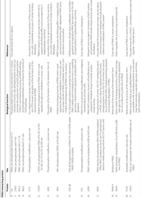
could regulate the composition ratio between PKM2 dimer and tetramer, and then regulate the glucose metabolism of tumor cells [53]. PKM2 binds to CD44 to inhibit phosphorylation of ser-ine at a certain position in PKM2, inhibit pro-moting aerobic glycolysis and propro-moting glucose metabolism to biosynthesis [125].
In addition to phosphorylation, proteins such as pro-tein tyrosine phosphatase 1B (PTP1B) inhibit the phos-phorylation of PKM2, and PTP1B inhibits the biological activity of PKM2 by phosphorylating the Tyr105 and Tyr148 site of PKM2 [71].
Acetylation
Acetylation is another pre-translational modification of PKM2 that reduces the enzymatic activity of PKM2, increases glycolysis intermediates, and supports biosyn-thesis. One of the important features of the tumor micro-environment is hypoxia. Hypoxia inducible factor-1α (HIF-1α) and hypoxia inducible factor-1β (HIF-1β) are
under hypoxic conditions, and the expression will be increased when tumor and normal cells is under hypoxic stress [144]. The same hypoxia transcription factor CBP/ P300 can be used as an effector molecule downstream of HIF-1α and HIF-1β, so that it can play an important biological role in the nucleus and cytoplasm. P300 can be used as an acetyltransferase to catalyze exon 10 of PKM2 [145]. PKM2 is acetylated by P300 in Lys433, and the PKM2 Lys433 is acetylated to regulate cell prolifera-tion and transform the biological behavior of PKM2 from cytoplasmic metabolic kinase to nuclear protein kinase activity [146]. Parkin can ubiquitinate the Lys186/206 site of PKM2, and then regulate the ratio of tetram-ers and dimtetram-ers of PKM2. In addition, a complex can be formed between the PKM2/P300/PHD3/HIF-1α/HIF-1β in the nucleus, and finally regulate the tumor fine glu-cose metabolism, O2 consumption and CO2 production
[147]. Acetylation is also present in heat shock cognate protein70 (HSC70) and P300/CBP-associated factor (PCAF), which acetylates the Lys305 site of PKM2. By inhibiting the biological effects of PKM2 by acetylation of the PKM2 Lys305 site, this kind of acetylation behav-ior mediates the binding of PKM2 to HSC70 and attenu-ates the degradation of PKM2 via the lysosomal pathway [148]. In addition to HSP70, there is similar acetylation, but the site is unknown. After acetylation of PKM2, HSP70 can acetylate PKM2, which mediates intracellular localization of lysosomes, such maintaining intracellular homeostasis [142]. Table 3 summarizes the various post-translational modification sites mentioned above and the biological effects they exert.
Other interprotein interactions
The activity of PKM2 is also regulated by many proteins in contact with it, and the binding sites and mechanisms are of action vary. These special regulatory sites are also summarized in Tables 2, 3 and Fig. 6.
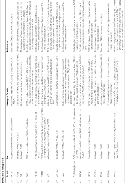
1. (1) Mucin-1 (MUC-1), death-associated protein binding to PKM2 can increase enzyme activity and promote glycolysis; hybrid double yeast technology studies show that PIAS3 (protein inhibitor of acti-vated STATA3) regulates enzyme activity through binding of carboxy terminus to PKM2 to promote glycolysis [149]. (2) O-GlcNAcylation can block the Thr405 and Ser406 sites of PKM2, and the stability of PKM2 in tetrameric form is reduced to enhance the aerobic glycolysis, namely Warburg Effect [150]. (3) The binding of histone demethylation enzyme Jumonji-C (JmjC) domain-containing protein 5 (JMJD5) [151], l-cysteine [152] and HSP40 [153] to PKM2 can reduce the enzyme activity. (4) 5-Amino-4-succinic acid carboxamide imidazole
ribonu-Table 3 Post‑translational modification of PKM2 protein and its related biological effects
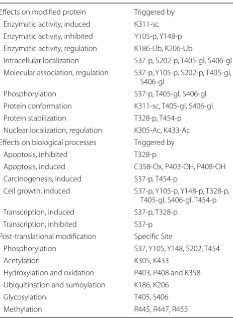
PTM (post-translational modification) effects of PKM2
Effects on modified protein Triggered by
Enzymatic activity, induced K311-sc
Enzymatic activity, inhibited Y105-p, Y148-p
Enzymatic activity, regulation K186-Ub, K206-Ub
Intracellular localization S37-p, S202-p, T405-gl, S406-gl
Molecular association, regulation S37-p, Y105-p, S202-p, T405-gl,
S406-gl
Phosphorylation S37-p, T405-gl, S406-gl
Protein conformation K311-sc, T405-gl, S406-gl
Protein stabilization T328-p, T454-p
Nuclear localization, regulation K305-Ac, K433-Ac
Effects on biological processes Triggered by
Apoptosis, inhibited T328-p
Apoptosis, induced C358-Ox, P403-OH, P408-OH
Carcinogenesis, induced S37-p, T454-p
Cell growth, induced S37-p, Y105-p, Y148-p, T328-p,
T405-gl, S406-gl, T454-p
Transcription, induced S37-p, T328-p
Transcription, inhibited S37-p
Post-translational modification Specific Site
Phosphorylation S37, Y105, Y148, S202, T454
Acetylation K305, K433
Hydroxylation and oxidation P403, P408 and K358
Ubiquitination and sumoylation K186, K206
Glycosylation T405, S406
cleotides activate PKM2 by direct contact allosteric regulation. (5) Parkin promotes the ubiquitination of Lys186 and Lys206 sites in PKM2, which inhibits the biological activity of PKM2 and regulates glucose metabolism by promoting ubiquitination of Lys186 and Lys206 sites of PKM2 [154]. (6) PKM2 can bind to SAICAR, and mutation of PKM2 Gly415 site will make PKM2 unable to bind to SAICAR [55]. Prolyl hydroxylase 3 (PHD3) can hydroxylate the Pro403 and Pro408 sites of PKM2 [155]. While PKM2 with the help of methylglyoxal (MG) can glycosylate the Arg399 site of PKM2, the result of which can change [156]. (7) Similarly, in the nucleus PKM2 exon 10 can be methylated by co-activator associated arginine methyltransferase 1 (CARM1), but the specific loca-tion is unknown. After being methylated by CARM1, PKM2 can promote aerobic glycolysis and malignant transformation of tumors [157].
2. Because PKM2 can enter the nucleus to act as a tran-scriptional regulator, the intermodulation between proteins is also present in the nucleus, and NAD-dependent deacetylase sirtuin6 (SIRT 6) can deacety-late the 433th lysine of PKM2 in the nucleus [158]. It inhibits PKM2 transfer out of the nucleus and inhib-its tumor growth. Proline hydroxylation of PKM2 at 403 and 408 by the PHD3 enzyme favors the inter-action of PKM2 with the HIF-1α transcription com-plex, which results in recruitment of P300-acetyl-transferase to facilitate the transactivation of HIF target genes.
3. Although this mode of regulation of intermolecular modification is more common in cells, for PKM2 proteins with three-dimensional structure, the mode of regulation involved is not limited to the modes we mentioned above. For example, for certain pro-tein molecules, it does not have propro-tein kinase activ-ity, but it can bind to a specific domain on PKM2, thereby regulating the biological activity of PKM2 [159]. For example: (1) probable E3 ubiquitin-protein ligase (HERC-1) can bind to the amino acid sequence of paragraphs 406 to 531 of PKM2. HERC-1 can ubiquitinate PKM2 to induce MET processes in tumor cells [160]. (2) SUMO E3-ligase (PIAS-3) can bind to the amino acid sequence of paragraphs 1 to 348 of PKM2. The sumoylation of PKM2 can pro-mote its nuclear localization [161]. (3) Oct-4 can bind to the amino acid sequence of paragraphs 307 to 531 of PKM2. It can work with PKM2 to regu-late gene transcription in cells, thereby regulating cell cycle and cell division [161]. (4) Opa can bind to the amino acid sequence of paragraphs 367 to 531 of PKM2. The interaction between OPA and PKM2 can promote the MET process of the free tumor cells that
have undergone MET conversion, and then adhere to the metastatic site to invade [162]. (5) This inter-action also occurred between HIF-1α/β, HCV, HPV, PANK-4, PML, SOCS-3, TEM-8, etc. protein and PKM2. In addition to promoting the formation of PKM2 dimer, its role is to promote the nuclear locali-zation of PKM2. This in turn induces transcription and translation of downstream genes of PKM2 and exacerbates the malignancy of tumor cells [46, 163–
165].
4. Co-activator-associated arginine methyltransferase 1 (CARM1) also known as PRMT4 methylates specifi-cally the dimeric form of PKM2 at Arg445, Arg447, Arg455 residues in the C domain which is PKM2 exon 10 located. TEPP-46 and FBP, the allosteric activators that induce PKM2 tetramerization lim-its PKM2 methylation. Importantly, PKM2 activity remains unaltered by methylation; however, meth-ylated PKM2 reprograms the metabolic phenotype toward aerobic glycolysis from oxidative phosphoryl-ation to support tumor cell proliferphosphoryl-ation, migrphosphoryl-ation, and metastasis [161].
PKM2 can not only exert the activity of PK enzyme in the form of tetramer, but also can enter the nucleus as a transcription factor to mediate the transcription of other genes when convert to dimer, can also regulate each other in the cytoplasm and other proteins. It has an impact on many different biological effects.
The application of PKM2 in Clinical
Fig. 7 Kaplan–Meier Curves for Survival of Four Most Relevant Cancers. Kaplan–Meier curves for survival of four most relevant cancers according to
PKM2 special transcript (NP-002645.3 refer to Fig. 5a and Table 1) expression in cancer tissues. Patients were divided into high and low PKM2 special