University of Pennsylvania
ScholarlyCommons
Publicly Accessible Penn Dissertations
1-1-2015
The Connection of Composition, Structure, and
Dynamic Processes to Tendon Mechanics:
Structure-Function Relationships in Collagen V
Deficient Tendons
Brianne Kathryn Connizzo
University of Pennsylvania, [email protected]
Follow this and additional works at:
http://repository.upenn.edu/edissertations
Part of the
Biomechanics Commons
This paper is posted at ScholarlyCommons.http://repository.upenn.edu/edissertations/1666
For more information, please [email protected].
Recommended Citation
Connizzo, Brianne Kathryn, "The Connection of Composition, Structure, and Dynamic Processes to Tendon Mechanics: Structure-Function Relationships in Collagen V Deficient Tendons" (2015).Publicly Accessible Penn Dissertations. 1666.
The Connection of Composition, Structure, and Dynamic Processes to
Tendon Mechanics: Structure-Function Relationships in Collagen V
Deficient Tendons
Abstract
Tendons are able to withstand the broad range of stresses and strains via their finely tuned composition and
structure. In addition, tendons undergo a coordinated set of dynamic responses, specifically collagen
uncrimping, re-alignment, sliding and deformation, within the matrix. To date, a complete understanding of
the hierarchical structure-function relationships in tendon is lacking. Therefore, the overall goal of this thesis
was to measure tendon structure and function in a mouse supraspinatus model of altered structure, and to
analyze links between mechanical properties, dynamic processes and composition/structure using a series of
statistical analyses. In the studies presented here, we used novel and established methods to measure the
multi-scale composition, structure and mechanical function of mouse supraspinatus tendons from wild type,
collagen V heterozygous and collagen V null mice. Overall, we found that the experimental groups were
mechanically inferior to the wild type group, with larger changes in both macroscale function and the
dynamic responses (re-alignment, crimp, deformation, sliding). In addition, while fibril morphology was
altered at both locations, the insertion site also exhibited alterations in cell and fiber morphology as well as
extracellular matrix composition. Finally, using a novel regression approach, we found that the contribution of
composition and structure as well as the contribution of dynamic processes to determining macroscale
mechanical function was highly dependent on location and that the dynamic processes were significant
mediators of the relationship between composition/structure and mechanical properties. Overall, we
conclude that although collagen V is a quantitatively minor component in mature tendon/ligament, it is a
major regulator of composition and structure during development which ultimately leads to mechanical
function. Furthermore, we conclude that the dynamic responses to load are crucial factors in ultimately
determining regionally-dependent mechanical function. This information will help to guide clinicians in
developing preventative techniques and appropriate rehabilitation strategies, as well as help to define the
appropriate and important parameters on which to base tissue engineering efforts for tendon augmentation or
replacement. Finally, this work presents a strong foundation on which to develop future experimental and
modeling efforts in order to fully understand the complex structure-function relationships present in tendon.
Degree Type
Dissertation
Degree Name
Doctor of Philosophy (PhD)
Graduate Group
Bioengineering
First Advisor
Louis J. Soslowsky
Keywords
collagen V, composition, dynamic, mechanics, structure, tendon
Subject Categories
Biomechanics
THE CONNECTION OF COMPOSITION, STRUCTURE, AND DYNAMIC PROCESSES
TO TENDON MECHANICS: STRUCTURE-FUNCTION RELATIONSHIPS IN
COLLAGEN V DEFICIENT TENDONS
Brianne Kathryn Connizzo
A DISSERTATION
In
Bioengineering
Presented to the Faculties of the University of Pennsylvania
In
Partial Fulfillment of the Requirements for the
Degree of Doctor of Philosophy
2015
Supervisor of Dissertation Graduate Group Chairperson
__________________________ _________________________
Louis J. Soslowsky, Ph.D. Jason A. Burdick, Ph.D.
Fairhill Professor of Orthopaedic Surgery Professor of Bioengineering
Dissertation Committee
Robert L. Mauck, Ph.D. (Committee Chair)
Associate Professor of Orthopaedic Surgery and Bioengineering, University of Pennsylvania
Andrew F. Kuntz, M.D.
Assistant Professor of Orthopaedic Surgery, University of Pennsylvania
X. Sherry Liu, Ph.D.
Assistant Professor of Orthopaedic Surgery, University of Pennsylvania
David E. Birk, Ph.D.
Distinguished Professor, Molecular Pharmacology and Physiology, University of South Florida
Abbas F. Jawad, Ph.D.
THE CONNECTION OF COMPOSITION, STRUCTURE, AND DYNAMIC PROCESSES
TO TENDON MECHANICS: STRUCTURE-FUNCTION RELATIONSHIPS IN
COLLAGEN V DEFICIENT TENDONS
COPYRIGHT
2015
iii
ACKNOWLEDGEMENTS
Without the generous assistance and counsel of my many mentors, colleagues, friends,
and family, the completion of this thesis would have been immeasurably more difficult. I’d like to
first thank Dr. Louis Soslowsky, my advisor and mentor, for his continued guidance, commitment
and support. I’d also like to thank Dr. David Birk for his support of our many collaborative studies
over the past five years, as well as his insight and knowledge in the planning and execution of
this thesis. Thank you Dr. Rob Mauck, my committee chair, for continuing to support me from the
first day I stepped into McKay. I’d also like to thank Dr. Sherry Liu for her professional and
personal support throughout my career. I also thank Dr. Andy Kuntz for his clinical perspective
and Dr. Abbas Jawad for his assistance with planning and executing my statistical regressions.
Finally, I’d like to thank all of my committee members for always providing valuable commentary
and suggestions throughout my studies.
I’d like to also express my gratitude to the many people who contributed directly to this
thesis work. I extend my deepest gratitude to the entire laboratory staff at the University of South
Florida for all of their hard work and hospitality during my several visits. I’m incredibly grateful for
Mei Sun and Qingmei (Chris) Yao for all of their work breeding and caring for all of the mice
necessary for this work. I’d also like to thank Thomas and Sheila Adams for their dedication and
attention to detail in collecting and analyzing data in this thesis. I can’t thank these colleagues
and friends enough for the many hours we worked together on my marathon day trips to Florida.
I’d also like to thank Dr. Lin Han and Dr. Joseph Sarver for their mentorship, patience, and
guidance in development of the fibril deformation assay. Additionally, I thank Jennica Tucker,
Carrie Barnum, Benjamin Freedman, and Pankti Bhatt for always having a positive attitude while
helping out on long days of assay development and execution.
Of course this work would not have been possible without a lot of hard work from
members in the McKay Orthopaedic Research Laboratory as well. Thank you to all of the office
staff who do all of the behind-the-scenes work for the lab. I’d especially like to thank Susan
iv
hardworking individuals I’ve ever met. I’d also like to thank Kelly McGinnis for always being
available for a chat and constantly having snacks to fuel us. I’d also like to thank Dr. Mike Hast
and Dr. Snehal Shetye for their help with my several hard drive failures and frozen computers.
I would not be able to accept full credit for this work without acknowledging several of the
opportunities that lead me to be able to do this work. I’d like to thank my high school volleyball
coach and close friend, Beth Powell, who always encouraged and challenged me to be my best
against all adversity. I’d also like to thank all of the incredible women before me at Smith College
who paved the way for me in the first all women engineering program. I am truly proud to be a
part of such an incredible group of alumni. Thank you to Dr. Kristin Miller who sparked my interest
in this work at the very beginning of my career and to Dr. Katie Reuther for her continued
friendship and support. I’m so lucky to have been part of such an incredibly talented and
collaborative community. It would not have been such a fun journey without the encouragement
and friendship from my fellow labmates and friends. In particular, I’d like to thank Sarah Rooney
and Jennica Tucker for being with me, sometimes literally next to me, every day since the very
beginning. I’d also like to express my gratitude to the Graduate Association of Bioengineering
(GABE) and the Graduate Student Engineering Group (GSEG) for all of the opportunities I’ve
been given and the friends I’ve made during my four years of service.
Finally, none of this work would have been possible without the love and support of my
family and friends. Thank you to my mom, Barbara, who is my confidant, my number one
supporter and my best friend. Thank you to my dad, Al, who challenged me to be the best I could
be no matter what I was doing. Thank you to my older brother Alex for teaching me new things
and always reminding me that there’s much more to life than science. Thank you also to my
oldest brother Nick and his wife Kate for inspiring me to keep learning in hopes of someday
surpassing them in trivia knowledge. Finally, I thank Nick Trojanowski for his endless patience,
understanding and love. I am so thankful that we’ve been able to share this experience and I
v
ABSTRACT
THE CONNECTION OF COMPOSITION, STRUCTURE, AND DYNAMIC PROCESSES TO
TENDON MECHANICS: STRUCTURE-FUNCTION RELATIONSHIPS IN COLLAGEN V
DEFICIENT TENDONS
Brianne Kathryn Connizzo
Dr. Louis J. Soslowsky
Tendons are able to withstand a broad range of stresses and strains via their finely tuned
composition and structure. In addition, tendons undergo a coordinated set of dynamic responses,
specifically collagen uncrimping, re-alignment, sliding and deformation, within the matrix. To date,
a complete understanding of the hierarchical structure-function relationships in tendon is lacking.
Therefore, the overall goal of this thesis was to measure tendon structure and function in a
mouse supraspinatus model of altered structure, and to analyze links between mechanical
properties, dynamic processes and composition/structure using a series of statistical analyses. In
the studies presented here, we used novel and established methods to measure the multi-scale
composition, structure and mechanical function of mouse supraspinatus tendons from wild type,
collagen V heterozygous and collagen V null mice. Overall, we found that the experimental
groups were mechanically inferior to the wild type group, with larger changes in both macroscale
function and the dynamic responses (re-alignment, crimp, deformation, sliding). In addition, while
fibril morphology was altered at both locations, the insertion site also exhibited alterations in cell
and fiber morphology as well as extracellular matrix composition. Finally, using a novel regression
approach, we found that the contribution of composition and structure as well as the contribution
of dynamic processes to determining macroscale mechanical function was highly dependent on
location and that the dynamic processes were significant mediators of the relationship between
composition/structure and mechanical properties. Overall, we conclude that although collagen V
is a quantitatively minor component in mature tendon/ligament, it is a major regulator of
vi
Furthermore, we conclude that the dynamic responses to load are crucial factors in ultimately
determining regionally-dependent mechanical function. This information will help to guide
clinicians in developing preventative techniques and appropriate rehabilitation strategies, as well
as help to define the appropriate and important parameters on which to base tissue engineering
efforts for tendon augmentation or replacement. Finally, this work presents a strong foundation on
which to develop future experimental and modeling efforts in order to fully understand the
vii
TABLE OF CONTENTS
ACKNOWLEDGEMENTS ... III
ABSTRACT ... V
TABLE OF CONTENTS ... VII
LIST OF TABLES ... X
LIST OF FIGURES ... XI
CHAPTER 1: INTRODUCTION ... 1
A. Introduction ... 1
B. Background ... 1
B-1. Tendon Mechanics ... 1
B-2. Tendon Composition and Structure... 2
B-3. Tendon Dynamic Parameters ... 4
C. Significance of Studies ... 8
C-1. Multiscale Structure-Function Relationships in Tendon ... 8
C-2. Classic Ehlers-Danlos Syndrome ... 9
D. Specific Aims ... 10
E. Study Design ... 12
E-1. Animal Model ... 12
E-2. Animal Use and Sample Size Justification ... 13
F. Chapter Overview ... 14
G. References ... 14
CHAPTER 2: IN SITU FIBRIL STRETCH AND SLIDING IS
LOCATION-DEPENDENT IN MOUSE SUPRASPINATUS TENDONS ... 21
A. Introduction ... 21
B. Methods ... 23
B-1. Sample Preparation ... 23
B-2. Atomic Force Microscopy (AFM) ... 24
B-3. Data Analysis ... 25
B-4. Statistical Analysis ... 26
C. Results ... 27
D. Discussion ... 28
E. References ... 32
CHAPTER 3: COLLAGEN V-DEFICIENT TENDONS EXHIBIT ALTERED
DYNAMIC MECHANICAL BEHAVIOR AT MULTIPLE HIERARCHICAL
SCALES ... 36
A. Introduction ... 36
B. Methods ... 38
B-1. Dynamic Viscoelastic Testing ... 39
viii
B-3. Collagen Fiber Re-Alignment ... 40
B-4. Collagen Fiber Uncrimping ... 42
B-5. Fibril Deformation and Sliding ... 42
C. Results ... 44
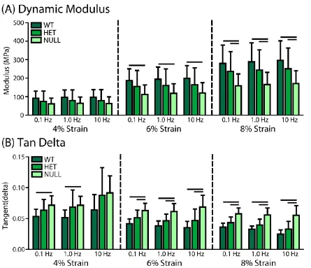
C-1. Tissue-level Quasi-static and Dynamic Viscoelastic Mechanics. ... 44
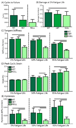
C-2. Tissue-level Fatigue Mechanics ... 47
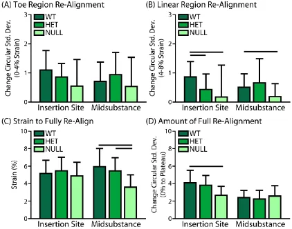
C-3. Collagen Fiber Re-Alignment ... 48
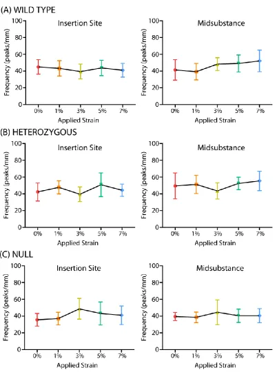
C-4. Collagen Fiber Uncrimping ... 50
C-5. Fibril Deformation ... 51
C-6. Fibril Sliding ... 52
D. Discussion ... 53
E. References ... 57
CHAPTER 4: COLLAGEN V EXPRESSION IS CRUCIAL FOR PROPER
DEVELOPMENT OF SUPRASPINATUS INSERTION SITE STRUCTURE AND
COMPOSITION ... 65
A. Introduction ... 65
B. Methods ... 66
B-1. Animals and Sample Collection ... 66
B-2. Collagen Fiber Crimp Morphology ... 67
B-3. Fibril Morphology ... 67
B-4. Cell Morphology ... 68
B-5. Biochemistry ... 69
B-6. Elastin Immunofluorescence ... 69
B-7. Statistical Analysis ... 70
C. Results ... 70
C-1. Joint Morphology ... 70
C-2. Fiber Morphology ... 71
C-3. Fibril Morphology ... 72
C-4. Cell Morphology ... 75
C-5. ECM Composition ... 76
D. Discussion ... 77
E. References ... 82
CHAPTER 5: MULTISCALE REGRESSION MODELING IN MOUSE
SUPRASPINATUS TENDONS REVEALS THAT DYNAMIC PROCESSES ACT
AS MEDIATORS IN STRUCTURE-FUNCTION RELATIONSHIPS ... 88
A. Introduction ... 88
B. Methods ... 90
B-1. Data Collection ... 90
B-2. Multiple Regression Analysis ... 92
B-3. Mediator Modeling ... 93
C. Results ... 95
C-1. Correlations Between Independent Variables ... 95
C-2. Regression of Mechanical Parameters on Dynamic Processes ... 96
C-3. Regression of Dynamic Processes on Composition and Structure ... 98
C-4. Mediator Modeling ... 100
D. Discussion ... 101
E. References ... 107
ix
A. Introduction ... 114
B. Chapter Two Conclusions ... 114
C. Chapter Three Conclusions ... 116
D. Chapter Four Conclusions ... 118
E. Chapter Five Conclusions ... 119
F. Overall Study Conclusions ... 121
G. Future Directions ... 122
G-1. Joint Mechanics ... 123
G-2. Injury and Healing ... 124
G-3. Aging ... 125
G-4. Gender ... 126
G-5. Detection and Prevention of Injury ... 126
G-6. Alternative Models of Structure-Function Relationships ... 128
G-7. Alternative Loading Conditions ... 130
G-8. Recovery, Biological Adaptation and Remodeling ... 132
H. Final Conclusions ... 133
I. References ... 133
APPENDIX A: EXPERIMENTAL PROTOCOLS ... 142
x
LIST OF TABLES
CHAPTER 2
Table 2-1: Fibril Strains………..28
CHAPTER 5 Table 5-1: Parameters in Statistical Analysis……….91
Table 5-2: Independent Parameter Correlations………95
Table 5-3: Mechanical Properties Regressed on Dynamic Processes………..………96
Table 5-4: Dynamic Processes Regressed on Composition and Structure………...98
xi
LIST OF FIGURES
CHAPTER 1
Figure 1-1: Tendon Composition and Structure………2
Figure 1-2: Dynamic Re-Organizations of Collagen Fiber Structure……….5
Figure 1-3: Mouse Models of Classic Ehlers-Danlos Syndrome………9
Figure 1-4: Overall Study Design………..13
CHAPTER 2 Figure 2-1: Testing Schematic for AFM Method……….24
Figure 2-2: AFM Analysis………..……….25
Figure 2-3: AFM Study Fibril Deformation………..……….27
Figure 2-4: AFM Study Fibril Sliding…….………..……….28
CHAPTER 3 Figure 3-1: Viscoelastic Testing and Analysis………..………..39
Figure 3-2: Fatigue Testing and Analysis………40
Figure 3-3: Re-Alignment Analysis………...…………41
Figure 3-4: Fiber Uncrimping and Fibril Deformation Protocol……….42
Figure 3-5: Animal and Tendon Morphology………..44
Figure 3-6: Quasi-Static Mechanical Parameters………..45
Figure 3-7: Dynamic Mechanical Parameters……….46
Figure 3-8: Fatigue Parameters………47
Figure 3-9: Collagen Fiber Re-Alignment………..……….49
Figure 3-10: Collagen Fiber Uncrimping………..50
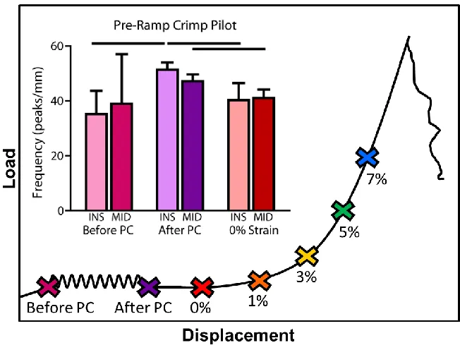
Figure 3-11: Pre-Ramp to Failure Crimp Pilot Study………...…..51
xii CHAPTER 4
Figure 4-1: Joint Morphology………70
Figure 4-2: Crimp Morphology……….……….71
Figure 4-3: Representative Fibril Micrographs...………...….72
Figure 4-4: Fibril Diameter Distribution...……….73
Figure 4-5: Fibril Morphology……….74
Figure 4-6: Cell Morphology……….….75
Figure 4-7: Biochemical Quantification………...….76
Figure 4-8: Elastin Immunofluorescence……….77
Figure 4-9: Humerus Histomorphometry……….80
1
CHAPTER 1: INTRODUCTION
A. Introduction
Tendons function primarily as mechanical, load-bearing structures to transmit forces from
muscle to bone, thus allowing for motion. To perform these tasks efficiently and to withstand the
broad range of stresses and strains, tendon composition and organization are finely tuned. When
focusing on the initial response of tendon to load, there is not enough time to synthesize new or
remodel the existing matrix and thus we consider the structure and composition to be unchanging
during this early time period. However, during this initial loading, tendons do undergo a
coordinated set of responses which we term ‘dynamic’, specifically fiber uncrimping and
re-alignment and fibril sliding and deformation within the matrix. To date, a complete understanding
of these unique structure-function relationships in tendon is lacking. Recent statistical models
have been used to correlate some compositional/structural measures, such as fibril morphology,
glycosaminoglycan (GAG) content, and collagen content with tendon mechanical properties.
However, the dynamic response to loading is often overlooked. Advances in methodology have
recently allowed measurement of these dynamic properties and evidence suggests that the
dynamic response is critical for proper tendon function. Therefore, the overall goal of this thesis
was to measure tendon structure and function in a mouse supraspinatus model of altered
structure, and to analyze links between mechanical properties, dynamic processes and
composition/structure using a series of statistical analyses.
B. Background
B-1. Tendon Mechanics
Tendon is a compliant, anisotropic material which has a high modulus under tension, but
collapses under compression. This primary uniaxial tensile function along the longitudinal axis of
2
exhibits nonlinear biomechanical behavior as exhibited by a typical stress-strain curve with an
initial, non-linear “toe-region” followed by the “linear-region” (Rigby et al., 1959). Clinically, the
ability of tendon to demonstrate these properties allows for its ability to both guide movement (low
stiffness) and provide stability (high stiffness). The toe-region is thought to be caused by gradual
recruitment of crimped collagen fibers (Hansen et al., 2002; Woo et al., 2000). After the
toe-region, all collagen fibers are believed to be supporting load and thus, the stress-strain curve
becomes linear. In addition to their anisotropic, non-linear behavior, tendons exhibit viscoelastic
properties identified as stress relaxation, hysteresis, and creep (Woo et al., 2000). Stress
relaxation refers to a non-linear decrease in stress over a period of time when a tendon is held
under constant tension. This process is both static and dynamic, as demonstrated by a similar
decrease in peak stress over time with repetitive, cyclic tensile loading. Hysteresis represents the
energy loss within the tendon with dynamic testing, accounting for a gradual change in load
elongation curves with tendon loading and unloading. Creep occurs when a tendon is held under
constant tension, and a measurable increase in tendon length along its longitudinal axis over time
is observed. These viscoelastic properties emphasize the ability of tendon to structurally adapt to
constant or cyclical loads in order to reach biomechanical equilibrium (Einhorn et al., 2007).
B-2. Tendon Composition and Structure
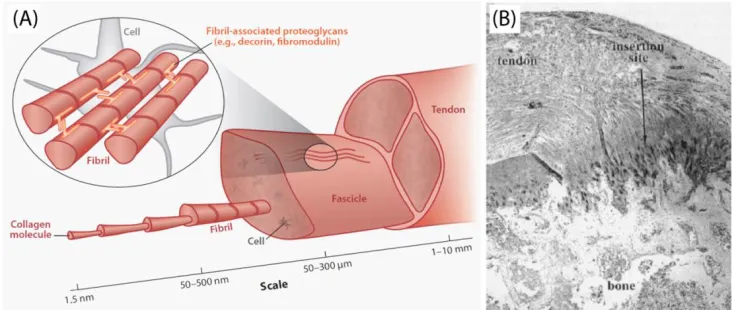
3
proteoglycans (Reproduced from Voleti et al. 2012) (B) Histological (4x) section of the rat supraspinatus tendon-to-bone insertion site, highlighting the transition zone (Reproduced from
Thomopoulos et al. 2002)
Tendon’s mechanical function is tuned by the unique composition and structure of the
tissue, primarily by its extracellular matrix. This matrix, comprised predominantly of collagen type
I, is organized in a hierarchical manner parallel to the mechanical axis of the tendon (Fig. 1-1).
Collagen microfibrils organize by lateral and longitudinal stacking, thus forming a lattice-type
configuration, called a fibril. Fibrils then associate together to form fibers, which then further
combine to form fascicles, which then bundle to form full tendon (Birk et al., 1995; Birk et al.,
1997). The process of fibril association and growth, called fibrillogenesis, is modulated mainly by
proteoglycans (decorin, biglycan, fibromodulin, lumican) and minor collagens, such as collagen
types V, XI, XII, and XIV. The unique structure of the collagen fibrils is suggested to play a role in
the mechanical function of the tendon. This collagen orientation promotes very high strength in
the direction of fiber alignment, which is dependent on the underlying organizational structure of
collagen molecules. In addition, literature suggests that collagen fiber crimp is responsible for
some of the nonlinearity of tendon by acting as a shock absorber (Hansen et al., 2002).
In addition to collagen I, the extracellular matrix is composed of minor collagens, elastin,
proteoglycans (PGs), glycolipids, and cellular material (Woo et al., 2005). Proteoglycans attach to
collagen fibrils in an orthogonal manner (Cribb and Scott, 1995; Scott et al., 1981), and extend
their specialized carbohydrate chains, known as glycosaminoglycans (GAGs), into the
inter-fibrillar space. GAGs have a negative charge, thus attracting and binding to water molecules
(Woo et al., 2005). The role of PGs and GAGs in tendon’s response to load has been highly
debated over the past decade (Fessel et al., 2012; Fessel and Snedeker, 2009; Lujan et al.,
2007; Lujan et al., 2009; Screen et al., 2005). It has been speculated that GAGs may
mechanically interconnect adjacent collagen fibrils, specifically, the GAG chains linked to decorin.
Relative movements of stained GAGs during mechanical relaxation tests have been quantified,
which led to the hypothesis of interfibrillar force transfer through a ratchet mechanism (Cribb and
4
provided conclusive evidence to support this mechanism. There have been a few studies to
suggest that GAGs and PGs may play a role in dynamic mechanical properties (Rigozzi et al.,
2009). Most recently, the influence of decorin on the patellar tendon mechanical properties was
investigated in a dose dependent manner. This study found no differences in elastic or
compressive properties but a dependence on strain rate and frequency, necessitating further
exploration into the dynamic behavior of decorin and biglycan knockout tendons (Dourte et al.,
2012). In addition, proteoglycan removal was found to have a significant effect on collagen fiber
re-alignment and aging in the mouse supraspinatus tendon, suggesting a relationship between
composition and the dynamic processes (Connizzo et al., 2013).
In vivo, the structure and composition of tendon is capable of adapting to altered loading,
overuse, injury and healing via changes in gene and protein expression following the event.
However, when focusing on the initial response of tendon to load, there is not enough time to
synthesize new or remodel the existing matrix and thus we can consider these parameters to be
unchanging during this early time period. This period, the focus of these studies, is when direct
relationships between structure/composition and function can be defined.
B-3. Tendon Dynamic Parameters
There are a number of ‘dynamic’ processes that occur during the mechanical test, specifically
fiber uncrimping, fiber re-alignment, fibril deformation and fibril sliding.
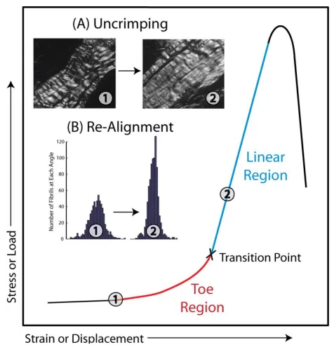
B-3-1. Fiber Uncrimping
Crimp is a periodic waveform configuration visualized within the collagen fibers and has
been implicated in the mechanical behavior of collagen (Woo et al., 2000). Particularly, the
flattening or disappearance of the crimp morphology has been implicated in the non-linear
behavior observed in the toe-region of the stress-strain curve (Fig. 1-2A) (Atkinson et al., 1999;
5
testing as visualized by polarized light microscopy has been well documented (Hansen et al.,
2002; Jozsa and Kannus, 1997), but few have been able to quantify crimp during the mechanical
test. Using a custom freeze-spraying technique and more recently using an integrated polarized
light testing setup, our lab has measured changes in crimp frequency throughout mechanical
testing. A recent analysis of mouse supraspinatus tendon demonstrated that the uncrimping of
collagen fibers along the entirety of the tendon was confined to the toe-region (Miller et al.,
2012b).
B-3-2. Fiber Re-Alignment
Figure 1-2. Typical stress-strain curve of tendon shows an elongated low stiffness toe region transitioning to a high stiffness linear region. Inset are two mechanisms of tendon’s response to load within the toe region, (A) the uncrimping and (B) re-alignment of collagen fibers, represented
by a decreased in the variance of the distribution of fiber angles from the toe to linear region.
The native collagen alignment of the tissue varies along the length of a tendon, with a
more disorganized fiber matrix located at the tendon-to-bone insertion (Lake et al., 2009; Lake et
6
towards the axis of loading, decreasing the distribution of fiber angles (Fig. 1-2B). This
mechanism is called collagen fiber re-alignment. Recent efforts in our lab have focused on the
ability to quantify collagen fiber re-alignment using a novel polarized light setup that allows for
simultaneous measurement and mechanical testing (Lake et al., 2009; Lake et al., 2010; Miller et
al., 2012a; Miller, 2012; Miller et al., 2012b). This approach has determined that re-alignment
occurs differently when measured at various points throughout the mechanical test, with the
majority of re-alignment occurring during preconditioning and the toe region of the mechanical
test. In addition, measurement of local re-alignment has determined that changes in collagen fiber
alignment throughout loading are location-dependent (Miller, 2012; Miller et al., 2012b).
B-3-3. Fibril Deformation and Sliding
A number of researchers have studied the behavior of collagen fibrils individually in
addition to fibril-fibril interactions (Gautieri et al., 2011, 2012; Heim et al., 2007; Svensson et al.,
2010; Szczesny and Elliott, 2014; van der Rijt et al., 2006; Veres and Lee, 2012). Collagen fibrils
have been likened to nanostructural biological cables and with techniques such as atomic force
microscopy, the isolated study of their mechanical properties is possible. Although mechanical
properties of single fibrils have been investigated by one-dimensional tensile testing using optical
tweezers (Wang et al., 2005) and atomic force microscopy (AFM) (Graham et al., 2004; Yang et
al., 2007), the mechanical behavior of the collagen fibrils themselves has not been able to
account for full tendon mechanics. Recent studies have been directed toward understanding the
behavior of single collagen fibrils in native arrangement (bundled) in response to the application
of macroscopic load. Collagen fibril deformation has been measured in situ recently by measuring
changes in d-period spacing with different level strains, providing evidence that d-period spacing
increases with increased applied load, thus supporting stretch of individual collagen fibrils
(Connizzo et al., 2014; Rigozzi et al., 2011). Macroscopic tendon extension is enabled by
7
fibrils, which is partially regulated by matrix components such as proteoglycans (Rigozzi et al.,
2009; Rigozzi et al., 2011; Screen et al., 2005). Fibril sliding has been cited as one contributor to
tendon viscoelastic behavior (Silver et al., 2002), and is thought to occur during the toe region of
the mechanical test. Recent evidence also suggests that fibril deformation and sliding are
location-dependent, alluding to structure-function relationships that are unique even along the
length of the tendon (Connizzo et al., 2014).
B-3-4. Regression Modeling of Mechanical Properties
Recent studies have addressed the relationships of quasi-static mechanical behavior of
tendon to composition/structure. One study measured the mechanical properties of rat tail tendon
fascicles and seven structural and compositional variables, attempting to find evidence of direct
structure-function relationships using a multiple regression model (Robinson et al., 2004). The
most prevalent predictors of mechanical behavior in this study were GAG content and collagen
fibril area fraction, suggesting that both GAGs and collagen fibril size can predict mechanical
properties. Interestingly, mean collagen fibril diameter was not a significant predictor. This
contrasts several studies where fibril diameter was correlated with mechanical properties (Derwin
and Soslowsky, 1999; Hansen et al., 2010; Parry, 1988), but agrees with other studies observing
the lack of correlation in developing tendon or self-assembled collagen fibers (Birk et al., 1991;
Christiansen et al., 2000; Hansen et al., 2010). A study in human patellar tendon found
differences in collagen fibril distribution between the posterior and anterior aspect of the tendon,
but no correlation with fascicle mechanical properties (Hansen et al., 2010). Another study
investigated fibril morphology and mechanical properties in the Achilles tendon, noting that larger
fibrils may be associated with a stiffer tendon but that this could be due to fibril-fibril interactions
or fibril-non-fibrillar matrix interactions (Rigozzi et al., 2010). Confusion in the literature suggests
8
structure and that there must be alternative mechanisms of load transfer such as re-alignment,
uncrimping, sliding, etc.
Recent advances in technology have allowed for the measurement of dynamic
parameters, but only a limited number of studies have investigated the relationship between
dynamic measures and mechanical properties. In a study of collagen fiber re-alignment in the
human supraspinatus tendon, initial fiber alignment was found to be significantly correlated with
linear region modulus (Lake et al., 2010). This was confirmed in the mouse supraspinatus tendon,
showing that increased mechanical properties were associated with increased alignment (Miller et
al., 2012a). Recent evidence also suggests a dynamic relationship between collagen crimp and
mechanical parameters from fatigue loading (Freedman et al., 2015). In addition, changes in
dynamic properties have been noted to occur with changes in composition and structure as well
(Connizzo et al., 2013; Miller et al., 2012a; Miller et al., 2012c), suggesting a complex, yet
unidentified, relationship between the three hierarchical levels of properties.
C. Significance of Studies
C-1. Multiscale Structure-Function Relationships in Tendon
Tendons function in a complex environment and are subjected to a variety of multi-axial
loads. The ability to respond effectively under these conditions is directly tuned by the unique
structure of the tissue. Characterization of structure-function relationships in tendon has been
mostly limited to comparisons between macroscopic (full tissue) mechanical properties and
nanoscale composition and structure. These studies will greatly enhance the fundamental
knowledge of structure-function relationships at several hierarchical levels and elucidate the
relationships between them. An improved understanding of these relationships also provides
insight on the mechanisms driving normal tissue adaptation, aging, and degeneration. Further,
9
comprehensive than that of any previous study of tendon, can be used as a paradigm to
thoroughly evaluate tendon and other fiber-reinforced soft biological tissues.
Previous work has used linear correlations to assess the independent, univariate effects
of a single organization or a single biochemical variable on tendon mechanics. Unfortunately, in
vivo systems are much more complicated than can often be addressed with such an approach,
particularly when considering the complex structural hierarchy in tendon. Two recent studies in
our lab have utilized a method for evaluating structure-function relationships in tissue taking into
consideration multiple independent variables that may be present (Ansorge et al., 2012; Robinson
et al., 2004). These studies build on these previous studies by investigating if the dynamic
processes (re-alignment, uncrimping, deformation, and sliding) act as mediators in the
relationship between composition/structure and mechanical properties. Given that the dynamic
processes take place at a larger hierarchical scale (nano/micro) than the compositional and
structural parameters, this unique modeling technique is able to determine if there is a mediating
relationship between these parameters and to determine which dynamic processes act as
mediators for various mechanical parameters.
C-2. Classic Ehlers-Danlos Syndrome
Figure 1-3. (A) Classic EDS mouse model exhibits hyperelastic skin and (B) tendon/ligament-specific collagen V null mouse exhibits extreme joint hypermobility.
In addition to the advantages this work has for the basic study of tendon biomechanics,
10
Syndrome (EDS) patients and the physicians that treat them. The classic form of EDS, defined by
haploinsufficiency for COL5A1 which is present in ~67% of affected individuals (Malfait et al.,
2005; Symoens et al., 2012), is characterized by hyperextensible skin, joint hypermobility and
instability, as well as abnormal wound healing and scarring (Steinmann, 2002). These functional
deficiencies can often lead to recurrent joint dislocations, particularly at the knee and shoulder
(Ainsworth and Aulicino, 1993; Stanitski et al., 2000), as well as to injury and disease. Our
established haploinsufficient Col5a1+/- classic EDS mouse model recapitulates the human
clinical phenotype (Wenstrup et al., 2006), and our tendon-specific col5a1 null mouse exhibits
even more severe effects (Fig. 1-3). In addition to the mechanical studies presented here, we
also examined the macroscopic tissue morphology and quantified changes in cell morphology
and many key matrix proteins (total collagen, GAGs, pyridinoline, elastin). Investigation into the
alterations in soft tissue structure and function, specifically in the tendon studied here, provide
more insight into the functional limitations and abnormalities in this severely understudied patient
population. In addition, this greatly improves the ability to treat those affected as well as help to
better prevent injuries.
D. Specific Aims
The overall goal of this thesis was to measure tendon structure and function in a mouse
supraspinatus model of altered structure, and to analyze links between mechanical properties
dynamic processes, and composition/structure using a series of statistical analyses. To
accomplish this objective, transgenic animal models with a reduction in collagen V expression
were analyzed for changes in multi-scale composition, structure, and mechanical function (Aim
1). Our global hypothesis was that alterations at the fibril level due to reduced collagen V
expression would cause alterations in composition and structure at increasing hierarchical levels
and ultimately to decreased dynamic and quasi-static mechanical function. We then used this
11
analyses followed by a causal analysis using single and multiple mediator models (Aim 2). Our
global hypothesis was that dynamic processes are mediating the relationship between
composition/structure and mechanical properties, mimicking the native structural hierarchy in
tendon.
Specific Aim 1: Define the structural, compositional, and mechanical properties of
mouse supraspinatus tendons from wild type, collagen V heterozygous and collagen V null mice.
Hypothesis 1a: Based on the reduction in fibril nucleation sites, collagen V heterozygous
and null tendons will have increased fibril diameters and decreased fibril density with similar
collagen content when compared to controls. Collagen V heterozygous and null tendons will also
have reduced cross-linking and GAG content due to the changes in diameter and intrafibrillar
spacing.
Hypothesis 1b: Based on previous and pilot mechanical data, collagen V heterozygous
and null tendons will have inferior quasi-static, dynamic, and fatigue properties, when compared
to control tendons. Since collagen V does not play a major role in fibrocartilage, these changes
will be more pronounced at the midsubstance than at the insertion site.
Hypothesis 1c: Collagen V heterozygous and null tendons will have increased crimp but
reduced alignment compared to control tendons, allowing for a longer toe region and later
transition to the linear region. They will also have reduced uncrimping, later re-alignment, and
increased fibril sliding. Finally, collagen V deficient tendons will have reduced fibril properties due
to altered collagen fibrillogenesis.
Specific Aim 2: Use correlation and multiple linear regression to investigate relationships
between (1) mechanical properties and dynamic processes and (2) dynamic processes and
composition/structure and determine if dynamic processes are mediators of the relationship
between composition/structure and mechanical properties.
Hypothesis 2a: As in previous studies, tendon mechanical properties will be correlated
12
determinant of overall mechanics, mechanical properties will also be strongly correlated to
dynamic measurements.
Hypothesis 2b: Dynamic processes will be strongly correlated with composition/structure,
since these parameters control structure and subsequently structural responses to load.
Hypothesis 2c: Dynamic processes will act as mediators in the relationship between
composition/structure and mechanical properties, due to the ability to mimic native tendon
structure and account for relationships between hierarchical levels.
E. Study Design
E-1. Animal Model
This study investigated the compositional, structural and mechanical properties in the
collagen V heterozygous and null mouse model. The collagen V heterozygous mouse model
(Col5a1+/-) has been well characterized as a model of classic Ehlers-Danlos syndrome, exhibiting
many clinical phenotypes (Wenstrup et al., 2006; Wenstrup et al., 2000; Wenstrup et al., 2011).
Due to the critical role of collagen V in fibril nucleation, the traditional collagen V null mouse
model exhibits embryonic lethality. Therefore, we used a tendon-specific collagen V null mouse,
Col5a1Δten/Δten. Col5a1flox/flox mice were created by flanking exons 3 and 4 of the Col5a1 gene with
loxP elements. The tendon/ligament-specific scleraxis-Cre (Scx-Cre) transgenic mice were gifts
from Dr. Ronen Schweitzer (Shriners Hospital, Portland, Oregon). Tendon/ligament–specific
expression was characterized by breeding Scx-Cre mice with Cre reporter mT/mG mice (The
Jackson Laboratory).To generate tendon specific type V collagen conditional mice Scx-Cre
transgenic mice were cross-bred with Col5a1flox/flox mice two generations to create
scx-Cre/Col51a1flox/flox (Col5a1Δten/Δten) mice. These mice were bred at the University of South Florida
13
Figure 1-4. Overall study design.
E-2. Animal Use and Sample Size Justification
Three hundred mice from three different groups were used in this study: control (WT),
Col5a1+/- (het), and Col5a1Δten/Δten (null). All animal procedures were IACUC approved prior to the
start of work. Mice were bred to 120 days of age and sacrificed humanely. The overall study
design is shown in Figure 1-4. Supraspinatus (SST) tendons from shoulders designated for
biological assays were dissected immediately following sacrifice and stored appropriately for the
particular assay. All other limbs were frozen at -20°C until dissection for mechanical testing.
Based on the correlation coefficients and R2 values from previous multiple regression analyses
performed in our lab, as well as knowledge of the variance in the specific parameters measured
here, we determined that a sample size of 20 tendons would provide us with sufficient power
(80%) to test the hypotheses presented above with significance set at p<0.05. Although not
explicitly stated in the aims of the study, qualitative or semi-quantitative measurements were also
performed to better characterize changes with the reduction of collagen V, specifically histology to
obtain a macroscopic view of tendon structure and immunofluorescence staining of elastin. For
14
F. Chapter Overview
Chapter two will describe the development of an AFM-based method to measure
instantaneous in situ collagen fibril deformation and sliding in mouse supraspinatus tendons.
Chapter three will describe the methods, results, and discussion for the experimental studies
performed to investigate the multi-scale mechanical function of mouse supraspinatus tendons
from collagen V wild type, heterozygous, and null mice. Chapter four will describe the methods,
results, and discussion for the experimental studies performed to investigate the multi-scale
composition and structure of mouse supraspinatus tendons from collagen V wild type,
heterozygous and null mice. Chapter five will describe the statistical studies investigating the
relationship between composition/structure, dynamic processes, and mechanical function.
Chapter six will summarize the conclusions of the previous chapters and provide future directions
for this area of research.
G. References
Ainsworth, S.R., Aulicino, P.L., 1993. A survey of patients with Ehlers-Danlos syndrome. Clin
Orthop Relat Res, 250-256.
Ansorge, H.L., Adams, S., Jawad, A.F., Birk, D.E., Soslowsky, L.J., 2012. Mechanical property
changes during neonatal development and healing using a multiple regression model. J Biomech
45, 1288-1292.
Atkinson, T.S., Ewers, B.J., Haut, R.C., 1999. The tensile and stress relaxation responses of
human patellar tendon varies with specimen cross-sectional area. J Biomech 32, 907-914.
Birk, D.E., Nurminskaya, M.V., Zycband, E.I., 1995. Collagen fibrillogenesis in situ: fibril segments
undergo post-depositional modifications resulting in linear and lateral growth during matrix
15
Birk, D.E., Silver, F.H., Trelstad, R.L., 1991. Matrix Assembly, in: Hay, E.D. (Ed.), Cell biology of
extracellular matrix. Plenum Press, New York.
Birk, D.E., Zycband, E.I., Woodruff, S., Winkelmann, D.A., Trelstad, R.L., 1997. Collagen
fibrillogenesis in situ: fibril segments become long fibrils as the developing tendon matures. Dev
Dyn 208, 291-298.
Christiansen, D.L., Huang, E.K., Silver, F.H., 2000. Assembly of type I collagen: fusion of fibril
subunits and the influence of fibril diameter on mechanical properties. Matrix Biol 19, 409-420.
Connizzo, B.K., Sarver, J.J., Han, L., Soslowsky, L.J., 2014. In situ fibril stretch and sliding is
location-dependent in mouse supraspinatus tendons. J Biomech 47, 3794-3798.
Connizzo, B.K., Sarver, J.J., Iozzo, R.V., Birk, D.E., Soslowsky, L.J., 2013. Effect of age and
proteoglycan deficiency on collagen fiber re-alignment and mechanical properties in mouse
supraspinatus tendon. J Biomech Eng 135, 021019.
Cribb, A.M., Scott, J.E., 1995. Tendon response to tensile stress: an ultrastructural investigation
of collagen:proteoglycan interactions in stressed tendon. J Anat 187 ( Pt 2), 423-428.
Derwin, K.A., Soslowsky, L.J., 1999. A quantitative investigation of structure-function
relationships in a tendon fascicle model. J Biomech Eng 121, 598-604.
Diamant, J., Keller, A., Baer, E., Litt, M., Arridge, R.G., 1972. Collagen; ultrastructure and its
relation to mechanical properties as a function of ageing. Proc R Soc Lond B Biol Sci 180,
293-315.
Dourte, L.M., Pathmanathan, L., Jawad, A.F., Iozzo, R.V., Mienaltowski, M.J., Birk, D.E.,
Soslowsky, L.J., 2012. Influence of decorin on the mechanical, compositional, and structural
properties of the mouse patellar tendon. J Biomech Eng 134, 031005.
Einhorn, T.A., Buckwalter, J.A., O'Keefe, R.J., American Academy of Orthopaedic, S., 2007.
Orthopaedic basic science : foundations of clinical practice. American Academy of Orthopaedic
Surgeons, Rosemont, IL.
Fessel, G., Gerber, C., Snedeker, J.G., 2012. Potential of collagen cross-linking therapies to
16
Fessel, G., Snedeker, J.G., 2009. Evidence against proteoglycan mediated collagen fibril load
transmission and dynamic viscoelasticity in tendon. Matrix Biol 28, 503-510.
Freedman, B.R., Zuskov, A., Sarver, J.J., Buckley, M.R., Soslowsky, L.J., 2015. Evaluating
changes in tendon crimp with fatigue loading as an ex vivo structural assessment of tendon
damage. J Orthop Res.
Gautieri, A., Vesentini, S., Redaelli, A., Buehler, M.J., 2011. Hierarchical structure and
nanomechanics of collagen microfibrils from the atomistic scale up. Nano Lett 11, 757-766.
Gautieri, A., Vesentini, S., Redaelli, A., Buehler, M.J., 2012. Viscoelastic properties of model
segments of collagen molecules. Matrix Biol 31, 141-149.
Graham, J.S., Vomund, A.N., Phillips, C.L., Grandbois, M., 2004. Structural changes in human
type I collagen fibrils investigated by force spectroscopy. Exp Cell Res 299, 335-342.
Hansen, K.A., Weiss, J.A., Barton, J.K., 2002. Recruitment of tendon crimp with applied tensile
strain. J Biomech Eng 124, 72-77.
Hansen, P., Haraldsson, B.T., Aagaard, P., Kovanen, V., Avery, N.C., Qvortrup, K., Larsen, J.O.,
Krogsgaard, M., Kjaer, M., Peter Magnusson, S., 2010. Lower strength of the human posterior
patellar tendon seems unrelated to mature collagen cross-linking and fibril morphology. J Appl
Physiol 108, 47-52.
Heim, A.J., Koob, T.J., Matthews, W.G., 2007. Low strain nanomechanics of collagen fibrils.
Biomacromolecules 8, 3298-3301.
Jozsa, L., Kannus, P., 1997. Histopathological findings in spontaneous tendon ruptures. Scand J
Med Sci Sports 7, 113-118.
Lake, S.P., Miller, K.S., Elliott, D.M., Soslowsky, L.J., 2009. Effect of fiber distribution and
realignment on the nonlinear and inhomogeneous mechanical properties of human supraspinatus
tendon under longitudinal tensile loading. Journal of Orthopaedic Research 27, 1596-1602.
Lake, S.P., Miller, K.S., Elliott, D.M., Soslowsky, L.J., 2010. Tensile properties and fiber alignment
of human supraspinatus tendon in the transverse direction demonstrate inhomogeneity,
17
Lujan, T.J., Underwood, C.J., Henninger, H.B., Thompson, B.M., Weiss, J.A., 2007. Effect of
dermatan sulfate glycosaminoglycans on the quasi-static material properties of the human medial
collateral ligament. J Orthop Res 25, 894-903.
Lujan, T.J., Underwood, C.J., Jacobs, N.T., Weiss, J.A., 2009. Contribution of
glycosaminoglycans to viscoelastic tensile behavior of human ligament. J Appl Physiol 106,
423-431.
Malfait, F., Coucke, P., Symoens, S., Loeys, B., Nuytinck, L., De Paepe, A., 2005. The molecular
basis of classic Ehlers-Danlos syndrome: a comprehensive study of biochemical and molecular
findings in 48 unrelated patients. Hum Mutat 25, 28-37.
Miller, K., Connizzo, B., Soslowsky, L., 2012a. Collagen Fiber Re-Alignment in a Neonatal
Developmental Mouse Supraspinatus Tendon Model. Annals of Biomedical Engineering 40,
1102-1110.
Miller, K.S., Connizzo, B.K., Feeney, E., Soslowsky, L.J., 2012b. Characterizing local collagen
fiber re-alignment and crimp behavior throughout mechanical testing in a mature mouse
supraspinatus tendon model. Journal of Biomechanics 45, 2061-2065.
Miller, K.S., Connizzo, B.K., Feeney, E., Tucker, J.J., Soslowsky, L.J., 2012c. Examining
differences in local collagen fiber crimp frequency throughout mechanical testing in a
developmental mouse supraspinatus tendon model. J Biomech Eng 134, 041004.
Miller, K.S., Edelstein, L., Connizzo, B. K., and Soslowsky, L. J... 2012. Effect of Preconditioning
and Stress Relaxation on Local Collagen Fiber Re-Alignment: Inhomogeneous Properties of Rat
Supraspinatus Tendon. Journal of Biomechanical Engineering In press.
Parry, D.A., 1988. The molecular and fibrillar structure of collagen and its relationship to the
mechanical properties of connective tissue. Biophys Chem 29, 195-209.
Rigby, B.J., Hirai, N., Spikes, J.D., Eyring, H., 1959. The Mechanical Properties of Rat Tail
18
Rigozzi, S., Muller, R., Snedeker, J.G., 2009. Local strain measurement reveals a varied regional
dependence of tensile tendon mechanics on glycosaminoglycan content. J Biomech 42,
1547-1552.
Rigozzi, S., Muller, R., Snedeker, J.G., 2010. Collagen fibril morphology and mechanical
properties of the Achilles tendon in two inbred mouse strains. J Anat 216, 724-731.
Rigozzi, S., Stemmer, A., Muller, R., Snedeker, J.G., 2011. Mechanical response of individual
collagen fibrils in loaded tendon as measured by atomic force microscopy. J Struct Biol 176, 9-15.
Robinson, P.S., Lin, T.W., Jawad, A.F., Iozzo, R.V., Soslowsky, L.J., 2004. Investigating tendon
fascicle structure-function relationships in a transgenic-age mouse model using multiple
regression models. Ann Biomed Eng 32, 924-931.
Scott, J.E., Orford, C.R., Hughes, E.W., 1981. Proteoglycan-collagen arrangements in developing
rat tail tendon. An electron microscopical and biochemical investigation. Biochem J 195, 573-581.
Screen, H.R., Shelton, J.C., Chhaya, V.H., Kayser, M.V., Bader, D.L., Lee, D.A., 2005. The
influence of noncollagenous matrix components on the micromechanical environment of tendon
fascicles. Ann Biomed Eng 33, 1090-1099.
Silver, F.H., Ebrahimi, A., Snowhill, P.B., 2002. Viscoelastic properties of self-assembled type I
collagen fibers: molecular basis of elastic and viscous behaviors. Connect Tissue Res 43,
569-580.
Stanitski, D.F., Nadjarian, R., Stanitski, C.L., Bawle, E., Tsipouras, P., 2000. Orthopaedic
manifestations of Ehlers-Danlos syndrome. Clin Orthop Relat Res, 213-221.
Steinmann, B., 2002. The collagen family: structure, assembly, and organization in the
extracellular matrix, Connective Tissue and Its Heritable Disorders. Wiley-Liss, New York, pp.
431-523.
Svensson, R.B., Hassenkam, T., Hansen, P., Peter Magnusson, S., 2010. Viscoelastic behavior
of discrete human collagen fibrils. J Mech Behav Biomed Mater 3, 112-115.
Symoens, S., Syx, D., Malfait, F., Callewaert, B., De Backer, J., Vanakker, O., Coucke, P., De
19
over 90% of patients with classic EDS and allows to refine diagnostic criteria. Hum Mutat 33,
1485-1493.
Szczesny, S.E., Elliott, D.M., 2014. Interfibrillar shear stress is the loading mechanism of collagen
fibrils in tendon. Acta Biomater 10, 2582-2590.
van der Rijt, J.A., van der Werf, K.O., Bennink, M.L., Dijkstra, P.J., Feijen, J., 2006.
Micromechanical testing of individual collagen fibrils. Macromol Biosci 6, 697-702.
Veres, S.P., Lee, J.M., 2012. Designed to fail: a novel mode of collagen fibril disruption and its
relevance to tissue toughness. Biophys J 102, 2876-2884.
Wang, X., Li, X., Yost, M.J., 2005. Microtensile testing of collagen fibril for cardiovascular tissue
engineering. J Biomed Mater Res A 74, 263-268.
Wenstrup, R.J., Florer, J.B., Davidson, J.M., Phillips, C.L., Pfeiffer, B.J., Menezes, D.W.,
Chervoneva, I., Birk, D.E., 2006. Murine model of the Ehlers-Danlos syndrome. col5a1
haploinsufficiency disrupts collagen fibril assembly at multiple stages. J Biol Chem 281,
12888-12895.
Wenstrup, R.J., Florer, J.B., Willing, M.C., Giunta, C., Steinmann, B., Young, F., Susic, M., Cole,
W.G., 2000. COL5A1 haploinsufficiency is a common molecular mechanism underlying the
classical form of EDS. Am J Hum Genet 66, 1766-1776.
Wenstrup, R.J., Smith, S.M., Florer, J.B., Zhang, G., Beason, D.P., Seegmiller, R.E., Soslowsky,
L.J., Birk, D.E., 2011. Regulation of collagen fibril nucleation and initial fibril assembly involves
coordinate interactions with collagens V and XI in developing tendon. J Biol Chem 286,
20455-20465.
Woo, S.L., Debski, R.E., Zeminski, J., Abramowitch, S.D., Saw, S.S., Fenwick, J.A., 2000. Injury
and repair of ligaments and tendons. Annu Rev Biomed Eng 2, 83-118.
Woo, S.L., Thay, Q.L., Abramowitch, S.D., Gilbert, T.W., 2005. Structure and Function of
Ligaments and Tendons, Basic orthopaedic biomechanics & mechano-biology, 3 ed. Lippincott,
20
Yang, L., van der Werf, K.O., Koopman, B.F., Subramaniam, V., Bennink, M.L., Dijkstra, P.J.,
Feijen, J., 2007. Micromechanical bending of single collagen fibrils using atomic force
21
CHAPTER 2: IN SITU FIBRIL STRETCH AND SLIDING IS LOCATION-DEPENDENT
IN MOUSE SUPRASPINATUS TENDONS
A. Introduction
Tendon’s primary function is to transmit mechanical load and displacement from muscle
to bone (Lichtwark and Barclay, 2010). It is able to perform this function due to its finely tuned
hierarchical structure, composed of collagen fibrils organized into fibers or fascicles and further
bundled to form tendon proper (Kastelic et al., 1978). Macroscopic structure-function studies of
tendon have shown that mechanical changes occurring at lower scale levels are likely
responsible for the complex non-linear and viscoelastic response of full tendon (Derwin and
Soslowsky, 1999; Fessel and Snedeker, 2009; Lake et al., 2009; Lake et al., 2010; Rigozzi et al.,
2009; Robinson et al., 2004). Recent evidence suggests that tendons are able to withstand high
forces by employing a number of unique mechanisms occurring at many of the fibril and fiber
length scales, including uncrimping, re-alignment, sliding, and deformation or stretch (Connizzo et
al., 2013b; Gupta et al., 2010; Miller et al., 2012b; Screen et al., 2013). While collagen fiber
uncrimping and re-alignment have been studied extensively in recent literature (Connizzo et al.,
2013a; Connizzo et al., 2013b; Miller et al., 2012a; Miller et al., 2012b; Miller et al., 2012c; Miller
et al., 2012d), the quantification of collagen fiber and fibril sliding and stretch has been studied
less due to the experimental difficulties, particularly the inability to visualize individual collagen
fibrils in vivo during mechanical loading.
Mechanical properties of single collagen fibrils have recently been investigated using
several different technologies (Eppell et al., 2006; Graham et al., 2004; Tang et al., 2010; Yang et
al., 2007). While these studies substantially improved our understanding of mechanics of
individual collagen fibrils, they do not replicate the in vivo environment of collagen, where
collagen fibrils are interacting with other collagen fibrils and with the surrounding extracellular
matrix proteins. Furthermore, it has been reported that fiber-level elongation cannot be solely
attributed to the deformation of the individual collagen fibrils, suggesting fibril-fibril and fibril-matrix
22
Recent investigations utilizing atomic force microscopy have successfully measured d-period
length changes as a quantitative measure of collagen fibril stretch in situ (Li et al., 2013; Rigozzi
et al., 2011). This work introduces a significant advancement in the literature, allowing for the
ability to study fibril stretch under various types of mechanical loading as well as with cases of
altered structure, such as disease, aging, or injury (Li et al., 2013). However, these studies have
primarily investigated fiber sliding during or following stress relaxation or creep events (Gupta et
al., 2010; Li et al., 2013; Rigozzi et al., 2011; Screen et al., 2013). The strain rate dependence of
tendon identifies that the timing and rate of loading, in addition to the magnitude, is extremely
important to tendon’s response. Furthermore, since tendons have been known to rupture clinically
due to a single traumatic event or impact (Moller et al., 1996), the instantaneous response to
load, as well as the ability to repetitively undergo that impact stress, is critical to the overall
function and has not been investigated.
In addition, fibril sliding and deformation have primarily been studied during the linear
region of the mechanical test. Due to the prevalence of collagen uncrimping and re-alignment
during the initial toe region (Miller et al., 2012b; Miller et al., 2012c), it is likely that these other
fibrillar responses are also occurring. Finally, while many studies have demonstrated that the
specific transition in composition, structure and collagen organization from the midsubstance to
the insertion site contributes significantly to the full tendon’s mechanical response (Lake et al.,
2009; Lake et al., 2010; Shaw and Benjamin, 2007), the location-dependent response to
mechanical load at the fibril level has not been studied. Therefore, the purpose of this study was
to quantify the instantaneous response of collagen fibrils throughout a mechanical loading
protocol both in the insertion site and midsubstance of the mouse supraspinatus tendon. We
hypothesized that more fibril stretch will occur at the insertion site than the midsubstance (higher
strains) and that more fibril sliding will occur at the midsubstance than at the insertion site.
23
B. Methods
B-1. Sample Preparation
Fifteen C57BL/6 mice at 150 days of age were used in this study (IACUC approved).
Supraspinatus tendons from both shoulders of each mouse were used for this study, but no two
tendons from the same animal were used in the same testing group to ensure independence of
samples. All soft tissues were removed from around the tendon, leaving the supraspinatus tendon
attached to the humerus. Tendon cross-sectional area was then measured using a custom
laser-based device (Peltz et al., 2009). The humerus was then embedded in an acrylic tube with
PMMA. A second coating of PMMA was applied to prevent failure at the growth plate. The
proximal end of the tendon was glued between two pieces of sandpaper with an initial gauge
length of 2.5mm and both the tendon and the acrylic pot were placed in custom grips for tensile
testing (Fig. 2-1A,B), as described previously (Connizzo et al., 2013a).
All samples were kept hydrated using phosphate buffered saline (PBS) and were then
loaded in a tensile testing system (Instron, Norwood, MA) for mechanical testing. A 10 N load cell
was used for all tests with a resolution of 0.01 N. All tendons underwent a preload to 0.02N and
ten cycles of preconditioning from 0.02N to 0.04N followed by a 60 second hold before the ramp
to failure. Tendons were then divided into 6 groups and stretched to a randomly assigned
grip-to-grip strain value (0, 1, 3, 5, 7, or 10%) at a rate of 0.1% strain per second. Tendons were then
frozen using freezing spray (McMaster-Carr Electrical Cleaning and Maintenance Aerosol,
Product #7437K43), removed from the mechanical testing setup, and placed in a specimen dish
with tissue freezing medium. The sample was kept frozen during this process using freezing
spray and the dish was then submerged in liquid nitrogen to complete the flash-freezing phase.
Tendons were then stored at -20°C until they were sectioned in a cryostat microtome in the
coronal anatomical plane at 20 microns and sections were then again kept frozen at -20°C until
further processing. Frozen sections were then immersed in cold 10% neutral buffered formalin for
24
Figure 2-1. (A) Testing image of supraspinatus tendon prepared for mechanical testing. (B) Zoomed-in view of supraspinatus tendon. (C) Diagram depicting insertion site and midsubstance
regions for d-period analysis. (D) Microscope images depicting typical scan regions for the midsubstance (top image) and insertion site (bottom images).
B-2. Atomic Force Microscopy (AFM)
Tendons were imaged in air using Peak Force Quantitative Nanomechanical Mapping
mode using a Dimension Icon AFM (BrukerNano, Santa Barbara, CA). Imaging of 2 µm × 2 µm
regions was performed using Bruker ScanAsyst Fluid+ probes (nominal spring constant k ≈ 0.70
N/m, radius R ≈ 2 nm). Tendons were scanned at 2-3 regions across the width of the insertion
site and midsubstance of the tendon. The insertion and midsubstance locations were determined
consistently by taking images within the bottom quarter (about 0.5mm) of the specimen for the
insertion site and the top quarter of the specimen for the midsubstance region (a single sample
region shown in Figure 2-1C). Scans were also taken from 4-6 sections throughout the depth of
25
Figure 2-2. (A) Sample scan of collagen fibrils in tendon with line highlighting the measurement of d-period in a single fibril. (B) Intensity versus length plot for line in panel (A) which is used to
measure d-period length using custom software.
B-3. Data Analysis
Several parameter maps were produced from imaging, including Height, Peak Force,
Peak Force Error, Modulus, LogModulus, Dissipation, and Adhesion. Analysis on approximately
two tendons (~20 images) showed no difference between the different parameter maps for
measurement of d-period. Due to the strength of contrast from the LogModulus and Adhesion
maps, these two maps were used to analyze all specimens. Custom MATLAB software
(MathWorks, Natick, MA) was written to allow for the measurement of d-period length for many
fibrils in a single image and for the ability to enhance the processing technique in the future.