Diagnosing Signi
fi
cant PDA Using
Natriuretic Peptides in Preterm
Neonates: A Systematic Review
Madhulika Kulkarni, MDa, Ganga Gokulakrishnan, MDa, Jack Price, MDb, Caraciolo J. Fernandes, MDa, Mariska Leeflang, PhDc, Mohan Pammi, MD, PhDa
abstract
BACKGROUND AND OBJECTIVES:Echocardiogram is the gold standard for the diagnosis ofhemodynamically significant patent ductus arteriosus (hsPDA) in preterm neonates. A simple blood assay for brain natriuretic peptide (BNP) or amino-terminal pro-B-type natriuretic peptide (NT-proBNP) may be useful in the diagnosis and management of hsPDA. Our objectives were to determine the diagnostic accuracy of BNP and NT-proBNP for hsPDA in preterm neonates and to explore heterogeneity by analyzing subgroups.
METHODS:The systematic review was performed as recommended by the Cochrane Diagnostic Test Accuracy Working Group. Electronic databases, conference abstracts, and cross-references were searched. We included studies that evaluated BNP or NT-proBNP (index test) in preterm neonates with suspected hsPDA (participants) in comparison with echocardiogram (reference standard). A bivariate random effects model was used for meta-analysis, and summary receiver operating characteristic curves were generated.
RESULTS:Ten BNP and 11 NT-proBNP studies were included. Studies varied by methodological quality, type of commercial assay, thresholds, age at testing, gestational age, and whether the assay was used to initiate medical or surgical therapy. Sensitivity and specificity for BNP at summary point were 88% and 92%, respectively, and for NT-proBNP they were 90% and 84%, respectively.
CONCLUSIONS:The studies evaluating the diagnostic accuracy of BNP and NT-proBNP for hsPDA varied widely by assay characteristics (assay kit and threshold) and patient characteristics (gestational and chronological age); therefore, generalizability between centers is not possible. We recommend that BNP or NT-proBNP assays be locally validated for specific patient population and outcomes, to initiate therapy or follow response to therapy.
aSection of Neonatology andbDivision of Cardiology, Department of Pediatrics, Texas Children’s Hospital & Baylor College of Medicine, Houston, Texas; andcDepartment of Clinical Epidemiology, Biostatistics and Bioinformatics, Academic Medical Center, University of Amsterdam, Amsterdam, Netherlands
Dr Kulkarni participated in the design, data acquisition, and interpretation and wrote the manuscript; Dr Gokulakrishnan participated in the design, data acquisition, analysis, and interpretation and assisted in writing the manuscript; Drs Price, Fernandes, and Leeflang provided critical intellectual input and revised the manuscript; and Dr Pammi conceived the project, participated in the design, data acquisition, analysis, and interpretation, and assisted in writing the manuscript.
www.pediatrics.org/cgi/doi/10.1542/peds.2014-1995
DOI:10.1542/peds.2014-1995 Accepted for publication Nov 21, 2014
Address correspondence to Mohan Pammi, MD, PhD, 6621 Fannin, MC: WT 6-104, Houston, TX 77030. E-mail: [email protected]
PEDIATRICS (ISSN Numbers: Print, 0031-4005; Online, 1098-4275).
Copyright © 2015 by the American Academy of Pediatrics
FINANCIAL DISCLOSURE:The authors have indicated they have nofinancial relationships relevant to this article to disclose.
FUNDING:No external funding.
A symptomatic hemodynamically significant PDA (hsPDA) is often treated medically or by surgical closure. Echocardiogram is the gold standard for the diagnosis of hsPDA, but it is expensive and not always available in resource-limited settings. A simple blood assay that can diagnose hsPDA reliably will be useful to clinicians in such settings.
Brain natriuretic peptide (BNP) and amino-terminal pro-B-type
natriuretic peptide (NT-proBNP) are synthesized and released into the circulation by the cardiac ventricular myocytes in response to pressure overload, volume expansion, and increase in myocardial wall stress. Within the myocytes, the precursor pro-BNP (108 amino acids) is converted to the biologically active form, BNP (77–108 amino acids) and the inactive NT-proBNP fragment (1–76 amino acids). BNP improves myocardial relaxation and regulates responses to acute increases in ventricular volume by opposing the vasoconstriction, sodium retention, and antidiuretic effects of the activated
renin-angiotensin-aldosterone system.1Plasma BNP and NT-proBNP are cleared by the kidneys and hence elevated in patients with renal failure. NT-proBNP has a longer half-life (60–120 minutes vs
22 minutes2,3) and is more stable in vitro. BNP and NT-proBNP are well-established markers of heart failure in adults4,5and children.6
Many commercial kits are available for assessment of BNP and
NT-proBNP, and levels reported vary with chronological age, gestational age (GA), PDA, and renal function. The normative values of BNP and NT-proBNP in neonates from various studies are presented in
Supplemental Table 3. BNP and NT-proBNP values are highest in the
first 3 days of life and decline progressively thereafter.7,8Levels in preterm neonates are higher than in full-term neonates and are similar by 1 month of life.9,10NT-proBNP values
are 6 to 20 times higher than BNP values, and the ratio between NT-proBNP and BNP is influenced by age.11–17
BNP and NT-proBNP assays have been reported in the management of multiple neonatal conditions
including hsPDA.9,18–51BNP and NT-proBNP have been used in preterm neonates both for diagnosis and to initiate medical or surgical treatment of hsPDA. The diagnostic accuracy of BNP and NT-proBNP in the management of hsPDA in neonates has not been systematically reviewed.
Our primary objective was to determine the diagnostic accuracy of cardiac biomarkers BNP and NT-pro-BNP in the diagnosis of hsPDA in preterm neonates.
The secondary objective was to explore heterogeneity among studies evaluating BNP and NT-proBNP by analyzing the following subgroups: commercial assay, test threshold, age of the patient at testing, GA at birth, and whether the test was used to initiate medical or surgical treatment.
METHODS
The method recommended by the Cochrane Diagnostic Test Accuracy Working Group was followed (http:// srdta.cochrane.org/). The title has been registered with the Cochrane Neonatal Review Group.
Criteria for Consideration of Studies for Review
Prospective and retrospective studies that evaluated blood BNP or
NT-proBNP (index tests) in the diagnosis of PDA (target condition) in preterm neonates in conjunction with an echocardiogram (reference standard) were eligible for inclusion. Studies were excluded in which a threshold was not reported or could not be obtained from the authors.
There is no consensus for
echocardiographic criteria for hsPDA. We chose 2 echocardiographic
criteria to define hsPDA in preterm infants based on the best available evidence: LA/Ao ratio.1.5 and ductal diameter.1.5 mm (as measured by color Doppler), which are early predictors of hsPDA.52 LA/Ao ratio is estimated from the diameter of the left atrium (LA) and from the aortic root diameter (Ao) in the parasternal long-axis view at the level of the aortic valve.52–54
Search Strategy for Identification of Studies
We searched the following sources in April 2014 without any language restriction:
1. Electronic bibliographic databases were searched: Medline (1966 to present) and Cumulative Index to Nursing and Allied Health Litera-ture (CINAHL) (1982 to present).
2. Abstracts of conferences: pro-ceedings of pediatric academic societies (American Pediatric Society, Society for Pediatric Research, and European Society for Pediatric Research) were searched from 1990 from the journalPediatric Researchand abstracts2view.com online.
3. The Web of Science was searched.
4. PubMed’s related citations feature and relevant identified articles were searched.
5. The Science Citation Index was used to identify relevant articles by using previously identified articles.
6. Additional searches were made from reference lists in identified studies.
The search strategy is described in Supplemental Appendix 1.
Data Collection and Analyses
Selection of Studies
inclusion eligibility by M.P. and M.K. independently. The results were compared and disagreements were resolved by mutual discussion.
Data Extraction and Management
Data extracted included author, year of publication and journal, study design, study population, reference standard and performance of the reference standard, index tests and performance of the index tests, information about quality assessment items based on Quality Assessment of Diagnostic-Accuracy Studies
(QUADAS-2),55and data for 232 tables. Additional information to clarify the study design and data was sought from the authors via e-mail for
$3 attempts. All the data were entered electronically on Microsoft Excel (Microsoft Corp, Redmond, WA) spreadsheets by M.P. and M.K. independently. The data extracted by each author were compared, and any discrepancies were resolved by mutual discussion and input from author C.J.F.
Assessment of Methodological Quality
The methodological quality of each study was assessed as recommended by the Cochrane Diagnostic Test Accuracy Working Group adapting from the QUADAS-2 by authors G.G. and M.K. The 4 domains assessed for risk of bias were patient selection, index test, reference test, andflow and timing. Applicability concerns were assessed in thefirst 3 domains, as described in Supplemental Appendix 2. The data were then entered into Review Manager Software (RevMan 5.3; The Cochrane Collaboration, Oxford, England) for meta-analysis and to generate
figures.56
Statistical Analyses and Data Synthesis
We constructed 232 tables for all studies with the reference standard and index tests and enumerated true-positives, true-positives, false-negatives, and true-negatives for all the reported thresholds. For the
studies that reported sensitivity and specificity at defined thresholds, reverse calculation was done to generate a 232 table. Data were entered in RevMan 5.3, and forest plots with 95% confidence intervals (CIs) for sensitivity and specificity for each study were created. Results were also plotted in the receiver operating characteristic (ROC) space with 95% confidence estimates, summary point, and the summary curve. The meta-analyses by bivariate random effects model were
performed by using the metandi package in the statistical software Stata 11 (Stata Corp, College Station, TX). The following covariates were evaluated: test assay kit, thresholds (as continuous), GA, chronological age
at the time of the test, and whether used to initiate medical or surgical therapy.
Investigations of Heterogeneity Among Studies
We investigated heterogeneity through subgroup analyses if data were available from$4 studies:
1. Type of commercial test assay used for BNP and NT-proBNP, be-cause measurements may vary with each commercial assay.
2. Effects of age at testing: BNP and NT-proBNP levels are higher at birth and then decline over time.
3. Effects of GA: BNP and NT-proBNP levels are higher in preterm infants than in term infants.
FIGURE 1
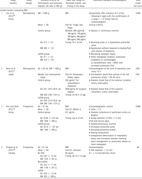
TABLE 1 Characteristics of Included Studies Study,
Country
Study Design
Inclusion Criteria for Participants’and Summary Statistic: GA (wk) or BW (g)
Index Test, Assay Kit, Threshold Cutoffs, and
Timing of the Assay
Reference Standard Echocardiogram Area Under the Curve
Included studies evaluating BNP 1 Lee et al
2013,20 Korea
Retrospective BW,1000 g BNP Symptomatic PDA: presence of 2 of the
following 5 signs with the confirmation of a large L→R ductalflow by
echocardiogram
0.830
Mean6SD Test kit: Triage, San
Diego
Clinical criteria
Control group Multiple: 864 pg/mL¥,
100 pg/mL, 150 pg/mL, 200 pg/mL, 400 pg/mL*, 600 pg/mL, 900 pg/mL
•Systolic or continuous murmur
GA: 27.362.3 Timing: 24 h of life •Bounding pulse or a hyperactive precordial pulse
BW: 8556112 •Hypotension without response to loadingfluid
and infusion of dopamine
hsPDA group: •Worsening ventilator status
GA: 27.162.2 •Chest radiograph evidence: pulmonary
congestion or cardiomegaly (a cardiothoracic ratio.60%) with increased pulmonaryflow BW: 8196123
2 Mine et al 2013,21 Japan
Retrospective GA,33 wk, BW,1500 g BNP Echocardiogram at the time of admission and
every 12 h
0.68
Median and interquartile range
Test kit: Shionospot, Osaka, Japan
•End-diastolic bloodflow velocity of the left pulmonary artery.30–40 cm/s
0.94
Control group 250 pg/mL* for
indomethacin treatment
•Diastolic bloodflow of the anterior cerebral artery interrupted
GA: 28.1 (25.5–29.2) wk 2000 pg/mL for surgical ligation
•Diastolic bloodflow of the superior mesenteric artery interrupted BW: 950 (799–1181) g Timing: 24–48 h of age
hsPDA group
GA: 28.0 (27.0–29.2) wk BW: 960 (735–1137) g
3 Kim and Shim
2012,19 Korea
Prospective GA,37 wk BNP Echocardiographic criteria 0.998
Mean6SD Test kit: Abbott, IL •LA/Ao.1.4
Control group 412 pg/mL •Diastolic turbulence of pulmonary artery on
Doppler
GA: 32.8362.22 wk Timing: day 4 of life •Ductal diameter of PDA.1.4 mm
BW: 19306490 g PLUS 3/6 clinical signs
hsPDA group •Systolic/continuous murmur
GA: 32.1861.67 wk •Increased precordial pulse
BW: 18506480 g •Bounding peripheral pulses
•Resting tachycardia
•Unexplained deterioration of respiratory status and increased vascular marking, heart enlargement, or pulmonary edema on chest radiograph
4 Elsayed et al 2012,58
Canada
Prospective GA,31 wk BNP Echocardiogram NA
Mean6SD Test kit: unknown •PDA diameter.1.5 mm
No PDA 90 pg/mL •L→R nonrestrictive shunt
GA: 29.361.0 wk Timing: 48–72 h of age BW: 13256261 g
Non-hsPDA GA: 28.261.7 wk BW: 11606257 g hsPDA
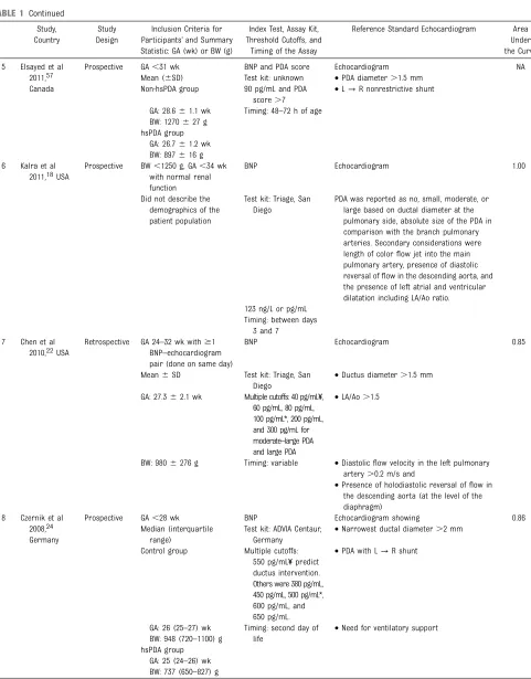
TABLE 1 Continued Study, Country
Study Design
Inclusion Criteria for Participants’and Summary Statistic: GA (wk) or BW (g)
Index Test, Assay Kit, Threshold Cutoffs, and
Timing of the Assay
Reference Standard Echocardiogram Area Under the Curve
5 Elsayed et al 2011,57
Canada
Prospective GA,31 wk BNP and PDA score Echocardiogram NA
Mean (6SD) Test kit: unknown •PDA diameter.1.5 mm
Non-hsPDA group 90 pg/mL and PDA
score.7
•L→R nonrestrictive shunt
GA: 28.661.1 wk Timing: 48–72 h of age BW: 1270627 g
hsPDA group GA: 26.761.2 wk BW: 897616 g 6 Kalra et al
2011,18USA
Prospective BW,1250 g, GA,34 wk with normal renal function
BNP Echocardiogram 1.00
Did not describe the demographics of the patient population
Test kit: Triage, San Diego
PDA was reported as no, small, moderate, or large based on ductal diameter at the pulmonary side, absolute size of the PDA in comparison with the branch pulmonary arteries. Secondary considerations were length of colorflow jet into the main pulmonary artery, presence of diastolic reversal offlow in the descending aorta, and the presence of left atrial and ventricular dilatation including LA/Ao ratio. 123 ng/L or pg/mL
Timing: between days 3 and 7
7 Chen et al 2010,22USA
Retrospective GA 24–32 wk with$1 BNP–echocardiogram pair (done on same day)
BNP Echocardiogram 0.85
Mean6SD Test kit: Triage, San
Diego
•Ductus diameter.1.5 mm
GA: 27.362.1 wk Multiple cutoffs: 40 pg/mL¥, 60 pg/mL, 80 pg/mL, 100 pg/mL*, 200 pg/mL, and 300 pg/mL for moderate–large PDA and large PDA
•LA/Ao.1.5
BW: 9806276 g Timing: variable •Diastolicflow velocity in the left pulmonary artery.0.2 m/s and
•Presence of holodiastolic reversal offlow in the descending aorta (at the level of the diaphragm)
8 Czernik et al 2008,24
Germany
Prospective GA,28 wk BNP Echocardiogram showing 0.86
Median (interquartile range)
Test kit: ADVIA Centaur, Germany
•Narrowest ductal diameter.2 mm
Control group Multiple cutoffs:
550 pg/mL¥predict ductus intervention. Others were 380 pg/mL, 450 pg/mL, 500 pg/mL*, 600 pg/mL, and 650 pg/mL.
•PDA with L→R shunt
GA: 26 (25–27) wk Timing: second day of life
•Need for ventilatory support BW: 948 (720–1100) g
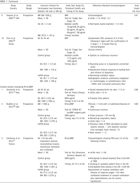
TABLE 1 Continued Study, Country
Study Design
Inclusion Criteria for Participants’and Summary Statistic: GA (wk) or BW (g)
Index Test, Assay Kit, Threshold Cutoffs, and
Timing of the Assay
Reference Standard Echocardiogram Area Under the Curve
9 Sanjeev et al 2005,25USA
Prospective BW 500–1500 g BNP Echocardiogram 0.91
Mean6SD Test kit: Triage, San
Diego, CA
•LA/Ao.1.4, LV/Ao.2.1
GA: 2662 wk Multiple cutoffs:
70 pg/mL¥,50 pg/mL, 60 pg/mL, 80 pg/mL, 90 pg/mL*, 100 pg/mL
•Narrowest ductal diameter.1.5 mm
BW: 873.56247 g Timing: variable 10 Choi et al
2005,26 Korea
Prospective GA 25–34 wk BNP Symptomatic PDA: presence of 2 of the
following 5 signs with the confirmation of a large L→R ductalflow by
echocardiogram
0.997
Mean6SD Test kit: Triage, San
Diego, CA
Clinical criteria
Control group Multiple cutoffs:
1110 pg/mL and 1295 pg/mL
•Systolic or continuous murmur
GA: 30.362.4 wk Timing: day 3 •Bounding pulse or a hyperactive precordial pulse
BW: 13966418 g •Hypotension without response to loadingfluid
and infusion of dopamine
hsPDA group •Worsening ventilator status
GA: 29.162.75 wk Radiographic evidence: pulmonary congestion
or cardiomegaly (a cardiothoracic ratio
.60%) with increased pulmonaryflow BW: 12836359 g
Included studies evaluating NT-proBNP 1 Occhipinti et al
2014,74Italy
Prospective GA 23–32 wk NT-proBNP •Ductal diameter/birth wt ratio.1.4 or 0.86
Mean6SD Test kit: Roche Elecsys,
Germany
•LA/Ao ratio.1.4 or
GA: 28.3 (62.6) wk 9854 pg/mL •Pulsatileflow pattern
BW: 1085 (6365) g Timing: 6–24 h of life 2 Buddhe et al
2012,60USA
Prospective BW,1500 g NT-proBNP PDA size.1 mm with$2 additional features of
PDA
0.98
Mean6SD Test kit: VITROS
NT-proBNP reagent pack
•Continuous murmur
Control group 5900 pg/mL •Pulse pressure.25 mm Hg
GA: 28.0 (62.4) wk Timing: day 3–5 of life •Worsening respiratory status
BW: 1053 (6278) g •LA/Ao ratio.1.4
hsPDA group •Resistive index of SMA calculated as peak
systolic velocity2end diastolic velocity/ time averaged mean velocity.6
GA: 26.6 (62.7) wk •Base excess.25
BW: 829 (6276) g 3 Cambonie et al
2012,27
France
Prospective GA,32 wk with respiratory distress necessitating invasive mechanical ventilation and surfactant
NT-proBNP Echocardiogram showing PDA and$2 of the
following criteria:
0.87
Mean6SD Test kit: RxL Dimension,
Siemens, Newark, NJ
•LA⁄Ao ratio.1.48
Control group 8500 pg/mL •Retrograde or absent diastolicflow in the ACA
or SMA
GA: 29.5 (62.2) wk Timing: 24–72 h of life •Growing or pulsatile patternflow in the DA
BW: 1256 (6397) g •End-diastolicflow velocity in the LPA.0.20 m⁄s
hsPDA group Requirement of invasive ventilation with
fraction of inspired oxygen.0.3 after surfactant treatment or invasive ventilation plus catecholamine use for severe hypotension
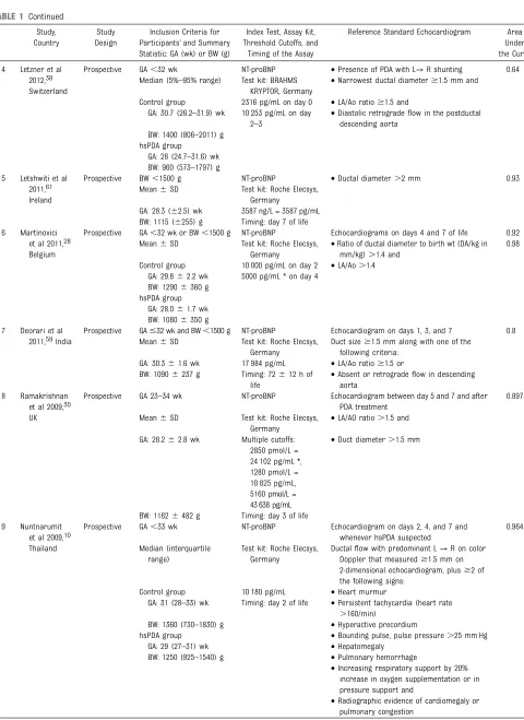
TABLE 1 Continued Study, Country
Study Design
Inclusion Criteria for Participants’and Summary Statistic: GA (wk) or BW (g)
Index Test, Assay Kit, Threshold Cutoffs, and
Timing of the Assay
Reference Standard Echocardiogram Area Under the Curve
4 Letzner et al 2012,38
Switzerland
Prospective GA,32 wk NT-proBNP •Presence of PDA with L→R shunting 0.64
Median (5%–95% range) Test kit: BRAHMS KRYPTOR, Germany
•Narrowest ductal diameter$1.5 mm and
Control group 2316 pg/mL on day 0 •LA/Ao ratio$1.5 and
GA: 30.7 (26.2–31.9) wk 10 253 pg/mL on day 2–3
•Diastolic retrogradeflow in the postductal descending aorta
BW: 1400 (806–2011) g hsPDA group
GA: 28 (24.7–31.6) wk BW: 960 (573–1797) g 5 Letshwiti et al
2011,61 Ireland
Prospective BW,1500 g NT-proBNP •Ductal diameter.2 mm 0.93
Mean6SD Test kit: Roche Elecsys,
Germany
GA: 28.3 (62.5) wk 3587 ng/L = 3587 pg/mL BW: 1115 (6255) g Timing: day 7 of life 6 Martinovici
et al 2011,28 Belgium
Prospective GA,32 wk or BW,1500 g NT-proBNP Echocardiograms on days 4 and 7 of life 0.92
Mean6SD Test kit: Roche Elecsys,
Germany
•Ratio of ductal diameter to birth wt (DA/kg in mm/kg).1.4 and
0.98
Control group 10 000 pg/mL on day 2 •LA/Ao.1.4
GA: 29.862.2 wk 5000 pg/mL * on day 4 BW: 12906360 g
hsPDA group GA: 28.061.7 wk BW: 10806350 g 7 Deorari et al
2011,59India
Prospective GA#32 wk and BW,1500 g NT-proBNP Echocardiogram on days 1, 3, and 7 0.8
Mean6SD Test kit: Roche Elecsys,
Germany
Duct size$1.5 mm along with one of the following criteria:
GA: 30.361.6 wk 17 984 pg/mL •LA/Ao ratio$1.5 or
BW: 10906237 g Timing: 72612 h of
life
•Absent or retrogradeflow in descending aorta
8 Ramakrishnan
et al 2009,30 UK
Prospective GA 23–34 wk NT-proBNP Echocardiogram between day 5 and 7 and after
PDA treatment
0.897
Mean6SD Test kit: Roche Elecsys,
Germany
•LA/AO ratio.1.5 and
GA: 28.262.8 wk Multiple cutoffs: 2850 pmol/L = 24 102 pg/mL *, 1280 pmol/L = 10 825 pg/mL, 5160 pmol/L = 43 638 pg/mL
•Duct diameter.1.5 mm
BW: 11626482 g Timing: day 3 of life 9 Nuntnarumit
et al 2009,10
Thailand
Prospective GA,33 wk NT-proBNP Echocardiogram on days 2, 4, and 7 and
whenever hsPDA suspected
0.964
Median (interquartile range)
Test kit: Roche Elecsys, Germany
Ductalflow with predominant L→R on color Doppler that measured$1.5 mm on 2-dimensional echocardiogram, plus$2 of the following signs:
Control group 10 180 pg/mL •Heart murmur
GA: 31 (28–33) wk Timing: day 2 of life •Persistent tachycardia (heart rate
.160/min)
BW: 1360 (730–1830) g •Hyperactive precordium
hsPDA group •Bounding pulse, pulse pressure.25 mm Hg
GA: 29 (27–31) wk •Hepatomegaly
BW: 1250 (925–1540) g •Pulmonary hemorrhage
•Increasing respiratory support by 20% increase in oxygen supplementation or in pressure support and
4. We determined whether the results of the test were used to initiate medical or surgical treat-ment of PDA.
5. Assay threshold cutoffs vary with the commercial assay and patient characteristics such as GA and chronological age.
RESULTS
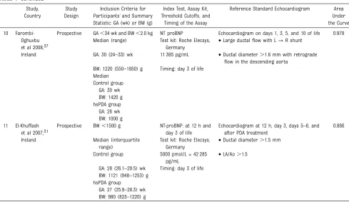
We identified 82 records through database searches and 21 additional articles from conference abstracts and cross-referencing. The inclusion process is detailed in the Preferred Reporting Items for Systematic Reviews and Meta-Analysesflow diagram (Fig 1). Ten studies that evaluated BNP and 11 studies that evaluated NT-proBNP in the
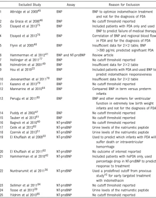
diagnosis of hsPDA met our inclusion criteria (Table 1). Excluded studies and reasons for exclusion are described in Table 2.
Methodological assessment of included studies revealed study
deficiencies in the following domains (Fig 2, Supplemental Figs 5 and 6):
1. Patient selection: Exclusion criteria in the individual studies were var-iable and were not defined in some studies, which may have in-troduced bias.30,57–59Some studies were retrospective in design.16–18 Inclusion criteria differed, and all studies except 119had restrictive inclusion criteria by birth weight (BW) and GA. Additional inclusion criteria included presence of re-spiratory distress necessitating mechanical ventilation and surfac-tant27and BNP echocardiogram performed on the same day,22 which might have excluded some eligible patients with hsPDA.
2. Index test: No study had a pre-defined threshold, and some studies did not blind the clinician.10,19,22,25,26,59–61Elsayed et al57,58used BNP in addition to a clinical PDA score as the index test. The PDA score incorporated echocardiographic parameters
reflective of both volume and pres-sure overload (maximum score 15), and clinical, radiologic, and labora-tory features of both pulmonary overcirculation and systemic hypo-perfusion (maximum score 13).
3. Reference standard: All studies used an acceptable reference standard, the echocardiogram, but echocardiogram criteria to define hsPDA were variable, which may introduce bias. There was absence of blinding of the cardiologists to the BNP assay in a few
studies.10,19,26,57,59–61
4. Flow and timing: We noted longer or unclear time intervals between the blood test (index test) and echocardiogram (reference stan-dard) in some studies, most nota-bly in the study by Kim and Shim,19 where time interval between the index and reference standard was as long as 48 hours.
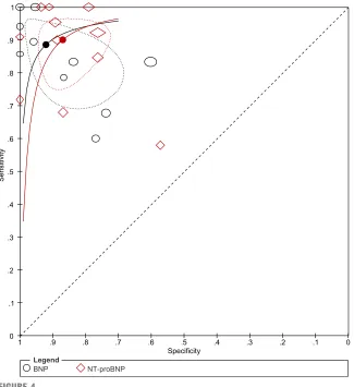
Estimated summary sensitivity for studies evaluating BNP was 0.88 (95% CI, 0.76–0.95) and for TABLE 1 Continued
Study, Country
Study Design
Inclusion Criteria for Participants’and Summary Statistic: GA (wk) or BW (g)
Index Test, Assay Kit, Threshold Cutoffs, and
Timing of the Assay
Reference Standard Echocardiogram Area Under the Curve
10 Farombi-Oghuvbu et al 2008,37
Ireland
Prospective GA,34 wk and BW,2.0 kg NT proBNP Echocardiogram on days 1, 3, 5, and 10 of life 0.978 Median (range) Test kit: Roche Elecsys,
Germany
•Large ductalflow with L→R shunt
GA: 30 (24–33) wk 11 395 pg/mL •Ductal diameter.1.6 mm with retrograde flow in the descending aorta
BW: 1220 (550–1950) g Timing: day 3 of life Median
Control group GA: 30 wk BW: 1420 g hsPDA group
GA: 26 wk BW: 1000 g 11 El-Khuffash
et al 2007,31 Ireland
Prospective BW,1500 g NT-proBNP: at 12 h and
day 3 of life
Echocardiogram at 12 h, day 3, days 5–6, and after PDA treatment
0.866
Median (interquartile range)
Test kit: Roche Elecsys, Germany
•Ductal diameter.1.5 mm
Control group 5000 pmol/L = 42 285
pg/mL
•LA/Ao.1.5
GA: 28 (26.1–29.5) wk Timing: day 3 of life BW: 1121 (948–1253) g
hsPDA group
GA: 27 (25.9–28.3) wk BW: 980 (823–1220) g
ACA, anterior cerebral artery; DA, ductus arteriosus; LPA, left pulmonary artery; SMA, superior mesenteric artery. * Threshold level used in the Forest plots and ROC analysis when using only one value per study;
NT-proBNP was 0.90 (95% CI, 0.79–0.96). Summary specificity for studies evaluating BNP was 0.92 (95% CI, 0.81–0.97) and for
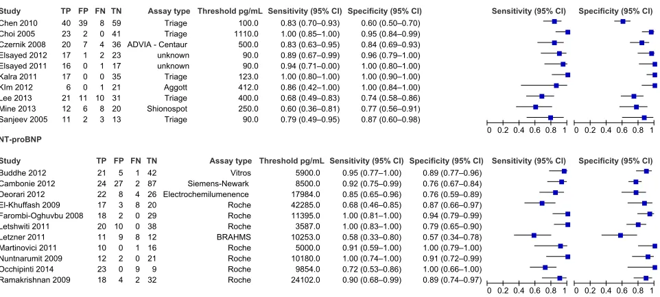
NT-proBNP 0.84 (95% CI, 0.77–0.90). Forest plots based on a single threshold from each study show that sensitivity across studies ranged from 0.60 to 1.0 for BNP and 0.58 to 1.0 for NT-proBNP, and specificity ranged from 0.6 to 1.0 for BNP and 0.57 to 1.0 for NT-proBNP (Fig 3). We also plotted the included studies in the ROC space to give a sense of the distribution of the sensitivity and specificity of the studies (Fig 4). We report the summary ROC curve, summary estimates of specificity and
sensitivity, and the 95% confidence regions.
We analyzed subgroups of studies evaluating BNP and NT-proBNP based on the type of test kit, age at testing, and whether used to initiate medical or surgical treatment, and summary estimates are shown in Supplemental Table 4 and
Supplemental Figs 7 and 8. Summary estimates by meta-analysis were possible only if$4 studies were available in the subgroup. Only 1 test kit and only day 3 of testing had.4 studies for both BNP and NT-proBNP. Only 2 studies evaluated BNP for surgical closure of the ductus and none for NT-proBNP. We could not
explore heterogeneity associated with gestational age or threshold cutoffs because of insufficient data and the large variability among studies, respectively.
DISCUSSION
We synthesized data from 21 studies by meta-analysis; 10 evaluated BNP and 1 evaluated NT-proBNP in the diagnosis of hsPDA in preterm neonates. Estimated summary sensitivity for BNP was 0.88 (95% CI, 0.76–0.95), and summary specificity was 0.92 (95% CI, 0.81–0.97). However, there was wide variation in sensitivity and specificity, and 2 of the 10 studies, at the reported thresholds, reported sensitivity,0.7.20,21Mine et al21described 2 thresholds in their study: 250 pg/mL for indomethacin treatment with sensitivity of 0.60 (95% CI, 0.36–0.81) and 2000 pg/mL for the surgical treatment of PDA with sensitivity of 1.00 (95% CI, 0.4–1.00), both with wide 95% CIs. Conversely, Lee et al20described sensitivity and specificity at a range of thresholds and, at a chosen threshold of 864 pg/mL, had a high specificity 0.95 (95% CI, 0.84–0.99) but low sensitivity at 0.55 (95% CI, 0.36–0.73). One outlier was the threshold given by Mine et al for indomethacin unresponsiveness and surgical indication with
100% sensitivity at a high level of 2000 pg/mL.21
The summary sensitivity for NT-proBNP was 0.90 (95% CI, 0.79–0.96), and summary specificity was 0.84 (95% CI, 0.77–0.90). In the 11 included studies that evaluated NT-proBNP, there was less variation in sensitivity and specificity of
NT-proBNP when compared with BNP in the detection of hsPDA. In 2 of the 11 studies, sensitivity was,0.734,38 because they used a higher threshold
.40 000 pg/mL. Letshwiti et al61 described a much lower threshold (3587 pg/mL) than El Khuffash and Molloy34(42 285 pg/mL) or
Ramakrishnan et al30(24 102 pg/mL) but had 100% sensitivity with narrow TABLE 2 Characteristics of Excluded Studies
Excluded Study Assay Reason for Exclusion
1 Attridge et al 200945 BNP BNP to optimize indomethacin treatment
and not for the diagnosis of PDA
2 da Graca et al 200646 BNP No cutoff threshold reported
3 Elsayed et al 201375 BNP Included patients with PDA only and used
BNP to predict failure of medical therapy
4 Elsayed et al 201376 BNP Correlation of BNP and regional bloodflow
in PDA and for the diagnosis of PDA
5 Flynn et al 200548 BNP Insufficient data for 232 table; BNP
.300 pg/mL predicted significant PDA 6 Hammerman et al 201241 BNP and NT-proBNP Review article
7 Hollinger et al 201177 BNP No cutoff threshold reported
8 Holmström et al 200149 BNP Insufficient data for 232 table
9 Hsu et al 201023 BNP Included patients with PDA and used BNP to
predict indomethacin responsiveness 10 Jeevananthan et al 201178 BNP Insufficient data for 232 table
11 Kazanci et al 201279 BNP No cutoff threshold reported
12 Mannarino et al 201044 BNP Compared BNP in term versus preterm
infants
13 Perugu et al 201180 BNP BNP and other markers for ventricular
function in extremely low birth weight infants and not for the diagnosis of PDA
14 Puddy et al 200247 BNP No cutoff threshold reported
15 Tauber et al 201381 BNP No cutoff threshold reported
16 Bagnoli et al 201040 NT-proBNP No cutoff threshold reported
17 Celik et al 201282 NT-proBNP Urine levels of the natriuretic peptide
18 Czernik et al 201351 NT-proBNP Urine levels of the natriuretic peptide
19 El Khuffash et al 200833 NT-proBNP Used to predict which infants with PDA will
suffer death or intraventricular hemorrhage
20 El Khuffash et al 201183 NT-proBNP No outcome of interest reported
21 Hammerman et al 201042 NT-proBNP Included patients with hsPDA only, used
percentage drop in NT-proBNP to predict response to treatment
22 Nuntnarumit et al 201136 NT-proBNP Used a predefined cutoff from previous study10for early targeted treatment with indomethacin
23 Sellmer et al 201184 NT-proBNP No cutoff threshold reported
24 Tosse et al 201239 NT-proBNP Urine levels of the natriuretic peptide
confidence intervals (95% CI, 0.83–1.00). The cutoffs in this study may be lower because of evaluation on day 7, when a physiologic decline in NT-proBNP levels is to be
expected.7,62,63
We assessed the methodological quality of studies by using the 4 domains of the QUADAS-2 checklist: patient selection, index test, reference standard, andflow and timing. Overall, most studies scored as low or unclear for risk of bias and
applicability concerns. All studies enrolled preterm neonates, but some of them were more restrictive than others. BNP levels vary by GA, and this could affect threshold values used to diagnose hsPDA. Some of the studies have additional inclusion criteria, such as ventilator support and surfactant,27and these criteria might introduce applicability concerns. Failure to report exclusion criteria or variable exclusion criteria may add bias.30,57–59Three studies
had a retrospective design but had comparable inclusion and exclusion criteria and definition for hsPDA.20–22 As described by Zonnenberg and de Waal,64there were wide variations in the definition of hsPDA in the included studies. This raises applicability concerns because a neonate with hsPDA in 1 study may be labeled as non-hsPDA in another. All the included studies evaluating NT-proBNP were prospective in design and more uniform in respect to inclusion criteria based on GA compared with studies evaluating BNP. One of the included studies (Kim and Shim19) reported the time interval of up to 48 hours between the index test and the reference standard. Because the half-life of BNP is 22 minutes and that of NT-proBNP is 60 to 120 minutes, changes in the physiologic status at such long time intervals may decrease the
comparability of the index and reference tests.2,3
Elsayed et al57,58used a clinical PDA score in addition to BNP levels for an ROC analysis. The clinical PDA score incorporated echocardiographic parameters reflective of both volume FIGURE 2
Risk of bias and applicability concerns graph based on QUADAS-2. Methodological assessment of included studies by QUADAS-2 method is presented. Review authors’judgments about each domain for risk of bias and applicability concerns presented as percentages across included studies: BNP (1st panel; 10 studies) and NT-proBNP (2nd panel; 11 studies). Studies with high risk of bias and appli-cability concerns are shown in red, those that are unclear in yellow, and those with low risk in green.
FIGURE 3
and pressure overload (maximum score 15) and clinical, radiologic, and laboratory features of both
pulmonary overcirculation and systemic hypoperfusion.65In addition, only 34 of 90 eligible neonates were recruited in the study. A sensitivity analysis excluding the 2 studies by Elsayed et al57,58with unknown test kits, unknown exclusion criteria, PDA scores, and unaccountable total patients did not change our summary estimates of sensitivity and specificity significantly (summary sensitivity [0.87 vs 0.88] and specificity [0.89 vs 0.92]).
We attempted to explore
heterogeneity on the basis of type of assay kit, age at testing, gestational
age, threshold cutoffs, and whether indicated for medical or surgical closure. Data were insufficient to make any meaningful comparisons. There were wide variations in the threshold cutoffs in studies on BNP as well as NT-proBNP, possibly because of differences in assay characteristics or patient characteristics, which preclude recommendation of a specific
threshold value of BNP or NT-proBNP for the diagnosis of hsPDA.
BNP and NT-proBNP assays are widely available and used in children and adults, both in hospitals and in the community, to diagnose or monitor cardiac failure in at-risk patients.66–68Many studies of BNP and NT-proBNP have been reported
from the developing countries, in resource-limited settings.10,38,59The easy availability and its potential to complement echocardiography in the diagnosis of hsPDA or cardiac dysfunction in patients with
bronchopulmonary dysplasia is likely to increase its usage in neonatal units, especially in resource-limited areas. In adults, serial NT-proBNP
estimations to guide therapy decrease mortality and hospitalization and decrease health care costs.68–71 However, studies that evaluated cost-effectiveness of BNP or
NT-proBNP have not been reported in the neonatal population.
Strength and Weaknesses of the Review
Strengths
Our systematic review follows the method recommended by the Cochrane Diagnostic Test Accuracy Working Group. We searched comprehensively for all eligible studies by using clinically relevant inclusion criteria. We used the bivariate random effects model for meta-analyses of the included studies and strove to explain the sources of heterogeneity by subgroup analyses based on type of commercial assay, test threshold, age of the patient at testing, gestational age, and whether used for medical or surgical
treatment of the PDA.
Weaknesses
Unlike meta-analyses of randomized control trials, heterogeneity is a well-recognized problem in reviews of diagnostic test accuracy.72Despite our extensive search strategy, we may have missed potential studies, because diagnostic accuracy studies are poorly tagged in electronic databases. Publication bias in studies reporting diagnostic test accuracy has been poorly studied.73Poor reporting of study design, method of
enrollment, and patient characteristics may hamper methodological assessment and external validity of the studies. FIGURE 4
Another limitation of our review might be that echocardiogram (reference standard) parameters to diagnose hsPDA were not consistent between studies. A systematic review of the definition of hsPDA by echocardiogram highlights this issue in detail.64An ideal reference standard in the diagnosis of hsPDA may be a composite of
echocardiographic, clinical, and radiographicfindings, which must be evaluated and validated.
Applicability of Findings to Clinical Practice and Policy
New diagnostic tests can assume the following roles in a diagnostic pathway: replacement of the existing test, triage, or add-on to an existing test. In the context of hsPDA, it is not reasonable to replace the
echocardiogram with BNP or NT-proBNP testing. The standard of care is to confirm the presence of hsPDA and rule out ductal-dependent lesions with an echocardiogram before initiating therapy. BNP and NT-proBNP testing may be useful to triage cases of suspected hsPDA to decrease the need for
echocardiograms, especially in resource-poor settings. A sensitivity of.85% would be preferable in this setting, where echocardiogram would be indicated if the BNP or NT-proBNP assay exceeds the threshold cutoff. In 100 preterm neonates with hsPDA, we would miss 15 neonates after BNP or NT-proBNP assay. Serial
estimations dictated by the clinical condition may diagnose hsPDA in the missed neonates. BNP or NT-proBNP levels do not decline with the worsening or persistence of hsPDA, and echocardiogram can be performed later. A decrease in the need for echocardiograms will decrease use of resources, including personnel and equipment, in addition to avoiding patient discomfort. This decrease can have a huge impact on the health care costs because the cost of 1 echocardiogram is.10 times the cost of a BNP or NT-proBNP test. BNP
can also be used as an add-on test to echocardiogram and has the
advantage of serial testing for trends before or after initiating medical therapy to guide management without the need for serial echocardiograms.10,23,26,28,30
Threshold cutoffs should be based on the normative values at different gestational and chronological ages, validated locally for the type of commercial assay used and the patient population being investigated. Costs of the BNP and NT-proBNP assays must be balanced with its ability to affect clinical outcomes before widespread acceptance in clinical practice.
CONCLUSIONS
We found wide variability in the diagnostic accuracy of BNP and NT-proBNP in the diagnosis of hsPDA in neonates. Heterogeneity in test results may result from both the characteristics of the assay (type of assay or thresholds used) and patient characteristics (gestational and chronological age), and therefore generalizability is limited. We recommend that the type of assay should be locally validated in the specified population for the specified outcome (to initiate therapy or follow response after therapy) for diagnostic accuracy before use to guide clinical decisions.
Future studies should be designed satisfying the methodological quality items expounded in the QUADAS-2 evaluation system, so that studies are of high methodological quality with minimal bias. Studies reporting diagnostic test accuracy should explicitly state the method of enrollment (prospective or
retrospective), characteristics of the population assessed (eg, gestational age, birth weight, comorbidity), blinding of reference standard and index tests, and explanation of withdrawals. There is a real need for international consensus on defining hsPDA based on echocardiographic
and objective clinical parameters. Studies should explicitly state the details of the clinical setting and patient characteristics so that clinicians can determine the
generalizability of the diagnostic test to their patient population. A composite scoring system that includes gestational and
chronological age-specific BNP values and clinical parameters may improve diagnostic accuracy and
generalizability but needs evaluation.
ACKNOWLEDGMENTS
We acknowledge the following investigators for providing data for their studies: Maria Pia de Carolis, Jan Miletin, Johannes Letshwiti, Gilles Cambonie, Sven Wellmann, J. B. Letzner, Henrik Holmstrom, Atsushi Ohashi (K Mine), Arun Sasi, and Afif El-Khuffash.
REFERENCES
1. Levin ER, Gardner DG, Samson WK. Natriuretic peptides.N Engl J Med. 1998; 339(5):321–328
2. Holmes SJ, Espiner EA, Richards AM, Yandle TG, Frampton C. Renal, endocrine, and hemodynamic effects of human brain natriuretic peptide in normal man.
J Clin Endocrinol Metab. 1993;76(1): 91–96
3. Vanderheyden M, Bartunek J, Goethals M. Brain and other natriuretic peptides: molecular aspects.Eur J Heart Fail. 2004;6(3):261–268
4. Clerico A, Fontana M, Zyw L, Passino C, Emdin M. Comparison of the diagnostic accuracy of brain natriuretic peptide (BNP) and the N-terminal part of the propeptide of BNP immunoassays in chronic and acute heart failure: a systematic review.Clin Chem. 2007; 53(5):813–822
5. Koulouri S, Acherman RJ, Wong PC, Chan LS, Lewis AB. Utility of B-type natriuretic peptide in differentiating congestive heart failure from lung disease in pediatric patients with respiratory distress.Pediatr Cardiol. 2004;25(4): 341–346
Presentation, diagnosis, and medical management of heart failure in children: Canadian Cardiovascular Society guidelines.Can J Cardiol. 2013;29(12): 1535–1552
7. Mir TS, Laux R, Hellwege HH, et al. Plasma concentrations of aminoterminal pro atrial natriuretic peptide and aminoterminal pro brain natriuretic peptide in healthy neonates: marked and rapid increase after birth.Pediatrics. 2003;112(4):896–899
8. Mir TS, Marohn S, Läer S, Eiselt M, Grollmus O, Weil J. Plasma
concentrations of N-terminal pro–brain natriuretic peptide in control children from the neonatal to adolescent period and in children with congestive heart failure.Pediatrics. 2002;110(6). Available at: www.pediatrics.org/cgi/content/full/ 110/6/e76
9. Mannarino S, Garofoli F, Cerbo RM, et al. Cord blood, perinatal BNP values in term and preterm newborns [published correction appears inArch Dis Child Fetal Neonatal Ed. 2010;95(3):f230].Arch Dis Child Fetal Neonatal Ed. 2010;95(1): f74
10. Nuntnarumit P, Khositseth A, Thanomsingh P. N-terminal probrain natriuretic peptide and patent ductus arteriosus in preterm infants.
J Perinatol. 2009;29(2):137–142 11. Koch A, Rauh M, Zink S, Singer H.
Decreasing ratio of plasma N-terminal pro-B-type natriuretic peptide and B-type natriuretic peptide according to age.
Acta Paediatrica. 2006;95(7):805–809 12. Costello-Boerrigter LC. Cardiac
natriuretic peptides: contributors to cardiac cachexia or possible anti-obesity agents or both?Diabetes. 2012;61(10): 2403–2404
13. Soldin SJ, Soldin OP, Boyajian AJ, Taskier MS. Pediatric brain natriuretic peptide and N-terminal pro–brain natriuretic peptide reference intervals.Clin Chim Acta. 2006;366(1–2):304–308
14. Sugimoto M, Manabe H, Nakau K, et al. The role of N-terminal pro-B-type natriuretic peptide in the diagnosis of congestive heart failure in children. Correlation with the heart failure score and comparison with B-type natriuretic peptide.Circ J. 2010;74(5):998–1005 15. Yeo KT, Wu AH, Apple FS, et al. Multicenter
evaluation of the Roche NT-proBNP assay
and comparison to the Biosite Triage BNP assay.Clin Chim Acta. 2003;338(1–2): 107–115
16. Vanderheyden M, Bartunek , Claeys G, Manoharan G, Beckers JF, Ide L. Head to head comparison of N-terminal pro-B-type natriuretic peptide and pro-B-type natriuretic peptide in patients with/ without left ventricular systolic dysfunction.Clin Biochem. 2006;39(6): 640–645
17. S¸ahin M, Portakal O, Karagöz T, Hasçelik G, Özkutlu S. Diagnostic performance of BNP and NT-ProBNP measurements in children with heart failure based on congenital heart defects and cardiomyopathies.Clin Biochem. 2010; 43(16-17):1278–1281
18. Kalra VK, DeBari VA, Zauk A, Kataria P, Myridakis D, Kiblawi F. Point-of-care testing for B-type natriuretic peptide in premature neonates with patent ductus arteriosus.Ann Clin Lab Sci. 2011;41(2): 131–137
19. Kim JS, Shim EJ. B-type natriuretic peptide assay for the diagnosis and prognosis of patent ductus arteriosus in preterm infants.Korean Circ J. 2012; 42(3):192–196
20. Lee JH, Shin JH, Park KH, Rhie YJ, Park MS, Choi BM. Can early B-type natriuretic peptide assays predict symptomatic patent ductus arteriosus in extremely low birth weight infants?Neonatology. 2013;103(2):118–122
21. Mine K, Ohashi A, Tsuji S, Nakashima J, Hirabayashi M, Kaneko K. B-type natriuretic peptide for assessment of haemodynamically significant patent ductus arteriosus in premature infants.
Acta Paediatr. 2013;102(8):e347–e352 22. Chen S, Tacy T, Clyman R. How useful are
B-type natriuretic peptide
measurements for monitoring changes in patent ductus arteriosus shunt magnitude?J Perinatol. 2010;30(12): 780–785
23. Hsu JH, Yang SN, Chen HL, Tseng HI, Dai ZK, Wu JR. B-type natriuretic peptide predicts responses to indomethacin in premature neonates with patent ductus arteriosus.J Pediatr. 2010;157(1):79–84 24. Czernik C, Lemmer J, Metze B, Koehne PS,
Mueller C, Obladen M. B-type natriuretic peptide to predict ductus intervention in infants,28 weeks.Pediatr Res. 2008; 64(3):286–290
25. Sanjeev S, Pettersen M, Lua J, Thomas R, Shankaran S, L’Ecuyer T. Role of plasma B-type natriuretic peptide in screening for hemodynamically significant patent ductus arteriosus in preterm neonates.
J Perinatol. 2005;25(11):709–713 26. Choi BM, Lee KH, Eun BL, et al. Utility of
rapid B-type natriuretic peptide assay for diagnosis of symptomatic patent ductus arteriosus in preterm infants.Pediatrics. 2005;115(3). Available at: www.pediatrics. org/cgi/content/full/115/3/e255
27. Cambonie G, Dupuy AM, Combes C, Vincenti M, Mesnage R, Cristol JP. Can a clinical decision rule help ductus arteriosus management in preterm neonates?Acta Paediatr. 2012;101(5): e213–e218
28. Martinovici D, Vanden Eijnden S, Unger P, Najem B, Gulbis B, Maréchal Y. Early NT-proBNP is able to predict spontaneous closure of patent ductus arteriosus in preterm neonates, but not the need of its treatment.Pediatr Cardiol. 2011;32(7):953–957 29. Sasi A, Deorari A. Patent ductus
arteriosus in preterm infants.Indian Pediatr. 2011;48(4):301–308
30. Ramakrishnan S, Heung YM, Round J, Morris TP, Collinson P, Williams AF. Early N-terminal pro–brain natriuretic peptide measurements predict clinically significant ductus arteriosus in preterm infants.Acta Paediatr. 2009;98(8): 1254–1259
31. El-Khuffash AF, Amoruso M, Culliton M, Molloy EJ. N-terminal pro-B-type natriuretic peptide as a marker of ductal haemodynamic significance in preterm infants: a prospective observational study.Arch Dis Child Fetal Neonatal Ed. 2007;92(5):f421–f422
32. El-Khuffash A, Molloy EJ. Are B-type natriuretic peptide (BNP) and N-terminal-pro-BNP useful in neonates?
Arch Dis Child Fetal Neonatal Ed. 2007; 92(4):f320–f324
33. El-Khuffash A, Barry D, Walsh K, Davis PG, Molloy EJ. Biochemical markers may identify preterm infants with a patent ductus arteriosus at high risk of death or severe intraventricular haemorrhage.
Arch Dis Child Fetal Neonatal Ed. 2008; 93(6):f407–f412
34. El-Khuffash A, Molloy E. The use of N-terminal-pro-BNP in preterm infants.
35. Cuna A, Kandasamy J, Sims B. B-type natriuretic peptide and mortality in extremely low birth weight infants with pulmonary hypertension: a retrospective cohort analysis.BMC Pediatrics. 2014; 14:68
36. Nuntnarumit P, Chongkongkiat P, Khositseth A. N-terminal-pro–brain natriuretic peptide: a guide for early targeted indomethacin therapy for patent ductus arteriosus in preterm Infants.Acta Paediatr. 2011;100(9): 1217–1221
37. Farombi-Oghuvbu I, Matthews T, Mayne PD, Guerin H, Corcoran JD. N-terminal pro-B-type natriuretic peptide: a measure of significant patent ductus arteriosus.Arch Dis Child Fetal Neonatal Ed. 2008;93(4):f257–f260
38. Letzner J, Berger F, Schwabe S, et al. Plasma C-terminal pro-endothelin-1 and the natriuretic pro-peptides NT-proBNP and MR-proANP in very preterm infants with patent ductus arteriosus.
Neonatology. 2012;101(2):116–124 39. Tosse V, Pillekamp F, Verde P, et al.
Urinary NT-proBNP, NGAL, and H-FABP may predict hemodynamic relevance of patent ductus arteriosus in very low birth weight infants.Neonatology. 2012; 101(4):260–266
40. Bagnoli F, Rossetti A, Casucci M, Mori A. Aminoterminal B-type natriuretic peptide (NT-proBNP) in the therapy of patent ductus arteriosus.Minerva Pediatr. 2010; 62(3 suppl 1):67–70
41. Hammerman C, Bin-Nun A, Kaplan M. Managing the patent ductus arteriosus in the premature neonate: a new look at what we thought we knew.Semin Perinatol. 2012;36(2):130–138
42. Hammerman C, Shchors I, Schimmel MS, Bromiker R, Kaplan M, Nir A. N-terminal-pro-B-type natriuretic peptide in premature patent ductus arteriosus: a physiologic biomarker, but is it a clinical tool?Pediatr Cardiol. 2010; 31(1):62–65
43. Mannarino S, Ciardelli L, Garofoli F, et al. Correlation between cord blood, perinatal BNP values and echocardiographic parameters in healthy Italian newborns.Early Hum Dev. 2009;85(1):13–17
44. Mannarino S, Garofoli F, Mongini E, et al. BNP concentrations and cardiovascular adaptation in preterm and fullterm
newborn infants.Early Hum Dev. 2010; 86(5):295–298
45. Attridge JT, Kaufman DA, Lim DS. B-type natriuretic peptide concentrations to guide treatment of patent ductus arteriosus.Arch Dis Child Fetal Neonatal Ed. 2009;94(3):f178–f182
46. da Graca RL, Hassinger DC, Flynn PA, Sison CP, Nesin M, Auld PA. Longitudinal changes of brain-type natriuretic peptide in preterm neonates.Pediatrics. 2006; 117(6):2183–2189
47. Puddy VF, Amirmansour C, Williams AF, Singer DR. Plasma brain natriuretic peptide as a predictor of
haemodynamically significant patent ductus arteriosus in preterm infants.
Clin Sci (Lond). 2002;103(1):75–77 48. Flynn PA, da Graca RL, Auld PA, Nesin M,
Kleinman CS. The use of a bedside assay for plasma B-type natriuretic peptide as a biomarker in the management of patent ductus arteriosus in premature neonates.J Pediatr. 2005;147(1):38–42 49. Holmström H, Hall C, Thaulow E. Plasma
levels of natriuretic peptides and hemodynamic assessment of patent ductus arteriosus in preterm infants.
Acta Paediatr. 2001;90(2):184–191 50. Holmstrom H, Omland T; Editorial Board.
Natriuretic peptides as markers of patent ductus arteriosus in preterm infants.Clin Sci (Lond). 2002;103(1): 79–80
51. Czernik C, Metze B, Müller C, Bührer C. Urinary NT-proBNP and ductal closure in preterm infants.J Perinatol. 2013;33(3): 212–217
52. Kluckow M, Evans N. Early echocardiographic prediction of symptomatic patent ductus arteriosus in preterm infants undergoing mechanical ventilation.J Pediatr. 1995;127(5): 774–779
53. Silverman NH, Lewis AB, Heymann MA, Rudolph AM. Echocardiographic assessment of ductus arteriosus shunt in premature infants.Circulation. 1974; 50(4):821–825
54. Iyer P, Evans N. Re-evaluation of the left atrial to aortic root ratio as a marker of patent ductus arteriosus.Arch Dis Child Fetal Neonatal Ed. 1994;70(2):f112–f117 55. Whiting PF, Rutjes AW, Westwood ME,
et al; QUADAS-2 Group. QUADAS-2: a revised tool for the quality assessment
of diagnostic accuracy studies.Ann Intern Med. 2011;155(8):529–536 56. RevMan [Computer program]. Version
5.3. Copenhagen, Denmark: The Nordic Cochrane Centre, The Cochrane Collaboration; 2014
57. Elsayed Y, Dakshinamurti S, Soni R, Buffo I, Seshia MM. Does serum brain natriuretic peptide (BNP) together with a novel PDA score improve PDA treatment selection?Pediatr Res. 2011; 70:643
58. Elsayed YN, Dakshinamurti S, Soni R, Baier J, Buffo I, Seshia M. Does bedside measurement of serum brain natriuretic peptide (BNP) at 48 to 72 hours of age improve PDA treatment selection? Pediatric Academic Societies Annual Meeting PAS2012:16756. 2012. Available at: http://www.abstracts2viewcom/pas/ 59. Deorari AKSA, Saxena A, Mani K,
Vivekanandan S, Agarwal R, Paul VK. N-terminal pro–brain natriuretic peptide (NTpBNP) in diagnosis of hemodynamically significant patent ductus arteriosus (hs-PDA) in preterm VLBW infants. Pediatric Academic Societies Annual Meeting E-PAS20111453597. 2011. Available at: http://www.abstracts2viewcom/pas/ 60. Buddhe S, Dhuper S, Kim R, Weichbrod L,
Mahdi E, Shah N, et al NT-proBNP levels improve the ability of predicting a hemodynamically significant patent ductus arteriosus in very low-birth weight infants.J Clin Neonatol. 2012;1(2):82–86 61. Letshwiti JB, Sirc J, O’Kelly R, Miletin J.
Serial N-Terminal Pro-Brain Natriuretic Peptide Measurement as a Predictor of Clinically Significant Ductus Arteriosus in Preterm Infants beyond the First Week of Life. 2011;E-PAS2011.3832.313 62. Nir A, Bar-Oz B, Perles Z, Brooks R,
Korach A, Rein AJ. N-terminal pro-B-type natriuretic peptide: reference plasma levels from birth to adolescence. Elevated levels at birth and in infants and children with heart diseases.Acta Paediatrica. 2004;93(5):603–607 63. Schwachtgen L, Herrmann M, Georg T,
Schwarz P, Marx N, Lindinger A. Reference values of NT-proBNP serum concentrations in the umbilical cord blood and in healthy neonates and children.Z Kardiol. 2005;94(6):399–404 64. Zonnenberg I, de Waal K. The definition of
randomized controlled trials: a systematic literature review.Acta Paediatr. 2012;101(3):247–251 65. Elsayed YN; NICU Hemodynamic Group.
PDA score at 48 to 72 hours of age (Manitoba score) predicts
a hemodynamically significant PDA.Arch Dis Child. 2012;97(suppl 2):a228 66. Kojima T, Fukuda Y, Hirata Y, Matsuzaki S,
Kobayashi Y. Changes in vasopressin, atrial natriuretic factor, and water homeostasis in the early stage of bronchopulmonary dysplasia.Pediatr Res. 1990;27(3):260–263
67. Cantinotti M, Vittorini S, Storti S, et al. Diagnostic accuracy and clinical relevance of brain natriuretic peptide assay in pediatric patients with congenital heart diseases.J Cardiovasc Med (Hagerstown). 2009;10(9):706–713 68. Dunlay SM, Gerber Y, Weston SA, Killian
JM, Redfield MM, Roger VL. Prognostic value of biomarkers in heart failure: application of novel methods in the community.Circ Heart Fail. 2009;2(5): 393–400
69. Ferrandis MJ, Ryden I, Lindahl TL, Larsson A. Ruling out cardiac failure: cost–benefit analysis of a sequential testing strategy with NT-proBNP before echocardiography.Ups J Med Sci. 2013; 118(2):75–79
70. Moertl D, Steiner S, Coyle D, Berger R. Cost–utility analysis of nt-probnp-guided multidisciplinary care in chronic heart failure.Int J Technol Assess Health Care. 2013;29(1):3–11
71. Khezri BS, Helmersson-Karlqvist J, Larsson A. Estimation of the possible economic effects of a sequential testing strategy with NT-proBNP before echocardiography in primary care.Clin Lab. 2014;60(6):881–886
72. Reitsma JB, Rutjes AWS, Whiting P, Vlassov VV, Leeflang MMG, Deeks JJ. Assessing methodological quality. In: Deeks JJ, Bossuyt PM, Gatsonis C, eds.
Cochrane Handbook for Systematic Reviews of Diagnostic Test Accuracy Version 100. Oxford, England: The Cochrane Collaboration; 2009. Available at: http://srdta.cochrane.org/
73. Leeflang MM, Deeks JJ, Gatsonis C, Bossuyt PM; Cochrane Diagnostic Test Accuracy Working Group. Systematic reviews of diagnostic test accuracy.Ann Intern Med. 2008;149(12):889–897
74. Occhipinti F, De Carolis MP, De Rosa G, et al Correlation analysis between echocardiographicflow pattern and N-terminal-pro–brain natriuretic peptide for early targeted treatment of patent ductus arteriosus.J Matern Fetal Neonatal Med. 2014;27(17): 1800–1804
75. Elsayed Y, Seshia MM, Buffo I, Soni NR, Baier RJ, Dakshinamurti S. Serial measurement of serum brain-type natriuretic peptide (BNP) predicts need for surgical PDA ligation. Pediatric Academic Societies Annual Meeting E-PAS2013:2923401. 2013. Available at: http://www.abstracts2view.com/pas/ 76. Elsayed Y, Baier RJ, Seshia MM,
Dakshinamurti S. Does serum brain-type natriuretic peptide (BNP) identify compromised regional bloodflow in infants with PDA? Pediatric Academic Societies Meeting 2013 E-PAS2013: 1532413. 2013. Available at: http://www. abstracts2view.com/pas/
77. Hollinger SM, Shah DS, Hothi J, et al. Beyond the bench: evaluating the clinical utility of B-type natriuretic peptide in the diagnosis of patent ductus arteriosus in preterm neonates. Pediatric Academic Societies Annual Meeting E-PAS2011:514. 2011. Available at: http://www.
abstracts2view.com/pas/
78. Jeevananthan K, Babla K, Paul J, Fields R, Jani B. Evaluation of near-patient B-type natriuretic peptide assay in predicting patent ductus arteriosus closure in preterm infants (#27+6 weeks) with low dose indomethacin prophylaxis regime.
Pediatr Res. 2011;70:657
79. Kazanci SY, Guzeltas A, Kavucuoglu S. The relation between brain natriuretic peptide and patent ductus arteriosus in premature infants.Turk Arch Ped. 2012; 47:92–96
80. Perugu S, Rehan V, Chow S. Baylen B. Serial echocardiograms and a multibiomarker approach for the assessment of ventricular function in very low birth weight infants. Pediatric Academic Societies Meeting 2011 E-PAS2011:3832319. 2011. Available at: http://www.abstracts2view.com/pas/ 81. Tauber K, Doyle R, Cummings JJ,
Clements KE, Granina E, Munshi UK. Early BNP levels predict PDA in preterm neonates. Pediatric Academic Societies Annual Meeting E-PAS2013:43405. 2013.
Available at: http://www.abstracts2view. com/pas/
82. Celik IH, Erdeve O, Demirel G, Canpolat FE, Dilmen U. Elevated urinary NT-proBNP after pharmacological closure of patent ductus arteriosus in very low birth weight infants.Early Hum Dev. 2012;89(3):187–189 83. El-Khuffash AF, Slevin M, McNamara PJ,
Molloy EJ. Troponin T, N-terminal pro natriuretic peptide and a patent ductus arteriosus scoring system predict death before discharge or neurodevelopmental outcome at 2 years in preterm infants.
Arch Dis Child Fetal Neonatal Ed. 2011; 96(2):f133–f137
84. Sellmer A, Bjerre J, Bech BH, et al. Increased plasma N-terminal pro-BNP in neonates less than 32 weeks gestation with open ductus arteriosus.Pediatr Res. 2011;70:276
85. Yildirim S, Gumus A, Gulcan H. N-terminal pro–brain natriuretic peptide levels and its relation with the patent ductus arteriosus in premature neonates.
Pediatr Res. 2010;68:113
86. Cantinotti M, Storti S, Parri MS, Prontera C, Murzi B, Clerico A. Reference intervals for brain natriuretic peptide in healthy newborns and infants measured with an automated immunoassay platform.Clin Chem Lab Med. 2010;48(5):697–700 87. Law YM, Hoyer AW, Reller MD, Silberbach
M. Accuracy of plasma B-type natriuretic peptide to diagnose significant cardiovascular disease in children: the Better Not Pout Children! Study.J Am Coll Cardiol. 2009;54(15):1467–1475 88. Koch A, Singer H. Normal values of B type
natriuretic peptide in infants, children, and adolescents.Heart. 2003;89(8): 875–878
89. Kunii Y, Kamada M, Ohtsuki S, et al. Plasma brain natriuretic peptide and the evaluation of volume overload in infants and children with congenital heart disease.Acta Med Okayama. 2003;57(4): 191–197
90. Yoshibayashi M, Kamiya T, Saito Y, et al Plasma brain natriuretic peptide concentrations in healthy children from birth to adolescence: marked and rapid increase after birth.Eur J Endocrinol. 1995;133(2):207–209
combined data from four studies.
Pediatr Cardiol. 2009;30(1):3–8 92. Albers S, Mir TS, Haddad M, Läer S.
N-terminal pro–brain natriuretic peptide: normal ranges in the pediatric population including method comparison and interlaboratory variability.Clin Chem Lab Med. 2006;44(1):80–85
93. Rauh M, Koch A. Plasma N-terminal pro-B-type natriuretic peptide concentrations in a control population of infants and children.Clin Chem. 2003;49(9): 1563–1564
94. Zhang SR, Zhang YH, Xu Q, Qiu HX, Chen Q. Values of brain natriuretic peptide and N-terminal pro–brain natriuretic peptide in evaluation of cardiac function in children with congenital heart disease [in Chinese].Zhongguo Dang Dai Er Ke Za Zhi. 2009;11(6):429–432
95. Bar-Oz B, Lev-Sagie A, Arad I, Salpeter L, Nir A. N-terminal pro-B-type natriuretic peptide concentrations in mothers just before delivery, in cord blood, and in newborns.Clin Chem. 2005;51(5): 926–927
96. Hammerer-Lercher A, Mair J, Tews G, Puschendorf B, Sommer R. N-terminal pro-B-type natriuretic peptide concentrations are markedly higher in the umbilical cord blood of newborns than in their mothers.Clin Chem. 2005; 51(5):913–915
DOI: 10.1542/peds.2014-1995 originally published online January 19, 2015;
2015;135;e510
Pediatrics
Mariska Leeflang and Mohan Pammi
Madhulika Kulkarni, Ganga Gokulakrishnan, Jack Price, Caraciolo J. Fernandes,
Systematic Review
Diagnosing Significant PDA Using Natriuretic Peptides in Preterm Neonates: A
Services
Updated Information &
http://pediatrics.aappublications.org/content/135/2/e510 including high resolution figures, can be found at:
References
http://pediatrics.aappublications.org/content/135/2/e510#BIBL This article cites 88 articles, 17 of which you can access for free at:
Subspecialty Collections
http://www.aappublications.org/cgi/collection/cardiology_sub
Cardiology
http://www.aappublications.org/cgi/collection/neonatology_sub
Neonatology
sub
http://www.aappublications.org/cgi/collection/fetus:newborn_infant_
Fetus/Newborn Infant
following collection(s):
This article, along with others on similar topics, appears in the
Permissions & Licensing
http://www.aappublications.org/site/misc/Permissions.xhtml in its entirety can be found online at:
Information about reproducing this article in parts (figures, tables) or
Reprints
DOI: 10.1542/peds.2014-1995 originally published online January 19, 2015;
2015;135;e510
Pediatrics
Mariska Leeflang and Mohan Pammi
Madhulika Kulkarni, Ganga Gokulakrishnan, Jack Price, Caraciolo J. Fernandes,
Systematic Review
Diagnosing Significant PDA Using Natriuretic Peptides in Preterm Neonates: A
http://pediatrics.aappublications.org/content/135/2/e510
located on the World Wide Web at:
The online version of this article, along with updated information and services, is
http://pediatrics.aappublications.org/content/suppl/2015/01/13/peds.2014-1995.DCSupplemental Data Supplement at:
by the American Academy of Pediatrics. All rights reserved. Print ISSN: 1073-0397.