Metal Complexes Of Compartmental Ligands, Synthesis, Spectroscopic Characterization And Chemotherapeutic Studies
Full text
Figure
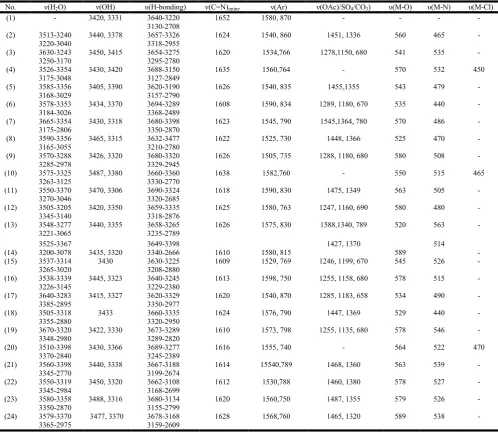
![Table 1. Analytical and physical data of the ligand [H 2L] (1) and its metal complexes](https://thumb-us.123doks.com/thumbv2/123dok_us/1218888.1625582/6.595.40.558.80.750/table-analytical-physical-data-ligand-h-metal-complexes.webp)

Related documents
But the pattern changes when we estimate erosivity with a differ- ent method: when erosivity is estimated from daily rainfall values, the multi-model mean indicates a decrease in
Figure 1 illustrates vascular NO signaling pathways, including the effect of NO on vasodilation, and a potential influence of garlic organosulfur compounds.. eNOS-derived
All partici- pants underwent a pulmonary function test (forced expiratory volume in fi rst second/forced vital capacity, FEV1/FVC; percentage value of predicted FEV1, FEV1%pred),
Table A31: Crystallographic data of α -CaSiN 2 [14] compared to calculated values (atomic param-. eters: (1) experimental coordinates, (2) coordinates from LDA calculation,
In the last two decades, Streptococcus pyogenes (group A streptococci [GAS]) has been identified as an emerging cause of severe infections: septic shock, organ failure,
maturation and germination. A- Leaf explant, B- Callus initiated from walls of leaf explant, C- Globular shape somatic embryos, D- Maturation stage of embryos, E- Mass of
Extracts 22 , 23 , 27 , 32 and 51 , and the standard anticancer drug cisplatin, were tested for cytotoxic activity with the resazurin assay in 12 additional
Combination tar- geting of both CD47 and PD-L1 resulted in synergistic inhibitory effect on tumor growth in the MPC-83 but not Panc02 syngeneic PDAC mouse model due to
