INTERNATIONAL RESEARCH JOURNAL OF PHARMACY
www.irjponline.com
ISSN 2230 – 8407
Research Article
BACTERIAL CHARACTERIZATION OF SILVER NANOPARTICLES FROM
TEMBAGAPURA SOIL SAMPLE ISOLATE, PAPUA, INDONESIA
Dani Prasetyo
1,2, Muhammad Fadli
1, Yuherman
3, Asiska Permata Dewi
4, Akmal Djamaan
5*
1
Department of Biotechnology, Graduate School of Andalas University, Padang, West Sumatera, Indonesia
2Faculty of Pharmacy, Kader Bangsa University, Palembang, South Sumatera, Indonesia
3
Animal Husbandry Faculty, Andalas University, Padang, West Sumatera, Indonesia
4
Department of Pharmacy, Faculty of Public Health, University of Abdurrab, Pekanbaru, Riau, Indonesia,
5
Department of Pharmaceutical Chemistry, Faculty of Pharmacy, Andalas University, Padang, West Sumatera, Indonesia
*Corresponding Author Email: [email protected]
Article Received on: 11/09/18 Approved for publication: 20/10/18
DOI: 10.7897/2230-8407.0910225
ABSTRACT
Characterization of silver nanoparticle-producing bacteria from Tembagapura, Papua, Indonesia soil samples isolates has been investigated. Bacteria characterization was carried out macroscopically, microscopically, biochemically and molecularly. Furthermore, the results of the formed silver nanoparticles were characterized using UV-VIS spectroscopy, Fourier transform infrared (FTIR) and scanning electron microscopy (SEM). The experimental results showed maximum absorbance at 414 nm in TP10-1 isolates in UV-Vis spectroscopy. FTIR spectra of silver nanoparticles samples of TP10-1 isolates showed strong peaks in wave numbers 1637.65 cm-1 and 3329.47 cm-1. SEM micrographs reveal the formation of well dispersed
silver nanoparticles. Silver nanoparticles of TP10-1 isolates that was measured by the imageJprogram had an average particle size of 16,991 nm. Bacterial isolates with TP10-1 sample code which are identical to the Bacillus cereus strain GCF1I2was able to synthesize silver nanoparticles.
Keywords: silver nanoparticle, characterization, Bacillus cereus strain GCF1I2
INTRODUCTION
Nanotechnology development is still developing by the researchers from academic world and from the industrial world. Nano particle synthesis means making particles with sizes less than 100 nm and at the same time changing their properties or functions1. One of the most popular nanoparticles is nanosilver
particles or nanosilver particles (NSPs). If the particle size is lowered, the surface area with the NSPs volume ratio will increases dramatically, which leads to significant changes in physical, chemical, and biological aspect. NSPs are most commonly used in health care systems for hundreds of years2.
Biosynthesis (green synthesis) of nanosilver has received widespread attention because of its growing need for environmentally friendly synthesis methods and the use of environmentally friendly reducing and capping agents, such as proteins, peptides, carbohydrates, from various species of bacteria, fungi, yeast and algae and plants2.
In this paper, we will be used to synthesize nanosilver particles (NSPs) from bacteria isolated from Tembaga Pura, Papua soil and reported the characterization was formed. Bacteria from Tembagapura which produce nanosilver will be identified molecularly.
MATERIAL AND METHOD
Isolation of Silver Nanoparticle-Producing Bacteria
Soil samples obtained from Tembagapura were used as a source for bacterial isolate. A total of 10 g of soil samples were put into 90 ml of NB medium, then incubated for 24 - 48 hours at 37 °C,
then it was diluted to 103,4. Isolation of metal-resistant bacteria
(Ag) was carried out by duplicate plating technique using a nutrient equipped with a 1 mM concentration of sterilized AgNO3
and using NA medium only. Then it was incubated at 37 °C for 48-72 hours, the Petri dish was observed to determine the bacterial growth5,6. Cultures of growing isolates were identified as silver
resistant bacteria. Separately grown bacterial colonies were then purified using the streak plate method on nutrient agar (NA) medium.
Conventional Identification of Bacteria
Conventional Identification Bacteria that produce silver nanoparticles were further identified by conventional methods. The main identification of bacterial isolates was carried out on the basis of colony, microscopic and biochemical characteristics which refer to the Bergey's Manual of Determinative of Microorganism3,7.
Molecular Identification of Silver Nanoparticles- Producing Bacteria
Molecular identification of bacterial isolates was done using the 16SrRNA gene. DNA products of bacterial isolates from amplification and purification were sequenced by Macrogen Inc. (South Korea). Sequence data were analyzed using the BLAST program (www.ncbi.nlm.gov.blast) and phylogenetic trees was built8.
Extracellular Biosynthesis of Silver Nanoparticles
given below. Isolated colonies were cultured in NB and incubated for 24 hours at 37 °C. Then, NB was centrifuged at 8000 rpm for 10 minutes to collect the supernatant. Silver nitrate solutionas much as 1 mM was prepared with distilled water. As much as 20 mL of 1 mM silver nitrate solution were mixed with 10 mL of the culture supernatant in a 50 mL Erlenmeyer flask. All samples were shaked using shaker tool at 150 rpm and maintained in dark conditions for 36-72 hours. Silver nitrate reduction was monitored with visible color changes from the solution5,9.
Synthesis Evaluation of Silver Nanoparticle
Synthesis evaluation of silver nanoparticle formed was done using UV-Vis spectrophotometer, Fourier transform infrared(FTIR) and Scanning Electron Microscopy (SEM)10,11,12.
RESULTS AND DISCUSSION
Isolation of Silver Nanoparticles-Producing Bacteria
Pure colonies grown on nutrient agar (NA) medium which had been added with 1mM AgNO3 solution were taken one of them
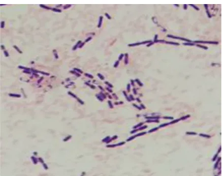
and given isolate code TP10-1. These colonies are considered as resistant bacteria to silver solution, then characterization of the bacteria and the products of silver nanoparticles formedwas done. Characterization of bacterial isolates obtained was done by comparing the isolate with standard literature. The result showed that the bacteria was Gram positive and has bacillary form (Fig. 1). Endospora tests was carried out on these isolates and showed that these bacteria can form endospores. Furthermore, biochemical tests were carried out, one of which was catalase test.
This test result showed that the bacterial isolates were positively catalase, characterized by the formation of bubbles. These results indicate that the isolate is likely to be Bacillus.
Figure 1: The results of Gram staining isolation of TP10-1 bacteria with 10x100 magnification
Molecular identification of silver nanoparticles-producing bacteria
The sequencing results of TP10-1 bacterial isolates were compared with GeneBank data using the BLAST program which was conducted online on the NCBI website.
Figure 2: Nucleotide sequencing results of TP10-1 bacterial isolates
TP10-1 bacterial isolates have an 99% similarity with Bacillus cereus strain GCF1I2. In the observation of phylogenetic trees, the distance between bacterial isolates of TP10-1 and Bacillus cereus strain GCF1I2 was very close (Fig. 3). This indicates that the bacterial isolate TP10-1 was found to be identical to the Bacillus cereus strain GCF1I2. This is in accordance with Hagström, Pinhassi and Zweifel (2000) whom stating that the isolates with
similar sequences of more than 97% can represent the same species. While the sequence similarity between 93% - 97% can represent the identity of bacteria at the genus level but from different species13. This is also consistent with Tamisier et al.
Figure 3: Phylogenetic tree of TP10-1 bacterial isolate
Extracellular Biosynthesis of Silver Nanoparticles
On visual observation, after the fifth day there was a change in the solution color from being initially yellow to ground brown. The supernatant culture without silver nitrate and 1 mM AgNO3
solution also observed for color changes and used as controls (Fig. 4). The appearance of brownish color in the solution which added with silver nitrate was identified as the formation of silver nanoparticles. This is in accordance with Agrawal and Kulkarni (2017) opinion that the color of the supernatant solution added with 1 mM sterile AgNO3 will turned brown after incubation6.
Kalishwaral et al. (2008) also stated that supernatant culture that incubated with silver nitrate showed color changes from yellow to brown while supernatant culture without silver nitrate and silver nitrate solution (as a control) were observed to have no color changes15.
Figure 4: Color change test on bacteria isolates after incubated for 5 days on 37 oC in a dark atmosphere; A. 1 mM AgNO3 solution, B.
Bacterial supernatant C. Bacterial supernatant + 1 mM AgNO3solution with color changes in TP 10-1 bacterial isolate.
Evaluation of the synthesis of silver nanoparticles
The result of UV-Vis spectrophotometer observation was indicated by the formation of the maximum absorption peak of isolate TP10-1 was 414 nm (Fig. 5). This is consistent with the
statement of Sileikaite et al. (2006) stated that silver nanoparticle colloids have typical absorption peaks in visible light with a range of 350 - 530 nm in spectrophotometer analysis16.
Figure 5: The maximum absorption peak of TP10-1 at 414 nm wavelength
Fourier Transform Infra Red (FTIR) was used to prove the interaction of TP 10-1 bacterial isolate protein during the process of formation of silver nanoparticles (Fig. 6). FTIR spectra of silver nanoparticles from TP10-1 isolates between wave numbers 400-4000 cm-1 showed strong peaks in wave numbers 1637.65 cm-1
and 3329.47 cm-1. This is consistent with the opinion of Kong and
Yu (2007) whom states that the spectrum region most sensitive to the secondary structural component of protein is the amide I band (1700-1600 cm-1) which is almost entirely caused by vibration of
C = O stretching connected to the peptide (around 80%). The frequency of amide component I was found to correlate closely with each of the secondary structural elements of the protein17.
Figure 6: FTIR result of TP10-1 isolate showed the formation of amide peak
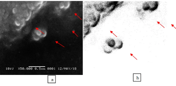
The particle size observation of silver nanoparticles was carried out using the imageJ program. This is consistent with Mazolli and Favoni (2012) that stated to evaluate particle size and size distribution, Scanning Electron Microscopy (SEM) and ImageJ
processing programs were used (Fig. 7). The number of analyzed nanoparticles was 5813 of the total 98765 with the average size of the nanoparticles formed was 16.991 nm19.
Figure7: SEM result of TP10-1 bacterial isolate, a. SEM results showed the formation of silver nanoparticles; b. The results of the particle size scanner for silver nanoparticles using ImageJ
The results showed that the TP10-1 bacterial isolate was able to synthesize AgNO3 solution into silver nanoparticles. This is in
accordance with Pugazhenthiran et al. (2009) who using Bacillus sp. to synthesize silver into silver nanoparticles and obtained a nanoparticle with size of 5-15 nm. Banu et al. (2014) was synthesized the nanoparticle from Bacillus thuringiensis and obtained nanoparticle with size of 43.52–142.97 nm. El-Shanshoury et al. (2011) also synthesized the nanoparticle with
Streptococcus thermophilus and obtained nanoparticle with size of 28–122 nm20. This is in accordance with Srikar et al. (2016) which
states that nanoparticle synthesis means making particles with sizes less than 100 nm and at the same time changing their properties or functions1.
CONCLUSION
This study showed the characterization of TP10-1 bacterial isolates that was tested molecularly had 99% similarity with Bacillus cereus strain GCF1I2. This results was confirmed using phylogenetic trees. Bacillus cereus strain GCF1I2 has the potential to synthesize silver nanoparticles. The average particle size
Red (FTIR) proved the interaction of TP 10-1 bacterial isolate protein during the process of forming silver nanoparticles.
REFERENCES
1. Srikar S K, Giri D D, Pal D B, Mishra P K, Upadhyay S N. Green synthesis of silver nanoparticles: A review. Green and Sustainable Chemistry 2016; 6(1):34-56. http://doi.org/ 10.4236/gsc.2016.61004
2. Ge L, Li Q, Wang M, Ouyang J, Li X, Xing MQM. Nanosilver particles in medical applications: synthesis, performance, and toxicity. Int. J. Nanomedicine, 2014; 9(1):2399–2407. http://doi.org/10.2147/IJN.S55015
3. Holt J G, Sneath P H A, Mair N S, Sharpe M E. Bergey’s manual of systematic bacteriology, Vol 2. Baltimore: Williams and Wilkins, 1986.
6. Agrawal P N, Kulkarni N S. Biosynthesis of silver nanoparticles from silver resistance bacteria isolated from metal contaminated soil. Sch. Acad. J. Biosci, 2017; 5(3):187-191. DOI: 10.21276/sajb.2017.5. 3.10
7. Sayuti I, Siregar Y I, Amin B, Agustien A, Djamaan A. Identification of Bacterial Hydrocarbonoclastic in Waste Tanks, Petapahan, Riau, Indonesia, using 16sr RNA, J. Appl. Microbiol, 2018; 12(2):671-677.
8. Tamura K, Stecher K, Peterson D, Filipski A, Kumar S. MEGA6: molecular evolutionary genetics analysis version 6.0. Molecular Biology Evolution 2013; 30(12): 2725-2729. https://doi.org/10. 1093/molbev/mst197
9. Shahverdi A R, Minaeian S, Shahverdi H, Jamalifar H, Nohi A A. Rapid synthesis of silver nanoparticles using culture supernatants of Enterobacteria: a novel biological approach. Process Biochem 2007; 42:919–923. http://doi.org/10.1016 /j.procbio.2007.02.005
10.Kalimuthu K, Babu R S, Venkataraman D, Bilal M, Gurunathan, S. Biosynthesis of silver nanocrystals by Bacillus licheniformis. Colloids and Surfaces B: Biointerfaces 2008; 65:150–153. http://doi.org/ 10.1016/j.colsurfb.2008.02.018 11.Rajeshkumar S, Malarkodi C. In vitro antibacterial activity
and mechanism of silver nanoparticles against foodborne pathogens. Bioinorganic Chemistry and Applications 2014; Article ID 581890, 1-10. http://dx.doi.org/10.1155 /2014/581890
12.Ajayi E, Afolayan A. Green synthesis, characterization and biological activities of silver nanoparticles from alkalinized
Cymbopogon citratus Stapf. Adv. Nat. Sci.: Nanosci. Nanotechnol, 2017; 8(01),1- 8. http://doi.org/10.1088/2043-6254/aa5cf7
13.Hagstrom A, Pinhassi J, Zweifel UL. Biogeographical diversity among marine bacterioplankton. Aquat. Microb. Ecol 2000; 21:231-244. http://doi.org/ 10.3354/ame021231 14.Tamisier M R, Benamar S, Raoult D, Fournier P E. Cautionary
tale of using 16S rRNA gene sequence similarity values in
identification of human-associated bacterial species. IJSEM Papers in Press. 2015; http://doi.org/10.1099/ijs.0.000161 15.Kalishwaralal K, Deepak V, Ramkumarpandian S, Nellaiah H,
Sangiliyandi G. Extracellular biosynthesis of silver nanoparticles by the culture supernatant of Bacillus licheniformis. Elsevier, Materials Letters, 2008; 62:4411– 4413. https://doi.org/10.1016/j.matlet.2008.06.051
16.Sileikaite A, Prosycevas I, Puiso J, Juraitis A, Guobiene A. Analysis of silver nanoparticles produced by chemical reduction of silver salt solution. Materials Science (Medžiagotyra) 2006;12 (4):287-291. [cited 2018 Apr 16] Available from: http://citeseerx.ist.psu.edu/ viewdoc/download?doi=10.1.1.
485.1461&rep=rep1&type=pdf
17.Kong J, Yu S. Fourier transform infrared spectroscopic analysis of protein secondary structures. Acta Biochimica et Biophysica Sinica, 2007;39(8):549–559. PMID: 17687489 18.Banker J. Amide modes and protein conformation. Biochim
Biophys Acta, 1992; 1120(2):123−143. PMID: 1373323 19.Mazzoli A, Favoni O. Particle size, size distribution and
morphological evaluation of airborne dust particles of diverse woods by Scanning Electron Microscopy and image processing program. Powder Technology 2012; 225:65–71. http://doi.org/10.1016/j.powtec.2012.03.033
20.Singh R, Shedbalkar U U, Wadhwani S A, Chopade B A. Bacteriagenic silver nanoparticles: synthesis, mechanism, and applications. Appl. Microbiol. Biotechnol. 2015; 99(11):4579-4593. DOI 10.1007/s00253-015-6622-1. http://doi.org/10.1007/s00253-015-6622-1
Cite this article as:
Dani Prasetyo et al. Bacterial characterization of silver nanoparticles from Tembagapura soil sample isolate, Papua, Indonesia. Int. Res. J. Pharm. 2018;9(10):53-57 http://dx.doi.org/10.7897/2230-8407.0910225
Source of support: Nil, Conflict of interest: None Declared