Functional and evolu tion ary analysis o f the m ou se M uc-1 gen e.
A ndrew Paul Spicer
A ugust 1993
A th esis p re s e n te d in p a rtial fu lfilm en t of th e re q u ire m e n ts for th e d e g re e of D octor of P h ilo so p h y of the U n iv e rsity o f L ondon.
M o lecu lar E p ith elial C ell Biology L aboratory,
Im p e ria l C an cer R esearch F und, P.O. Box 123,
L in c o ln ’s In n Fields, L o n d o n W C 2A 3PX UK.
D e p a rtm e n t of Biology, M e d a w a r B uild in g ,
U n iv e rs ity C o lleg e L o n d o n , G o w er Street,
ProQuest Number: 10046015
All rights reserved
INFORMATION TO ALL USERS
The quality of this reproduction is dependent upon the quality of the copy submitted. In the unlikely event that the author did not send a complete manuscript and there are missing pages, these will be noted. Also, if material had to be removed,
a note will indicate the deletion.
uest.
ProQuest 10046015
Published by ProQuest LLC(2016). Copyright of the Dissertation is held by the Author. All rights reserved.
This work is protected against unauthorized copying under Title 17, United States Code. Microform Edition © ProQuest LLC.
ProQuest LLC
789 East Eisenhower Parkway P.O. Box 1346
ABSTRACT
The m o u se h o m o lo g u e of th e h u m a n tu m o u r-asso ciated m ucin, M U C l, w as cloned an d full-length sequence w as d eterm in ed . This m ucin (p rev io u sly called p olym orphic epithelial m ucin) is expressed by the m ajo rity of sim ple secretory epithelial cells in b o th th e m o u se an d h u m a n a n d is also overexpressed in a large percentage of carcinom as. T he m o u se gene, M uc-1, encodes an integral m em b ran e p ro tein w ith 44% of its coding capacity m ade u p of serine, th reo n in e an d proline, a com position typical of a highly O -glycosylated protein. The Muc-1 core p ro te in consists of an am ino-term inal signal sequence, a rep etitiv e d o m ain encoding 16 repeats of 20-21 am ino acids, and u n iq u e sequence co n tain in g m em b ran e-sp an n in g a n d cytoplasm ic dom ains. A lth o u g h o v erall hom ology w ith the h u m an M U C l p ro tein is only 53%, th e
tran sm e m b ra n e an d cytoplasm ic dom ains exhibit hom ologies of 90% an d 87%, respectively. This level of sequence conservation w o u ld sug g est th at th ese reg io n s m ay be functionally im p o rtan t. Interestingly, the m ouse h o m o lo g u e, unlike its h u m an co u n te rp art does n o t exhibit a variable n u m b e r tan d em rep e at (VNTR) p o ly m o rp h ism . H ow ever, this ty p e of p o ly m o rp h ism w as fo u n d to be p resen t in all o th er m am m alian g ro u p s analysed. D ata is p resen ted , including sequence o b tain ed for the Muc-1 gene from a large n u m b er of species, to suggest how this gene has
ev o lv ed a n d to explain possible reasons w h y the m ouse Muc-1 gene does n o t exhibit m inisatellite characteristics.
N u m e ro u s functions have been su g g ested for this m olecule, yet it still rem ain s unclear w h a t role this p ro tein plays in the tissues an d tu m o u rs in w hich it is expressed. In an effort to learn m ore of the
ACKNOWLEDGEMENTS
I w ould like to acknowledge Dr. Sandy Gendler, in w hose lab I have been privileged to w ork these past four years. Sandy, you have been a w onderful research supervisor and friend over these past four years and have always given me continual encouragem ent and support. Your enthusiasm for the subject has been limitless and infectious.
There are num erous other people that I need to thank both in the UK and in the US. My friends at ICRF in London, especially Trevor D uhig an d Nigel Peat for their friendship and our num erous coffee break, lunch and 'other' interesting discussions. Between the tw o of them they taught m e a lot of m olecular biology, and a lot of 'life' in general. Discussions w ith them w ere always nothing b u t colourful! I w ould also like to thank num erous other ICRF staff, notably Lucy
Pem berton and Vania Braga. W ithout them , ICRF lab life w o u ld n 't have been the same. A t University College I w ould like to thank my internal research supervisor. Dr. Bill Richardson, for his helpful discussions of m y research and for his help through subm itting this thesis, and Peter King for his kind donation of the w ild m ouse tissue samples.
In the US I w ould like to especially thank Suresh Savarirayan, Bob Cichon, Anita Jennings and D awn Taylor at the Mayo Clinic. I thank Suresh for expertly carrying out all the ES animal w ork and teaching me the fine art of blastocyst microinjection and m anipulation. Bob I thank for the routine m aintenance and breeding of the mice described herein. W ithout Bob and Suresh, the germ line mice described w ithin w ould not have been possible. I thank Anita for her great stories, including the now infam ous "tomato-cake story", and for her outstanding histological preparations. I acknowledge Dawn Taylor and Kelly A nn Ross in the V isual C om m unications departm ent for their help in prep arin g the photographic plates. I acknowledge Dr. Stuart Patton for his gift of the m ilk fat globule proteins from the species studied herein. Dr. Pat
This acknow ledgem ents list w ould not be complete w ithout m ention of m y M um and Dad and m y brothers and sister w ho
TABLE OF CONTENTS
Title page 1
D edication 2
Abstract 3
A cknow ledgem ents 4
Table of contents 6
A bbreviations 13
CHAPTER ONE: GENERAL INTRODUCTION 15
l.lT h e hu m an M U Cl gene and protein 16 1.2 Gene targeting, by hom ologous recom bination, in 33
m ouse embryonic stem cells
1.3 M inisatellites and m ucin genes 45
1.4 Aims of the project 54
Figures
1.11 Cartoon representation of the M UCl 56 m em brane-associated m ucin glycoprotein
1.12 Expression of m ouse Muc-1 during 58 embryonic developm ent
1.21 The gene targeting strategy 60 1.22 Sequence replacem ent and insertion vectors 62 1.23 Screening for hom ologous recom binants 64 1.24 The Positive-Negative-Selection (PNS) system 66 1.31 The generation of new length VNTR alleles by 68
unequal crossing over
1.32 VNTR hypervariability at the M U Cl gene locus 70
Tables
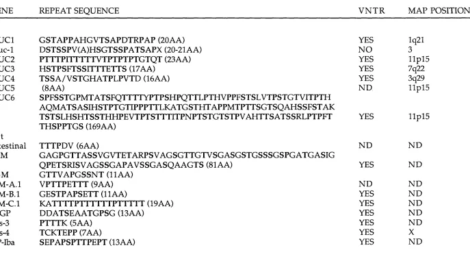
Table 1 M ucin tandem repeat sequences 72
CHAPTER TWO: MATERIALS AND METHODS 73
MATERIALS 73
2.1 Chemicals and solvents 73
2.3 Enzymes 74
2.4 M iscellaneous 74
2.5 Buffers 75
2.6 G row th m edia and tissue culture reagents 78
2.7 Histochemicals 81
2.8 M iscellaneous solutions 82
2.9 MOLECULAR BIOLOGY METHODS 84
2.91 Genomic DNA isolation 84
2.92 Restriction endonuclease digestion of plasm id DNA 86 and genomic DNA
2.93 Agarose gel electrophoresis of DNA 87 2.94 Isolation of DNA fragem ents from gels 87
2.95 Labelling DNA fragm ents 89
2.96 Southern blotting 90
2.97 DNA ligations 92
2.98 Bacterial transform ation 93 2.99 Preparation of plasm id DNA 95 2.910 Bacteriophage X library screening 98 2.911 Screening cosmid libraries 103 2.912 C hrom osom al localisation studies 107
2.913 Isolation of RNA 108
2.914 A garose gel electrophoresis of RNA 109
2.915 N orthern blotting 110
2.916 Polym erase chain reaction (PCR) 111 2.917 Reverse transcriptase-PCR (RT-PCR) 112 2.918 Direct cloning of PCR products into PCR T-vectors 112 2.919 Sequencing of plasm id DNA 114 2.920 SDS polyacrylam ide gel electrophoresis of proteins 115 2.921 Detection of m ilk-fat-globule associated Muc-1 protein 115
by silver staining
2.10 CELL METHODS 116
colonies
2.105 In vitro differentiation of ES cells 122 2.106 Karyotype analysis of embryonic stem cells 122 2.107 C alcium -phosphate transient transfection of 124
m am m alian cells
2.108 LacZ staining of transfected cells 124
2.11 ANIMAL METHODS 125
2.111 Production and recovery of m ouse blastocysts 125 2.112 Microinjection of em bryonic stem cells into blastocysts 125 2.113 Vasectomy, preparation of pseu d o p reg n an t females 126
and em bryo transfer
2.114 Breeding program m e of chim aeras 127 2.115 Sub-cutaneous injection of m ouse em bryonic stem 128
cells into athym ic nu d e mice
2.116 Histochem ical analysis of n u d e m ouse teratocarcinom as 128 2.117 Im m unohistochem ical analysis of spontaneous m ouse 130
m am m ary carcinom as
CHAPTER THREE: CLONING THE MOUSE HOMOLOGUE 131
OF THE HUMAN TUMOUR-ASSOCIATED M UCl GENE.
3.1 In tro d u ctio n 131
3.2 DNA probes 132
3.3 Screening ^gtlO library: cDNA cloning 133 3.4 Screening B alb/c cosmid library 135 3.5 5' cDNA cloning by RT-PCR 136 3.6 DNA and protein sequence analysis of the m ouse 137
Muc-1 gene
3.7 Prom oter and expression analysis 139
3.8 C onclusions 141
Figures
3.1 Genom ic structure of the hum an tum our-associated 146 M U Cl gene
3.2 Southern blot of m ouse and hum an genomic DNAs 148 hybridised to the hum an M UCl cDNA probe
pGEM-PEM16
3.31 A) N orthern blot of m ouse lactating m am m ary gland 150 RNA hybridised to three m ouse Muc-1 cDNA probes
B) M ouse Muc-1 cDNA cloning strategy and restriction m ap of full-length m ouse Muc-1 cDNA
3.32 Direct ^-plaque PCR assay 152
3.4 A) A utoradiogram displaying representative positive 154 m ouse Muc-1 cosmid clones, 1.21,1.22, and 1.23
B) Restriction endonuclease digestion of m ouse Muc-1 cosm id DNAs
3.61 Com plete nucleotide and predicted protein sequence 156 of the m ouse m ucin gene, Muc-1
3.62 Genomic structure of the m ouse Muc-1 gene 161 3.63 Dot-m atrix plot show ing hom ology betw een h u m an 163
and m ouse m ucin genomic DNA sequences
3.64 A com parison of m ouse and hum an Muc-1 am ino acid 165 sequences
3.65 Bar diagram to sum m arise the various levels of 167 hom ology at the DNA and protein levels
3.66 Tw o-dim ensional representation of the h u m an and 169 m ouse Muc-1 protein cores
3.71 A lignm ent of the prom oter sequences of the m ouse 171 an d hu m an Muc-1 genes
3.72 Expression of m ouse Muc-1 mRNA in 173 spontaneously arising m ouse m am m ary carcinom as
3.73 Expression of m ouse Muc-1 protein in 175 spontaneously arising m ouse m am m ary carcinom as
CHAPTER FOUR: EVOLUTION OF THE Muc-1 GENE LOCUS 176
4.1 In tro d u ctio n 176
4.2 Investigation of naturally occurring m inisatellite 177 polym orphism of the m ouse Muc-1 gene
4.3 Investigation of rodent Muc-1 evolution 180 4.4 C hrom osom al localisation of the m ouse Muc-1 gene 181 4.5 C loning the Muc-1 gene from diverse m am m alian 184
species
4.6 Evolution of the repeat unit of the Muc-1 gene 189
Figures
4.21 A lignm ent of the 16 m ouse Muc-1 repeats w ith the 197 derived consensus repeats at both the nucleic acid
an d protein level
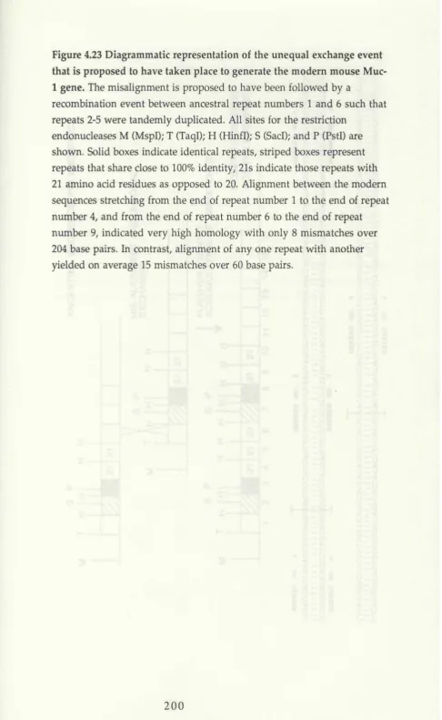
4.22 V ariation at the m ouse Muc-1 locus 199 4.23 D iagram m atic representation of the unequal exchange 201
event that is proposed to have taken place to generate the m odern m ouse Muc-1 gene
4.41 Chrom osom al localisation of the m ouse Muc-1 gene 203 through haplotype analysis of 114 [C3H/He]-gld x
Mus spretus)Fi x C3H/Hej-gld] interspecific backcross m ice
4.42 A pproxim ate localisation of the m ouse Muc-1 gene 205 w ith reference to a breakpoint in hom ology that has
been identified betw een m ouse chrom osomes 1 and 3 and h u m an chrom osom e 1
4.51 M ilk-fat-globule polym orphism of Muc-1 in a variety of 207 m am m alian species
4.52 W estern blot of Muc-1 m ilk-fat-globule proteins screened 209 w ith the m onoclonal antibody HMFG2
4.53 Sequencing gel displaying equivalent sequence of the 211 m ouse, ham ster, guinea-pig, cow and rabbit Muc-1
cytoplasmic tail dom ains
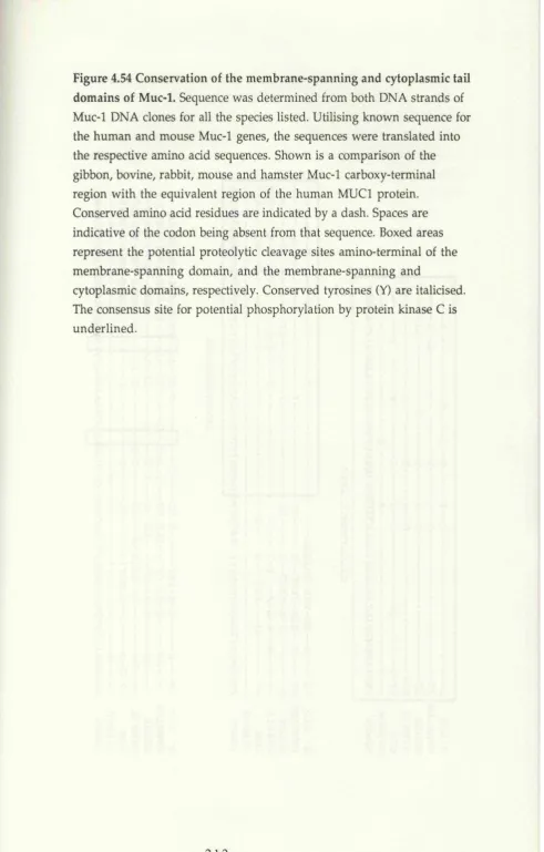
4.54 C onservation of the m em brane-spanning and 213 cytoplasmic tail dom ains of Muc-1
4.6 A lignm ent and com parison of the consensus Muc-1 215 tandem repeats obtained for hum an (H), gibbon (G),
cow (B), rabbit (R) and m ouse (M)
CHAPTER FIVE: TARGETED INACTIVATION OF THE 216 MOUSE Muc-1 GENE
5.1 In tro d u ctio n 216
5.2 B alb/c targeting vector design and construction 217 5.3 Establishm ent of conditions for use in gene targeting 220
experim ents
5.4 Targeted inactivation of the m ouse Muc-1 gene in 222 E14TG2a cells: Identification and analysis of targeted
clones
5.5 Re-isolation of the m ouse Muc-1 gene from 129 225 genomic library: 129 targeting vector design and
construction
5.6 Targeted inactivation of the m ouse Muc-1 gene in 228 E14TG2a and GK129 cells utilising an isogenic
M uc-1/LacZ fusion vector
5.7 Analysis of targeted ES cell clones 230 5.8 Germline transm ission analysis of a specific Muc-1 /L acZ 233
m u ta tio n
5.9 C onclusions 234
Figures
5.21 Structure of the m ouse Muc-1 gene locus 240 5.22 Cloning strategy for the construction of the B alb/c 242
Muc-1 targeting vectors pM uc-lGT Type I and Type II
5.41 Targeted inactivation of the m ouse Muc-1 gene w ith the 246 replacem ent vector pM uc-lG T Type I
5.42 Predicted structure of the Muc-1 locus after targeted 248 inactivation w ith the replacem ent vector
pM uc-lG T Type II
5.43 Southern analysis of an aberrantly targeted ES clone 251 #23.2
5.44 C hrom osom e analysis and em bryoid body form ation 253 assay of Muc-1 targeted clone #32.1
5.51 A) Double positive colonies obtained through screening 255 a 129Sv cosm id library w ith the m ouse Muc-1 cDNA
probe, pMuc2TR
B) RFLP investigation of the Muc-1 gene isolated from two m ouse strains
5.52 Cloning strategy for the construction of the 129 Muc-1 257 replacem ent vector, 129Muc-lGT
5.53 A) Sequence analysis of the M uc-1/LacZ ligation 261 ju n c tio n
B) Expression of the M uc-1/LacZ fusion protein in an HP-1, ham ster pancreatic carcinoma cell
5.61 Predicted structure of the Muc-1 gene locus after targeted 263 replacem ent by the vector 129Muc-lGT
vector
5.63 Southern analysis of aberrantly targeted ES clones 267 5.64 Targeted inactivation of the m ouse Muc-1 gene in 269
GK129 cells by an isogenic M uc-1/LacZ replacem ent vector
5.71 Histochem ical and im m unohistochem ical analysis of 271 ES-derived teratocarcinom as
5.72 C him aeras form ed through m icroinjection of the 273 targeted GK129 clone #56 into C57B1/6 blastocysts
5.8 Germline transm ission analysis of a M uc-1/LacZ 275 m utation in agouti offspring of GK129 Muc-1 chim aeras
Tables
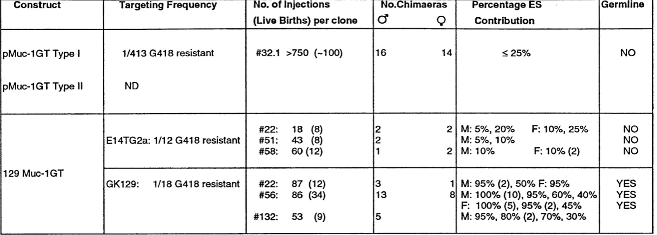
Table 5 Sum m ary of Muc-1 gene targeting results 277
CHAPTER SIX: DISCUSSION 278
6.1 The m ouse hom ologue of the hum an tum our- 278 associated m ucin gene, M UCl
6.2 A pproaching the function of the Muc-1 glycoprotein 282 6.3 Evidence for the evolution of the Muc-1 gene in 285
m a m m a ls
6.4 Future directions and concluding rem arks 290
APPENDIX 293
A ppendix 1: 293
Calculation of an approxim ate time for the duplication of a portion of the m ouse Muc-1 gene
A ppendix 2: 298
Oligonucleotides utilised in the construction of the B alb/c targeting vectors pM uc-lGT Type I and pM uc-lG T Type II, and in the PCR-based screening for hom ologous recom binants
REFERENCES 300
ADDENDA 333
ABBREVIATIONS
The hum an m ucin gene will be referred to as M U C l throughout,
w hereas the hom ologous m ouse m ucin gene will be referred to as Muc-1. This is to conform to the designated gene locus nam es as decided at the 1st International W orkshop on Carcinom a A ssociated M ucins, San Francisco 1990. As this thesis is centred around the m ouse m ucin gene, Muc-1, for sim plicity the hom ologous m ucin genes from the other species discussed will be referred to as ’Species N am e Muc-1, i.e bovine Muc-1 or rab b it M u c-1 . In addition, w hen tw o species hom ologues are being referred to they will be referred to collectively as Muc-1 genes.
bps base pairs of DNA
p-gal p-galactosidase gene of Escherichia coli BSA bovine serum album in
CAT chloram phenicol acetyl transferase cDNA copy deoxyribonucleic acid
CTAB cetyltrim ethylam m onium b rom ide DAB diam inobenzidine tetrahydrochloride DMEM Dulbecco's m odified Eagles m edium DMSO dim ethyl sulphoxide
DNA deoxyribonucleic acid ES cell em bryonic stem cell
EDTA ethylene diam ino-tetra-acetic acid FCS/FBS foetal calf seru m /fo etal bovine serum GanC gancyclovir
G418 geneticin
HSV-tk H erpes simplex virus 1 thym idine kinase gene ICM inner cell mass
i.p. intra-peritoneally
IPTG isopropyl-p-D -galactopyranoside kbps kilobase pairs of DNA
LacZ bacterial p-galactosidase gene LIF leukaem ia inhibitory factor p l/s m ic ro litre/s
m l/s m ililitre /s
nM n a n o m o la r m M m ilim o la r M m o la r
Myr m illion years n m /s n a n o m e te r/s
n eo neom ycin phosphotransferase OD optical density
PBS phosphate buffered saline
pBS-SKII+ pBluescriptSKII+ cloning vector pBS-KSII+ pBluescriptKSII+ cloning vector p .c post-coitum
pfus plaque form ing units PCR polym erase chain reaction PEM polym orphic epithelial m ucin
PGKneo neom ycin phosphotransferase gene driven by the prom oter of the m ouse phosphoglycerate kinase-1 gene
p m o le picom ole
PNS positive negative selection
RFLP restriction fragm ent length polym orphism RFLV restriction fragm ent length variant
R N A ribonucleic acid
rpm revolutions per m inute
SDS-PAGF sodium dodecyl sulphate polyacrylam ide gel electrophoresis SSC saline sodium citrate
SDS sodium dodecyl sulphate s.c sub-cutaneous/ly
TENS Tris-HCl, EDTA, sodium hydroxide, SDS T25 tissue culture flask w ith grow th area of 25cm2 T75 tissue culture flask w ith grow th area of 75cm2 T175 tissue culture flask w ith grow th area of 175cm2 V N T R variable num ber tandem repeat
(v /v ) volum e to volum e (w /v ) w eight to volum e
X-gal 5-brom o-4-chloro-3-indolyl-P-D-galactopyranoside
CHAPTER ONE:
GENERAL INTRODUCTION
Lumenal cells of the secretory epithelial organs of the body are in contact w ith a variety of external environm ental elements. As such, the apical or lum enal surface of these cells is covered by a protective
secretion which serves as a selective physical barrier betw een the extracellular m ilieu and the plasm a m em brane and cell interior. This secretion is often referred to as m ucous and is m ade up of a num ber of protein constituents, the m ost prom inent of w hich are the mucins. Mucins, or m ucin-type glycoproteins, are characterised as large extended molecules w ith a high percentage (50-90%) of their m olecular m ass m ade up of carbohydrate, which is attached via O-glycosidic linkage through N- acetylgalactosam ine to serine a n d /o r threonine.
M ucin-type glycoproteins can be sub-divided into the classical secretory, or soluble m ucins, and the m em brane-bound m ucin-like glycoproteins. The secretory, or soluble m ucins, constitute the viscous m ucous of the lungs, tracheo-bronchial tract, gut and reproductive tract, and typically form extrem ely large complexed oligomers through linkage of protein m onom ers via disulphide bonds. These proteins are secreted from the cell, and although they rem ain at the apical surface of the epithelial cells in the form of a mucous-gel, they are not m em brane bound. The m em brane-bound mucin-like glycoproteins, how ever, are intim ately associated w ith the plasm a m em brane through a hydrophobic m em brane-spanning dom ain and have not been observed to form
oligom eric complexes. It does, however, appear th at these m em brane- b ound m ucin-like glycoproteins can also be released from the cell m em brane by some sort of proteolytic cleavage event (Fig. 1.11).
hum an m ilk-fat-globule m em brane (Ceriani, 1977). A ttention was focused on this molecule prim arily d u e to the fact that antisera raised against elem ents of the hum an m ilk-fat-globule m em brane w ere also observed to react w ith proteins expressed by a num ber of hum an
m am m ary carcinom a cell lines in addition to tissue sections of hum an m am m ary tum ours (Arklie, 1981; Taylor-Papadim itriou, 1981).
This m ucin has been variously called epithelial m em brane antigen (EMA) (H eyderm an, 1979), C al antigen (Ashall, 1982), PAS-O (Schimizu, 1982), DU-PAN-2 antigen (Metzgar, 1982), peanut-reactive urinary m ucin (PUM) (Karlsson, 1983), non-penetrating glycoprotein (NPGP) (Ceriani, 1983), DF3 antigen (Kufe, 1984), MAM-6 (Hilkens, 1984), N C R C ll (Price, 1985); epitectin (Bramwell, 1986), polym orphic epithelial m ucin (PEM) (Gendler, 1988), H23 antigen (Keydar, 1989), and episialin (Ligtenberg, 1990) as a direct result of the large num ber of groups
w orldw ide that have been involved in the investigation of this
molecule. How ever, although it has had a large num ber of designated names, it w as found to be the product of a single gene locus, designated M UCl in the hum an. As such, this protein will herein be referred to as the M UCl protein.
1.1 The h u m an M U C l gene and protein
The derivation of antibodies directed to the deglycosylated m ucin of the hum an m ilk-fat-globule m em brane and sim ilarly to the
deglycosylated m ucin of the hum an pancreatic carcinom a cell line,
HPAF, allowed the isolation of cDNA clones for the hum an M U Cl gene through the screening of cDNA expression libraries constructed from m am m ary and pancreatic carcinoma cell line mRNAs (Gendler, 1987a; Gendler, 1988; Siddiqui, 1988; Gendler, 1990a; Ligtenberg, 1990; Lan, 1990; W reschner, 1990). A lignm ent of DNA sequences derived from the m ucin clones revealed that the respective cDNAs w ere the p roduct of the sam e gene locus.
Prior to the isolation of DNA clones for this gene, it h ad been show n that the peanut-reactive urinary m ucin (PUM), isolated from urine, kidney and lung, electrophoresed on SDS-polyacrylamide gels, and detected using lectin staining, exhibited a genetic polym orphism
characterised by the existence in most individuals of tw o co-m igrating
p ro tein b ands th at show ed person-to-person variation (Karlsson, 1983). The patterns of protein b ands observed w ere consistent w ith the sim ple M endelian inheritance of co-dom inant alleles at a single gene locus, b u t it w as n o t know n w h eth er this polym orphism w as the resu lt of variation of the gene encoding th e protein core or the glycan p a rt of the m olecule b eing analysed. A variety of tum our-binding antibodies, raised against th e h u m an m ilk-fat-globule m em brane, w ere dem o n strated to detect proteins on W estern blots w ith a p attern of polym orphic b an d s
rem iniscent of th e PUM polym orphism . Subsequently, it w as d educed th at the epitopes being recognised by these antibodies w ere carried on the sam e m olecules as the lectin-binding determ inants (Swallow, 1986).
Analysis of the DNA sequence obtained from the respective M U Cl clones revealed the presence of m ultiple copies of a highly GC-rich 60 base p air tandem repeat encoding a 20 am ino acid repeat m otif rich in serine, threonine and proline (Gendler, 1987a; G endler, 1988; Siddiqui, 1988; Ligtenberg, 1990; Lan, 1990; W reschner, 1990). Southern blots, utilising probes containing this repetitive sequence, yielded a resu lt that w as directly com parable to the p attern observed on protein gels for the PUM pro tein (Swallow, 1987a). It becam e ap p aren t th at the observed genetic polym orphism w as, therefore, the result of varying n um bers of a 60 base p air tandem repeat w ithin the coding dom ain. This variable n u m b er tandem rep eat (VNTR) polym orphism could be d em o n strated at the DN A (by Southern blot), the RNA (by northern blot) and p ro tein (by W estern blot) levels (Gendler, 1987b). A screen of 69 ran d o m individuals of N o rth ern E uropean origin identified 30 different allele lengths, and the variation in num ber of repeats p er allele has been observed to range from a low of betw een 20 to 30, u p to a high of over 100 rep eat units (G endler, 1990a). This variability in num bers of repeats p er allele m akes the M U C l locus one of the few expressed VNTR sequences th u s far identified in the h u m an genome. Repeat length differences a p p ea r to be generated by both intra and interallelic unequal recom bination events operating w ithin the repeat array, and this topic will be discussed in detail later.
The sequence of the repeat u n it encodes a m otif rich in serine, threonine an d proline, and it is this region of the protein th at is
length of the repetitive array is not critical for function, b u t that the repeat array functions prim arily as a scaffold for the attachm ent of O- linked carbohydrate. As early as 1973, it was postulated that the core protein of the bovine subm axillary m ucin w as com prised of repeating am ino acids (Pigman, 1973). Through the isolation of DNA clones for the genes encoding the h u m an MUC2 through MUC6 m ucin genes (Gum, 1989; Gum , 1990; Porchet, 1991; Van Cong, 1990; Aubert, 1991 and Toribara, 1993), the porcine (Timpte, 1988; Eckhardt, 1991) and bovine (Bhargava, 1990) subm axillary m ucin genes, three Xenopus
integum entary m ucin genes (Hoffmann, 1988; Probst, 1990 and Probst, 1992; H auser, 1992), tw o rat intestinal m ucin genes (Gum, 1991; Xu, 1992), and a gene coding for the apo-polysialoglycoprotein of rainbow trout eggs (Sorimachi, 1988), it has become apparent that the presence of a repetitive sequence dom ain coding for an expressed sequence rich in serine a n d /o r threonine and proline is a general characteristic of m ucin genes. Like the hu m an M U Cl gene, the repetitive dom ain of several of the other m ucin genes also displays the characteristic VNTR polym orphism (Sorimachi, 1988; Timpte, 1988; Griffiths, 1990; H auser, 1990; Eckhardt, 1991; Porchet, 1991; Toribara, 1991; Toribara, 1993). However, it should be pointed out th at although all the m ucin genes thus far characterised possess a repetitive dom ain rich in serine a n d /o r threonine and proline, apart from sim ilarity betw een hom ologues, the respective repeat units bear no hom ology to each other and also differ in the num ber of am ino acid residues per repeat (Table 1).
The hum an m ucin genes, M U Cl through MUC6, have been localised to chromosome lq21 (Swallow, 1987b; M iddleton-Price, 1988), llp l5 .5 (Griffiths, 1990), 7q22 (Gum, 1990), 3q29 (Porchet, 1991), l l p l 5 (Van Cong, 1990) and I lp l5 .4 - llp l5 .5 (Toribara, 1993), respectively. A lthough the genes for MUC2, 5 and 6 are located on the same
chrom osome, and have been m apped to lie in the sam e area, they show differing expression profiles and differ both in the length and sequence of their respective repeat units. It, therefore, rem ains to be seen w hether or not these three genes could have arisen from one ancestral m ucin gene through a series of duplications, and form ed a m ultigene family in which the m em bers subsequently diverged to take u p differing patterns of expression. How ever, the M U C l, MUC3 and MUC4 genes are perhaps less likely to have arisen through this type of duplication process as they
share no sim ilarity in sequence, repeat u n it length, gene structure or chrom osom e position.
From the deduced repeat sequence of the m ucin genes thus far characterised it appears that a large repetitive dom ain com prised of a high percentage of serine a n d /o r threonine interspersed by am ino acids w ith small side chains such as proline, glycine and alanine represents an efficient scaffold for O-linked glycosidic attachm ent, as this type of
sequence structure has been independently evolved and m aintained in a large num ber of m ucin genes in several different species. O-glycosidic bonds are form ed by the linkage of N-acetylgalactosam ine and the hydroxyl group of serine or threonine, and represent the first step in O- linked oligosaccharide synthesis. The action of the UDP-GalNAc:
polypeptide N -acetylgalactosam inyltransferase is thought to catalyse the following reactions (McGuire, 1967):
UDP-GalNAc + serine / threonine-polypeptide
—> G alN A c-serine/threonine-polypeptide + UDP
A UDP-GalNAc: polypeptide N-acetylgalactosam inyltransferase has recently been purified and characterised from porcine subm axillary glands (Wang, 1992). The porcine subm axillary enzym e was found to specifically incorporate N -acetylgalactosam ine from UDP-GalNAc into glycosidic linkages only w ith the hydroxyl groups of threonine in peptide substrates. M oreover, neither porcine, bovine, nor ovine subm axillary gland hom ogenates w ere able to incorporate N-acetylgalactosam ine into glycosidic linkages w ith serine residues in peptide substrates (Wang, 1992). These studies dem onstrated that different transferases catalyse the the form ation of O-glycosidic linkages on serine and threonine. The authors also exam ined the relative extent of glycosylation of threonines present in a variety of peptide substrates. A lthough, from this analysis, it is still not clear w hat the consensus am ino acid sequence is for the O- glycosylation of threonine, it was dem onstrated that some threonyl hydroxyl groups w ere more readily glycosylated than others, and some possible glycosylated products were sim ply not formed.
M ucin glycoproteins represent a class of proteins w ith possibly the highest level of O-linked carbohydrate observed (Jentoft, 1990). Therefore,
although the consensus am ino acid sequence for O -linked attachm ent is as yet not determ ined, from a stu d y of the sequences present in the repeat sequences of the various m ucin proteins it can be specified th at O-linked attachm ent to serines and threonines requires the presence of single or m ultiple serine or threonine residues in com bination w ith helix- disru p tin g residues such as prolines, and am ino acids w ith short side chains such as glycine and alanine.
A determ ination of the full-length cDNA sequence of the hum an M U C l gene revealed th at the sequence encoded a protein w ith four distinct dom ains; an am ino-term inal signal peptide dom ain, the large dom ain m ade up of variable num bers of the 20 amino acid repeat m otif flanked on either side by degenerate copies of the same repeat unit, and unique sequence w hich included a carboxy-term inal hydrophobic m em brane-spanning dom ain of 31 am ino acids, and a cytoplasmic dom ain of 69 amino acids (Gendler, 1990a; Ligtenberg, 1990; Lan, 1990; W reschner, 1990). Genom ic sequence of the gene revealed th at the coding sequence was represented on seven exons spanning betw een 4 and 7 kilobase pairs of DNA, depending upon the num ber of repeat units present (Lancaster, 1990; W reschner, 1990). The entire repeat dom ain was found to be located entirely w ithin the second exon. The deduced am ino acid sequence encoded a core protein w ith a predicted m olecular mass ranging from 120 kilodaltons up to 225 kilodaltons (Gendler, 1990a). The size observed on SDS-protein gels, for the m ature hum an m am m ary m ucin, is in the 300 to > 400 kilodalton range and this w ould im ply that as m uch as 50% or m ore of the w eight of the m ature glycosylated protein is m ade up of carbohydrate. From an analysis of the fully glycosylated M U Cl p roduct isolated from a variety of tissues it appears th at the M U Cl protein present in the pancreas has a m olecular mass closer to 1000
kilodaltons, m uch larger than the M U Cl protein identified in the milk- fat-globule. This ap p aren t organ-specific size variation of the m ucin m ay be sim ply a reflection of the spectrum of glycosyltransferases present w ithin a particular organ or, alternatively, m ay possibly be a reflection of a carbohydrate-associated function of the M UCl protein in each organ.
molecule made up of 40 repeat units could, therefore, potentially carry as many as 200 O-linked carbohydrate side chains. In addition to the
num erous sites for O-linked glycosylation, five potential sites for N- linked attachment of carbohydrate were also found, located between the repetitive and m em brane-spanning domains. Although the MUCl protein product has been demonstrated to carry N-Iinked sugar chains (Hilkens, 1984), it is not clear which of these sites are glycosylated.
The analysis of the sequence of the respective cDNA clones for the hum an MUCl gene resulted in the identification of two sites within the gene at which an alternative splicing mechanism may operate
(Ligtenberg, 1990; Wreschner, 1990). However, the alternative splice site identified by Ligtenberg, 1990, is the only one of these two sites that has been verified. At the amino-terminus of the molecule, it has been shown that an alternative splice variant, located at the three-prime end of the first intron, results in the addition of nine extra amino acids onto the signal peptide. It appears that this splice site recognition is based on a single A /G nucleotide difference in exon 2, eight nucleotides
downstream of the second splice acceptor site. The presence of an A at this position appears to result in the shorter of the two alternative splice products, whereas a G specifies the splicing in of the additional nine amino acid residues (Ligtenberg, 1991). It is yet to be discovered what if any functional significance this extra sequence might have, but it has been observed that in a number of cell lines and tissues expressing MUCl the ratio of the two alternative transcripts does vary. The authors
propose that, in an expressing cell line or tissue possessing alleles of two different lengths due to differing numbers of tandem repeats, the larger allele is in each case generally associated with the longer splice variant, whereas the small allele is generally found linked to the shorter splice variant. Their data show that in samples possessing only the so-called "small allele", the longer splice variant is nearly always undetectable. This observation would suggest that the majority of individuals
homozygous for the so-called "small allele" are incapable of expressing the longer splice variant of the MUCl message. Similarly, those
individuals homozygous for the "large allele" will almost invariantly express only the longer splice variant. It is, therefore, hard to imagine w hat possible differences in function, if any, the two respective splice variants may specify.
A second variant splice site, w hich w ould generate a transcript that incorporates the entire second intron into the mRNA, resulting in the generation of a p rem ature term ination signal and thus a transcript lacking the m em brane-spanning an d cytoplasm ic determ inants, was p roposed by W reschner, 1990. They suggested th at this alternative splice v arian t represents the transcript of the so-called secreted form of the m ucin. H ow ever, this alternative splice variant has rem ained undetected by all other investigators, and it is now generally accepted th at this
transcript and clone m ost probably arose through an incom pletely spliced m essenger RNA molecule. The M U Cl protein is, how ever, observed to be secreted from the cells that express it, b u t this secretion is m ost likely the result of a proteolytic cleavage event which releases the extracellular p o rtio n of the protein from its m em brane b ound and cytoplasmic
dom ains (Ligtenberg, 1992a; Boshell, 1992).
The post-translational m odifications of the M U Cl protein
rep resen t one of the m ost intriguing areas of the biology of this molecule. As m entioned previously, one of the prim ary reasons for the initial focus of interest in this molecule was d u e to the observation that several
antibodies directed to native and deglycosylated hum an m ilk-fat-globule proteins w ere found to react strongly w ith proteins present in sections of h u m an m am m ary tum ours. Indeed, som e antibodies, such as SM3, reacted very strongly w ith the tum our-associated M UCl product, yet show ed little or no reactivity w ith the M UCl p roduct of the
predictions and antigenicity predictions, th at this region of the tandem rep eat form s an antigenic p-turn (Fontenot, m anuscript subm itted; Taylor-Papadim itriou, 1992). Flanking the SM3 epitope are the two respective doublets of serine and threonine, i.e. ...V T S A P D T R P A P G S T A .... In m ost norm al secretory epithelia the presence of branched O- linked side-chains (Hanisch, 1989), attached to these sites, is enough to m ask the protein core and epitope. The M U C l protein p ro d u ct of carcinom as an d carcinom a cell lines has been show n to have less
complex, shorter carbohydrate side-chains w hich result in the exposure of novel core protein epitopes (Hull, 1989). Thus, antibodies raised to the deglycosylated m ucin recognise novel exposed epitopes on the
carcinom a-associated M U Cl product b u t are unable to recognise the p ro d u ct of the norm al tissue due to m asking by oligosaccharides. The expression of novel, tum our-associated m ucin epitopes by greater than 90% of h u m an breast, pancreatic, ovarian, colon and lung carcinom as has led to the exciting possibility for the use of the M UCl protein as a target in the specific active im m unotherapy of these cancers (H areuveni, 1990, Lalani, 1991).
Breast carcinom a cells that are found to express the M U Cl protein typically express levels higher than the level observed in the
corresponding norm al tissue (Zaretsky, 1990). In addition colon carcinom as also express this molecule at high levels (Boland, 1982; Irim ura, 1989; Hoff, 1989; Irim ura, 1991). It is, perhaps, the high levels of expression of the M U Cl protein in these cells that results in the addition of shorter carbohydrate side chains to the tum our-associated M UCl protein. O ne can envisage th at an over-expression of m ucin w ithout a parallel over-expression of the enzym es required for O-glycosylation may lead to under-glycosylated m ucin molecules reaching the cell surface (Carraway, 1992). A lternatively, the shorter side chains added to the tum our-associated M U Cl protein m ay be a reflection of a general or specific d isruption in the activity of certain glycosyltransferase enzym es present w ithin the Golgi apparatus of these cells.
A second post-translational m odification of the M U Cl protein that has been of great interest is its secretion. This molecule can be detected in the lum enal secretions of the epithelial cells that express it, and the
m olecule can also be detected circulating in the serum of patients w ith breast and pancreatic carcinoma (Burchell, 1984; M etzgar, 1984; Hayes, 1985; Hilkens, 1986; Tsarfaty, 1988). As m entioned previously,
W reschner, 1990, reported the isolation of a cDNA clone for the so-called secreted form of the hum an M UCl protein. How ever, as described
earlier, this clone has now been generally accepted to have been
generated from an incom pletely spliced m essenger RNA species. It has been assum ed th at the m ost likely m echanism for the secretion of M U Cl is som e kind of proteolytic cleavage event occurring betw een the
repetitive and m em brane spanning dom ain (Hilkens, 1988). Ligtenberg, 1992a, dem onstrated that this type of proteolytic cleavage-associated secretion of the molecule does indeed occur. The area of the proteolytic cleavage event w as found to reside w ithin 71 and 53 am ino acids
upstream of the transm em brane dom ain. In the region to which the cleavage event has been m apped, tw o phenylalanine-arginine doublets are present, putative substrates for a family of serine proteases, the kallikreins (Fiedler, 1987). The authors dem onstrated that the cleavage site m ay be cleaved w hile the protein is still w ithin the endoplasm ic reticulum and, thus, prior to the protein's passage to the apical surface of the cell. This im plies, how ever, that the extracellular and m em brane- spanning/cytoplasm ic portions (the tw o cleavage products) rem ain associated, as antibodies derived to sequences w ithin the cytoplasmic portion of the M U Cl protein efficiently im m unoprecipitate the entire m olecule (Ligtenberg, 1992a; Pem berton, 1992). The possible m echanism for this stable association is not known, b u t it is know n that a disulphide bond cannot be involved, as the only cysteine residues present w ithin the protein sequence are located w ithin the transm em brane dom ain.
To date the cleavage of the M UCl molecule and the subsequent stable association of the cleavage products prior to transport to the cell surface, rem ain points of contention. O ne exam ple in w hich a
m em brane-bound m ucin-like glycoprotein has been show n to be com prised of tw o cleavage products that rem ain stably associated at the cell surface is in the rat m am m ary carcinom a-associated m ucin
Cloning of regions encoding the the non-repetitive portions of the respective secreted m ucins has been extraordinarily difficult, and this is presum ably due to the fact that these genes are characterised by extremely large mRNAs, in the range of 11-14 kilobase pairs, com prised prim arily of m ultiple copies of the respective tandem repeat. In general, clones for the m ucin genes have been obtained through screening cDNA expression libraries w ith m onoclonal antibodies or polyclonal serum raised to the deglycosylated m ucin of the particular tissue or cell line being studied (Gendler, 1987a; Gum , 1989; Lan, 1990; Gum, 1991; Toribara, 1993). Due to the repetitive nature of the m ucins, the m ajority of antibodies raised to the deglycosylated m ucins react w ith epitopes present w ithin the repeats and, therefore, the use of antibodies such as these, in library screening, has yielded clones containing m ultiple copies of the tandem repeat. To date, the hum an M U Cl gene and its m ouse hom ologue, Muc-1, rem ain the only m ucin genes to be completely sequenced.
Cloning of the m ouse hom ologue (designated Muc-1) of the M U Cl gene (Spicer, 1991; Vos, 1991), and the derivation of a M UCl species cross reactive antiserum (Pemberton, 1992), has allowed a detailed
investigation of the pattern of expression of the m ouse Muc-1 gene du rin g m ouse embryogenesis and in the ad u lt m ouse (Braga, 1992;
Pem berton, 1992). The pattern of expression observed in the ad u lt m ouse reflects precisely the expression pattern previously reported for the
h u m an M U C l gene and for the hum an M U Cl gene in transgenic mice (Zotter, 1988; Peat, 1992). The pattern of expression of Muc-1 during m ouse em bryogenesis was investigated using northern analysis of mRNA, RT-PCR (reverse transcriptase-polym erase chain reaction), and im m unohistochem istry, utilising the polyclonal antiserum , C T l, raised to a peptide com posed of the last 17 amino acids of the hum an M UCl protein sequence (Braga, 1992; Pem berton, 1992). By
im m unohistochem istry, the Muc-1 protein w as first detectable in m ouse em bryonic stomach, pancreas and lung at gestational day 12 ( vaginal plug = day 1). In each case the protein was detected on the apical surface of the lum enal epithelial cells. A lthough the protein was detectable in several different organs of the developing m ouse, its expression w as observed not to be induced systemically, b u t according to the particular onset of epithelial polarisation and branching m orphogenesis in each individual organ (Fig 1.12). Muc-1 expression was observed to correlate well w ith the epithelial differentiation status of the stom ach, pancreas.
lu n g , trachea, k id n ey an d salivary glands an d w as detectable p rio r to the o n set of g la n d u lar activity of these organs. T he a u th o rs p ro p o se th at this p a tte rn o f exp ressio n m ay in d icate an im p o rta n t fu nction for the Muc-1 m o lecu le in th e early d e v elo p m e n t of these organs.
In terest in th e h u m an M U C l p ro tein w as in itiated by th e fact th at it w as d etected as b ein g highly ex p ressed by h u m a n carcinom as. The m olecule has been estim ated to be o verexpressed by at least an o rd e r of m a g n itu d e in carcin o m ato u s tissu e v ersu s th e n o rm al tissue. P e rh ap s th e h ig h est levels of expression of this m olecule are o b serv ed in the lu m en al cells of th e lactatin g m am m ary g lan d an d in m am m ary carcinom as. The tem p o rally an d spatially reg u la te d expression in th e d ev elo p in g em b ry o , th e o v erex p ressio n in h u m a n carcinom as, a n d th e u p -reg u latio n of expression a t lactation have g en erated a great deal of in te rest in the p ro m o te r an d reg u la to ry elem ents of th e M U C l gene. T ransgenic m o u se lines carrying th e h u m a n M U C l gene flanked b y as little as 1.6 kilobase pairs of 5' reg u lato ry sequence, w ere observed to express M U C l in th e correct tissue-specific m an n er, an d this suggested th a t m ost, if n o t all, of the elem ents req u ire d to define the precise
ex p ressio n p a tte rn of the M U C l gene w ere p resen t w ith in the 1.6 kilobase p a irs of 5' flanking sequence (Peat, 1992).
S ubsequently, tw o g ro u p s h av e analysed the reg u lato ry sequences of th e h u m a n M U C l gene in a m o re d etailed fashion. Both g ro u p s h av e u tilised deletio n frag m en ts of the h u m a n M U C l p ro m o te r region
w ithin 100 base pairs of the transcription start site, which appears to be binding specific transcription regulating factors responsible for
determ ining the tissue-specific expression of the M U Cl gene. This binding site seems to be binding one or m ore factors which operate in concert w ith Spl protein, binding to an adjacent Spl site. Specific m utation of the S p l or E-MUCl sequences in a CAT reporter construct containing an SV40 enhancer (pEnCAT) resulted in a m oderate increase in transcription of the CAT reporter gene in a cell line (HT1080 hum an fibrosarcoma) in w hich the M U Cl gene is not norm ally expressed.
M utation of both the Spl and E-MUCl sites resulted in a m arked increase of expression of the reporter gene in this cell line. The same constructs, analysed in the breast adenocarcinom a cell line, ZR-75-1, a cell line that has been show n to express the M UCl gene at high levels, indicated that m utation in the E-MUCl site alone resulted in no significant difference from w ild type in transcription levels of the CAT reporter. M utation of the Spl site alone resulted in a m oderate decrease in the rate of
transcription. In the ZR-75-1 cell line, the double m utant construct gave the sam e level of transcription as that observed for the Spl single
m utant. This observed level of expression was also sim ilar to the level observed in the HT1080 cell line w hen transfected w ith the double m u tan t construct. These results indicate that the E-MUCl site is binding factors involved in the repression of M U Cl transcription in cells that do not norm ally express M U Cl. Gel-shift assays, em ploying an E-MUCl oligonucleotide, generated w hat appeared to be three shifted DNA- protein complexes from nuclear extracts of the expressing cell line, ZR- 75-1, and only a single complex from extracts of the non-expressing cell line, HT1080.
Taken together, these results m ay suggest that non-expressing cells possess a single factor that binds im m ediately adjacent to the tata box, repressing transcription of the M UCl gene. Expressing cells, on the other h an d , m ay possess additional protein factors that could possibly bind to the E-MUCl recognition site and the repressor protein, form ing a m ulti protein DNA complex which relieves the repressor activity, thus
allow ing the initiation of transcription. It is interesting to note that the sequence of the E-MUCl binding site includes a direct repeat of 5' AGGTGA 3' separated by a single nucleotide. This sequence has been identified as a binding site for m em bers of the retinoic acid receptor
fam ily (Umesono, 1991; Cooney, 1992), some of which (e.g. Coup2) are able to act as negative regulators of transcription.
Recently, a second region has been identified, w ithin 550 base pairs of the M U C l transcription start, that also appears to be involved in the regulation of transcription of the M UCl gene (Abe, 1993). Em ploying a sim ilar stategy to th at of Kovarik, this group utilised the breast
adenocarcinom a cell line, MCF-7, in com bination w ith CAT reporter constructs to identify a specific sequence betw een -507 and -483 that appears to specifically bind a 45 kilodalton (kDa) protein present only in MCF-7 cells. The authors dem onstrated th at this sequence acts as an orientation independent enhancer in reporter constructs. It is interesting to note th at the sequence to which the 45 kDa protein binds, 5'-GGG AAG TGG TGG GGG GAG GGA-3', overlaps with a sequence that shares
hom ology w ith the binding site for a m ilk protein binding factor (MPBF), a factor th at has been show n to be abundantly present in both lactating sheep and m ouse m am m ary gland (Watson, 1991). Potential binding sites for the MPBF have been identified in a large num ber of milk protein genes and therefore it has been speculated that this protein m ay be a m am m ary gland-specific transcription factor w ith an essential role in m ilk protein synthesis. In this light, the fact that the M U Cl gene is expressed at its highest levels in both the lactating m am m ary gland and carcinom as is an intriguing observation. It will be of great interest to determ ine w hether or not the 45 kDa protein identified by Abe, 1993, is a protein factor generally present in tissues that express M U C l, i.e. both norm al and carcinoma, w hether the factor is present only in cells of the m am m ary gland th at express M U Cl, or w hether this factor is carcinoma- specific and is responsible for the general elevation of expression of M U C l observed in carcinomas.
w ith the upstream sequences of the M U Cl gene or w hether it operates via an interm ediate.
Recently, factors influencing the expression of the m ouse Muc-1 gene in m ouse m am m ary epithelial cell lines and in the differentiating m am m ary gland d u rin g pregnancy and lactation have been identified (Parry, 1992). These studies indicated that expression of Muc-1 builds from a m inim al level in the virgin m am m ary gland and increases dram atically in the late stages of pregnancy. Expression levels reached a m axim um at betw een 18 days of gestation and the first couple of days of lactation. CID-9 m am m ary epithelial cells expressed Muc-1 at the highest observed levels w hen cultured in the presence of insulin w ith prolactin and hydocortisone and w hen cultured on a basem ent-m em brane-like extracellular matrix. A 5 to 10 fold increase in expression levels of Muc-1 was observed w hen cells were cultured on EHS matrix as opposed to tissue culture plastic. A role for the presence of an extracellular m atrix in the regulation of gene expression has also been dem onstrated for a
num ber of other m ilk proteins, including the caseins (Li, 1987; Barcellos- Hoff, 1989). The culture of m ouse m am m ary epithelial cells on EHS m atrix resulted in them becom ing horm one-responsive. Parry, 1987, dem onstrated th at perm itting horm ones to interact w ith proteins present at the basal surface of the cells is crucial for the secretory differentiation of m am m ary epithelial cells.
It appears, therefore, that the regulation of expression of the M U Cl gene is controlled by a combination of several factors, including tissue- specific prom oters, enhancers, repressors and horm one responsive factors, and by the presence of an extracellular m atrix-like substratum . It is h oped th at through the continued investigation of different aspects of the regulation of expression of the M UCl gene, an overall picture will develop of how the various factors interact w ith each other and of the role each plays in the regulation of expression of this gene.
In the preceeding pages, an overview has been presented of the M U C l gene, its regulation and its protein product. Although, as can be seen, an enorm ous am ount of research has been carried o u t into the biology of this molecule, one of the biggest questions rem ains, w hat is the function of this m olecule in both the norm al and tum our-associated situations? N um erous functions have been proposed for this molecule.
and the following couple of pages will attem pt to collate some of the functions that have been attributed to M UCl.
The protein p roduct of the M U Cl gene is present on the apical cell surface of the m ajority of sim ple secretory epithelial cells. As described previously, the M U Cl protein is extensively glycosylated through O- glycosidic linkage to serine and threonine, w ith as m uch as 50 to 90% of its m olecular m ass m ade up of oligosaccharide side chains. According to Jentoft, 1990, an extensively O-glycosylated polypeptide of 28 amino acids is approxim ately 7 nanom eters (nm) in length. This w ould im ply that the hum an M UCl protein m ay extend as m uch as 200-500 nm above the cell surface, far above all the other m em brane-associated proteins.
Indeed, electron m icroscopic studies of the hum an M U Cl protein indicated that the molecule is present as a single strand w ith no discernible tertiary structure and had an average length of 270 nm (Bramwell, 1986). Ligtenberg, 1992b, dem onstrated that transfected
m elanom a cell lines expressing high levels of M UCl do not aggregate as efficiently as their control cells. Similarly, in m ouse L-cells, super
transfection of cells previously transfected w ith an expression vector for E-cadherin, the hom otypic cell adhesion molecule, w ith vectors
expressing M U C l, resulted in the blocking of E-cadherin prom oted cell adhesion and in m any cases also resulted in the loss of adherence of the cells to various m atrix com ponents (Wesseling, 1992). The anti-adhesion properties observed for M U Cl are m ost probably a reflection of its large size. M ost cell surface proteins rem ain w ithin the boundaries of the glycocalyx which is approxim ately 10 nm thick, w hereas the M UCl protein m ay extend up to 500 nm above the cell surface (Fig. 1.11).
Presum ably, a high concentration of the M UCl protein at the cell surface of the transfectants m asks or blocks cell-adhesion molecules. The
presence, on the molecule, of a high concentration of term inal sialic acid w hich is both bulky and carries a strong negative charge w ould
presum ably also contribute to the anti-adhesion properties of the M UCl protein. T reatm ent of M U Cl transfectants w ith neuram inidase, an
envisage that this type of property could play a role in both norm al and cancerous tissue w here M UCl is expressed.
The detection of Muc-1 in the m ouse em bryo at the apical surface of the developing lum ens d uring epithelial organogenesis suggests that the m ucin m ay be playing an im portant role in the developm ent of these organs. In particular, it can be im agined th at a concentration of an anti adhesion protein, such as Muc-1, on the apical surface of an aggregation of differentiating epithelial cells m ay have the effect of repelling adjacent cells. It can be envisaged that one of the ways cells expressing Muc-1 in a polarised m anner could escape from the repulsion effects of
neighbouring cells w ould be through the form ation of a lum en into w hich the extracellular portion of the proteins w ould project. The
hum an M U Cl protein has been show n to be associated w ith elem ents of the actin cytoskeletal netw ork, presum ably through interaction of its 69 am ino acid cytoplasmic tail (Parry, 1990). These studies also dem onstrated th at the M U Cl protein is found exclusively on the apical cell surface even in the absence of tight junctions. Thus, it is possible that early in the developm ent of the respective organs w here the m ucin is expressed, before the form ation of lum ens, the m ucin is restricted to the apical cell surface w here it m ay play an im portant role in the subsequent form ation of lum ens and glandular differentiation.
O verexpression of M UCl has been associated in particular w ith m alignant m etastatic carcinoma (Irim ura, 1991). In m any carcinom a cells, polarisation of the epithelial cells is lost and the M U Cl protein can be detected on all cell surfaces, including those facing the strom a and adjacent cells. U nder these circumstances, the anti-adhesive property of M U Cl m ay have the effect of destabilising cell-cell and cell-substratum interactions, thus prom oting the disaggregation of a tu m o u r site, leading to tum our spread and m etastasis. Electron microscopic studies of sections of hum an breast carcinom a have revealed th at in regions w here the m ucin protein is particularly abundant the adjacent m em branes m ake no direct contact (Hilkens, 1984). One of the steps thought to be involved in the invasive spread of tum ours is the low ering of intercellular adhesion th ro u g h the dow n-m odulation of E-cadherin (Behrens, 1989; Vleminckx, 1991). The increase in expression of M UCl in non-polarised epithelial cells m ay have the sam e effect as the functional dow n-m odulation of cell adhesion molecules.
In addition to the protein playing a possible role in the initial invasive spread of carcinomas, the presence of large am ounts of the protein on the surface of m etastatic carcinoma cells m ay also effectively shield them from im m une surveillance. This is th o u g h t m ost likely to be the result of m asking of cell-surface antigens involved in im m une recognition. Tum our-reactive cytotoxic T cells (CTLs) can be isolated from tum our-draining lym ph nodes of patients w ith breast and
pancreatic cancer. These CTLs have been found to recognise the M UCl protein as their target, in a non-MHC restricted m anner (Barnd, 1989). The T cell epitopes have been identified as being carried on the tandem repeats (Jerome, 1991). It is thought that num erous repetitive epitopes allow the M U Cl protein to crosslink the T cell receptors directly, thus activating CTLs independently of antigen processing and presentation in the context of self MHC molecules. The previously m entioned tum our- specific antibody, SM3, w as able to significantly inhibit lysis of tum our cells by cytotoxic T cells. More significantly, norm al breast epithelial cells expressing M UCl on their cell surfaces were not lysed by these CTLs nor could they act as "cold target inhibitors" of lysis of tu m our cells (Jerome, 1991). This suggests that the CTLs are recognising tum our-associated M U Cl only. In this respect, it m ay seem that a high level of expression of M U Cl by tum ours could be disadvantageous to the tum our cells, b u t this is not necessarily so. In patients w ith breast and pancreatic carcinoma, the M U Cl protein can be detected in the circulation (Burchell, 1984; M etzgar, 1984; Hayes, 1985; Hilkens, 1986; Tsarfaty, 1988), and free M UCl protein, produced by tum ours, has been dem onstrated to inhibit the CTL lysis of target cells (Barnd, 1989). H igh levels of circulating M U Cl might,
therefore, block the specific T cell activity and thus aid the cells in escaping from im m une surveillance. Thus, the cancer-associated functions of the M U Cl protein are two-fold; firstly, overexpression of M U Cl in non-polarised carcinoma cells m ay be associated w ith the low ering of cell-adhesion and the initial tum our spread and,
subsequently, high expression of M U Cl by tum our cells m ay effectively shield them from im m une surveillance and aid in their spread.
and m eningitis in neonates (Korhonen, 1985). Conversely, m ilk m ucin has been dem onstrated to enhance the grow th of the norm al,
constitutively present gut flora in the intestine of new born infants. In addition to num erous possible functions attributed through M U Cl in milk, M U Cl present on the endom etrial lining m ay even play a role in contributing to the receptiveness of the female reproductive tract to em bryo im plantation (Smith, 1989; Graham , 1990; H oadley, 1990; Kimber, 1990). It can be seen, therefore, that num erous functions have been
attributed to the M U Cl protein.
Through the advent of transgenic m ouse technology and the subsequent developm ent of pluripotent m ouse em bryonic stem (ES) cells it has become possible to create designed m utations in specific genes w ithin the m ouse genom e by gene targeting. The derivation of m ouse ES cells carrying such a m utation can subsequently lead to the generation of mice both heterozygous and eventually hom ozygous for the desired m utation. This technology is thus a pow erful w ay of analysing the possible function of a particular protein in both m ouse em bryo
developm ent and in the ad u lt mouse. The generation of mice carrying a m utation in the m ouse Muc-1 gene could lead to a better understanding of the role of this molecule in the tissues in which it is expressed. The follow ing section describes some of the param eters involved in gene targeting in embryonic stem cells.
1.2 Gene targeting, by hom ologous recombination, in mouse embryonic stem cells
W ithin the last decade, science has seen transgenic m ouse
technology become a sophisticated assay system to study a w ide range of diverse biological problem s. Transgenic m ouse technology has provided a pow erful tool for studying the effects of ectopic gene expression w ithin the intact animal. H ow ever, it has only been relatively recently, through the isolation of pluripotent m ouse em bryonic stem cells (Evans, 1981; M artin, 1981; Robertson, 1986; Gossler, 1986) and the developm ent of gene targeting procedures for 'knocking-out' genes (Smithies, 1985; Thomas, 1987, M ansour, 1988), that it has become possible to specifically m odify any endogenous gene of interest.
M ouse em bryonic stem cells are perm anent, pluripotent, euploid cell lines isolated from the inner cell mass of 3.5 day m ouse blastocysts (Evans, 1981; M artin, 1981). These cells, w hen m aintained u n d er the correct culture conditions, retain the ability, w hen injected back into a host blastocyst, of contributing to the form ation of all tissues of the norm al anim al, including its germ cells (Robertson, 1986; Gossler, 1986). Such an anim al is described as a chim aera due to its developm ent from tw o different origins. The developm ent of perm anent m ouse cell lines which could be utilised as a route into the m ouse germ line was a
necessary prerequisite for the developm ent of the gene targeting strategy (Fig 1.21). The advantage of the cell line approach to gene targeting lies in the ability to screen potentially millions of transform ed ES cells in
culture. The desired clones can thus be identified prior to injection into the host blastocyst and the subsequent derivation of chimaeric mice.
Gene targeting relies on the selection of particular ES clones carrying a specific m utation in the gene of interest. From these cells, carrying the designed m utation, germ line chim aeras can be derived which can be utilised to establish a perm anent m ouse line heterozygous for the m utation. Anim als heterozygous for the m utation can then be interbred in order to create mice hom ozygous for the m utation in the specific gene being analysed (Fig. 1.21). Mice lacking a particular protein, or possessing a designed m utation in that protein may, as a result, either i) exhibit specific phenotypic changes that can be attributed to the loss of function of the protein being analysed, ii) exhibit no phenotypic change or a cryptic phenotype which m ay be m asked by com pensation of
function by other sim ilar proteins, or iii) fail to develop properly due to the m utation being an embryonic lethal. This technology, therefore, represents a m eans by which the function of particular proteins can be studied in vivo and also by w hich m ouse m odels of specific hum an inherited genetic diseases can be generated. Such m ouse m odels will be invaluable in testing novel therapeutic strategies, including gene-therapy based approaches.