Copyright © 2002, American Society for Microbiology. All Rights Reserved.
Maintenance of Mating Cell Integrity Requires the Adhesin Fig2p
Mingliang Zhang, Daniel Bennett, and Scott E. Erdman*
Department of Biology, Syracuse University, Syracuse, New York 13244-1220Received 11 March 2002/Accepted 22 July 2002
Fungal adhesins represent a large family of serine/threonine-rich secreted glycoproteins. Adhesins have been shown to play roles in heterotypic and homotypic cell-cell adhesion processes, morphogenetic pathways and invasive/pseudohyphal growth, frequently in response to differentiation cues. Here we address the role of the
Saccharomyces cerevisiaemating-specific adhesin Fig2p. Cells lackingFIG2possess a variety of mating defects
that relate to processes involving the cell wall, including morphogenetic defects, cell fusion defects, and alter-ations in agglutination activities. We found that mating-specific morphogenetic defects caused by the absence ofFIG2are suppressible by increased external osmolarity and that, during mating,fig2⌬cells display reduced viability relative to wild-type cells. These defects result from alterations in signaling activated by the mating and cell integrity pathways. Finally, we show thatfig2⌬zygotes also have defects in zygotic spindle positioning that are osmoremedial, whereas the requirements forFIG2in normal cell-cell agglutination and cell fusion during mating are insensitive to changes in the extracellular osmotic environment. We conclude thatFIG2
performs distinct functions in the mating cell wall that are separable with respect to their ability to be sup-pressed by changes in external osmolarity and that a fundamental role ofFIG2in mating cells is the main-tenance of cell integrity.
Microbial cells regulate their adherence to one another and to components in their environment, including other cell types. In fungi, these interactions take the forms of regulated homo-typic adhesion during vegetative growth (flocculation) and reg-ulated heterotypic interactions during mating (agglutination). In symbiotic and pathogenic fungi, additional adhesive inter-actions occur between fungal cells and cells of plant or animal host tissues. Each of these adhesion interactions enables fungal cells to effectively persist in different environments and to colonize new ones. A general class of proteins, recently recog-nized for their similarity and termed “adhesins,” have been shown to control a number of these cell-cell and cell-environ-ment interactions (18, 20, 46, 58). Adhesins are often relatively large proteins containing an extensive number of residues modified by N-or O-linked glycosylation (25, 38). These pro-teins are typically maintained on the surface of the cell through a plasma membrane or cell wall-linked glycophosphatidyl ino-sitol (GPI) anchor (38).
An early step of mating in yeast involves the mating factor-induced expression of adhesins that are expressed both com-monly and in cell type-specific patterns (27, 56). Induction of agglutinins enables mating cells to modify their adhesion prop-erties so as to specifically adhere to potential mating partners.
Loss of any of the agglutinin genesAGA1,AGA2, or SAG1/
AG␣1from appropriate mating cell types leads to an inability
of cells to adhere to mating partners in liquid medium prior to
conjugation (reviewed in reference 27). The FIG2 gene
en-codes another adhesin-like protein, the expression of which occurs specifically in response to mating pathway activation and in both mating types (15, 47).
Yeast strains deleted forFIG2show a suite of phenotypes
related to cell surface changes occurring during mating: they
display increased agglutination and polarization and show a twofold increase in cell fusion defects relative to the number in wild-type cells (15). Recent studies also indicate that when
ectopically expressed, FIG2can modify vegetative cell
adhe-sion in liquid culture and during invasive growth (18).FIG2
encodes a predicted 1,609-amino-acid protein that in its pro-cessed form is secreted and becomes cell wall resident due to the presence of a GPI anchor addition site (9, 15). The GPI anchor of Fig2p has been experimentally shown to direct its
covalent linkage to the-1,6 glucan layer of the cell wall (60).
Many of the key features of adhesins relate to their cell wall residency. The fungal cell wall forms a dynamic extracellular matrix that is remodeled in response to growth signals and degraded during cell-cell fusion events. In addition to these morphogenetic roles, the cell wall also serves to localize man-noproteins, including adhesion proteins, which are involved in specialized functions of the cell wall during different phases of the fungal life cycle (25, 38, 55).
Activation of the mating pheromone signaling cascade in
Saccharomyces cerevisiaeleads to cell cycle arrest and repolar-ization of cell growth in the form of a mating projection, or
⬘shmoo’, toward the source of mating pheromone (the mating
partner) (7, 37, 52). A variety of proteins required for cell-cell fusion and subsequent nuclear fusion events are localized to the mating projection tip, permitting the rapid conversion of projections that have made contact into conjugation tubes be-tween the partnered cells (31, 56). The mating-specific
ad-hesins Aga1p, Aga2p, Sag1/Ag␣1p, and Fig2p all localize in a
polarized manner specifically to the plasma membrane and glucan layers of mating projections, presumably focusing ad-hesive interactions on this domain of the mating cell special-ized for contact-dependent events of mating (15, 18, 27, 56).
The cell wall begins to be remodeled with the onset of projection formation and continues to be remodeled in re-sponse to chemotactic and fusogenic signals emanating from mating partners (31, 56). This remodeling in response to
po-* Corresponding author. Mailing address: Department of Biology, Syracuse University, Syracuse, NY 13244-1220. Phone: (315) 443-3748. Fax: (315) 443-2012. E-mail: [email protected].
811
on September 8, 2020 by guest
http://ec.asm.org/
larized cell growth during mating is both responsive to and partially dependent on signaling through Rho1 and its effector proteins Pkc1 and, most likely, the glucan synthase subunits Fks1 and Gsc2/Fks2 (44, 56). The Rho1/Pkc1 cell integrity pathway functions to closely coordinate cell growth events and the monitoring and maintenance of cell integrity (6, 10, 14, 19, 26). Under conditions of cell wall stress, elements of the Rho1/ Pkc1 pathway are essential to the depolarization and subse-quent repolarization of the actin cytoskeleton from sites of polarized growth (14, 19). Polarity proteins localized in re-sponse to Cdc42p and Rho1p signaling also play important roles in localizing proteins such as Kar9p to sites of microtu-bule capture at the tips of mating and budding cells (33, 51). These sites are involved in orienting mating cell microtubules during karyogamy and subsequently orienting spindles during mitosis (3, 34, 39). Finally, a role of the Rho1/Pkc1 pathway during mating also appears to occur in the regulation of cell-cell fusion events that are prerequisite for nuclear fusion and zygotic division (42).
Studies of adhesins have revealed their requirements in cell-cell adhesion interactions and morphogenesis; however, little is known concerning the nature of these processes with regard to signaling and whether such functions are related or distinct. Here we report on a novel cell integrity role of the budding
yeast adhesin encoded byFIG2. We show that a specific subset
of the phenotypes shown by cells that lackFIG2are
osmore-medial under conditions that reduce cell wall stress. We also
demonstrate that fig2⌬ cells display reduced viability in
re-sponse to mating cell morphogenesis and that the activity levels of the Rho1/Pkc1 pathway are elevated in these cells. Our studies are interesting because they indicate the existence of a feedback relationship in the yeast mating pathway such that a product of a terminal differentiation gene of the pathway, Fig2p, influences signaling in the distinct, but functionally linked, Rho1/Pkc1 cell integrity pathway. This relationship may help to coordinate and ensure proper morphogenesis, cell in-tegrity, and cell fusion during mating under different environ-mental conditions.
MATERIALS AND METHODS
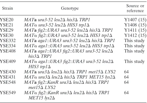
Yeast growth and manipulation.Yeast growth and sporulation media were as described in reference 54. The strains employed in this study were from either of two derivatives of the S288C background as indicated in Table 1. Bacterial manipulations involving plasmids were performed as described in reference 49. Yeast strains were transformed with plasmids by a modified one-step transfor-mation method (11). Replacements of theAGA1gene withURA3were made by using a PCR-mediated gene knockout technique (2). MATa and MAT␣
aga1::URA3 FIG2::URA3double mutants were constructed by mating haploid strains YSE29 and YSE334, selecting for the resulting diploid, sporulating, and selecting tetrad progeny in whichURA3segregated 2:2. The genotypes forMAT
and other markers were determined in the candidate double mutant strains, and the presence of disruptions was confirmed by PCR with flanking primers.
Measures of mating cell fusion, morphogenesis, and viability.Analyses of mating yeast cells were carried out as described in reference 15 with the following modifications. Cultures were mixed 1:1 and mated on nitrocellulose filters placed on different media for a period of 4 h. Following mating, cells were either immediately formaldehyde fixed, washed, and stored in 1⫻phosphate-buffered saline (PBS)–1 M sorbitol at 4°C, or an aliquot of live cells was removed for methylene blue staining to score cell viability, and the remainder of the cells were preserved as described above. Mating projection-conjugation bridge morphogen-esis was quantified by analyzing the widths of the parental cells relative to the width of the conjugation bridge joining them in zygotes as described in reference 15. To aid in quantitatively comparing this trait among different strains and under different mating conditions, over 100 zygotes were photographed for each
sample with a Nikon TE-300 microscope fitted with a⫻100 objective and dif-ferential interference contrast (DIC) optics. Images were captured with a Roper/ Princeton Instruments MicroMax 5-MHz charge-coupled device (CCD) camera. A ruler tool in Adobe Photoshop was used to determine the three relevant measurements for each zygote. These data were then tablulated and compared by spreadsheet analysis (Excel). A small number of zygotes were discarded from all sample analyses if they met the criterion that the two parental cells differed by more than 0.75⫻their mean width. These zygotes arise from matings between parental cells of significantly different size; thus, the signaling and growth dy-namics of the conjugation bridge joining them are likely to be more variable than those of two similarly sized parents. Typically, removal of these zygotes led to relatively small decreases in the variances of these data. While the morphoge-netic effects offig2⌬are most apparent in one of the parental strain backgrounds we have used for the present studies, qualitatively consistent results were found when these experiments were repeated with other S288C-derivedfig2⌬strains described in this study (Table 1). In this strain background (64), which is more resistant to the cell wall-disturbing effects of calcofluor white relative to the Y800 parental background of YSE21, YSE22, YSE29, and YSE30 (G. Huang and S. Erdman, unpublished observations), the morphogenetic effects of the loss of
FIG2are less pronounced.
For assays of cell viability in the presence of mating pheromone, strains were grown overnight, and their densities were standardized to an optical density at 600 nm (OD600of 0.4) as described previously (15). Cells were then treated with
10g of␣-factor per ml (Sigma T6901) and removed at subsequent times for staining with methylene blue. Staining was done by adding methylene blue to final concentration of 0.001% and staining the cells with shaking at room tem-perature for 30 min. Following staining, cells were washed once in synthetic complete (SC) medium and immediately scored by microscopy at a magnification of⫻100 for the fraction of cells staining. For each sample, at least 800 cells were scored. To examine cell viability in mating mixtures, cells were mated under the indicated conditions, after which, they were collected from the filters into SC medium and sonicated to disperse aggregated cells. Methylene blue was added to a final concentration of 0.001%, and the cells and zygotes were stained at room temperature for 30 min. We also quantified cell viability by measuring the colony-forming ability of cells plated to YPAD medium (yeast extract–peptone– dextrose [54] supplemented with 0.3 mM adenine) following treatments similar to a number of those described in the Results. The results of these experiments closely paralleled those we found by methylene blue assays. Cell fusion defects (partial and complete) were assayed by FM4-64 staining as described in refer-ence 15.
Analyses of Pkc1 pathway activity by anti-MAPK immunoblot and reporter gene constructs.Yeast cells of the indicated strains and carrying the Slt2/Mpk1 gene on a high-copy, YEp24 plasmid were grown to mid-log phase at 30°C in synthetic complete medium lacking uracil (SC⫺ura). Cells were then diluted to an OD600of 0.25 in fresh medium and allowed to grow until OD600of 0.5 at 30°C
before treatment with 10g of␣-factor per ml at 24°C for 4 h. Cells were pelleted and transferred to an Eppendorf tube in 200l of cold morpho-lineethanesulfonic acid (MES) buffer (25 mM MES [pH 6.5], 10% glycerol, 100 mM NaCl, 100 mM KCl, 1 mM EDTA, 1 mM dithiothreitol, 5 mM NaF, 1 mM sodium orthovanadate, 20 mM-glycerolphosphate, protease inhibitors). An equal volume of glass beads was added, and cells were lysed by vortexing. After
TABLE 1. Strains used in this study
Strain Genotype Source orreference
YSE20 MATaura3-52 leu2⌬his3⌬TRP1 Y1407 (15) YSE21 MAT␣ura3-52 leu2⌬HIS3 trp1⌬ Y1408 (15) YSE29 MATafig2::URA3 ura3-52 leu2⌬his3⌬TRP1 Y1411 (15) YSE30 MAT␣fig2::URA3 ura3-52 leu2⌬HIS3 trp1⌬ Y1412 (15) YSE332 MATaaga1::URA3 ura3-52 leu2⌬his3⌬TRP1 This study YSE334 MAT␣aga1::URA3 ura3-52 leu2⌬HIS3 trp1⌬ This study YSE408 MATaaga1::URA3 fig2::URA3 ura3-52 leu2⌬
his3⌬TRP1 This study
YSE409 MAT␣aga1::URA3 fig2::URA3 ura3-52 leu2⌬
HIS3 trp1⌬ This study
YSE430 MATaura3⌬leu2⌬his3⌬TRP1 met15⌬LYS2 64 YSE431 MAT␣ura3⌬leu2⌬his3⌬TRP1 MET15 lys2⌬ 64 YSE548 MATafig2::KanR ura3⌬leu2⌬his3⌬TRP1
met15⌬LYS2 64
YSE549 MAT␣fig2::KanR ura3⌬leu2⌬his3⌬TRP1
MET15 lys2⌬ 64
on September 8, 2020 by guest
http://ec.asm.org/
10 min of centrifugation at 4°C, supernatant of the extracts was collected. Lae-mmli buffer (15l) was added to 30l of extract, and the protein samples were denatured by heating at 95°C for 5 min and then separated on sodium dodecyl sulfate (SDS)–10% polyacrylamide gels. Western transfer to polyvinylidene di-fluoride Immobilon membranes was by semidry blotting. Blotted membranes were then preincubated for 1 h at room temperature in 1⫻PBST (PBS, 0.5% Tween 20) containing 5% nonfat dry milk. Following preincubation, the blot was incubated overnight at 4° in a 1/1,000 dilution of primary anti-p42/44 mitogen-activated protein kinase (MAPK) antibodies (New England Biolabs). The blot was then washed three times for 10 min each in 1⫻PBST and incubated for 2 h in the presence of a 1/4,000 dilution of a goat anti-rabbit horseradish peroxidase (HRP)-conjugated secondary antibody (Sigma). Detection of the HRP activity was by chemiluminescence via a Pierce SuperSignal detection kit. Blots were imaged by X-ray film or directly detected with a Kodak/NEN 440CF imaging system. Quantification of relative Mpk1 signals and total protein was determined with densitometry software associated with the Kodak/NEN 440CF imaging system. In the case of total protein, following development, the immunoblot was stained with Coomassie blue and imaged, and band intensities were integrated in each lane for all resolved proteins with the densitometry software.
To measure Rho1/Pkc1 pathway activity by Rlm1 binding site-drivenlacZ
reporter assay, cells of the indicated strains were transformed with a reporter plasmid, p2⫻Rlm1, or the parent, pLG178 (gifts of D. Levin) (22). Cells were grown SC⫺ura medium overnight and diluted into fresh YPAD medium at an OD600of 0.1, after which they were allowed to double until they reached an
OD600of 0.4 to 0.5. Cells were then treated with 10g of ␣-factor mating
pheromone per ml. Cells were harvested by centrifugation, washed 1⫻in Z buffer, and lysed and assayed for-galactosidase activity as described in refer-ence 15. The data presented represent the mean and standard error of three experiments.
Measurement of zygotic spindle alignment.Cells to be mated were grown, standardized for density, and mixed 1:1 in 18-mm-diameter culture tubes. One additional equivalent of fresh YPAD liquid medium was added (1 ml each of
MATa,MAT␣, and YPAD). Cells were mixed by vortex and immediately vacuum filtered onto a 0.45-m-pore-diameter nitrocellulose filter (Millipore). Mating cells were placed onto solid media under the indicated conditions for 4 h. After mating, cells were recovered by washing from the filters into dropout medium supplemented with 0.1 volume of 10 mM adenine to suppress background fluo-rescence. Aggregates of mating cells were dispersed by sonication, and green fluorescent protein (GFP) fusion proteins were immediately visualized in the strains with a TE-300 microscope fitted with a⫻100 Plan objective (Nikon) and GFP filter set (Chroma). Samples were not fixed or stored before viewing.
To quantify spindle alignment, zygotes viewed under the microscope were examined to determine the orientation of the Stu2::GFP fluorescence pattern. The orientation of this pattern with respect to the zygotic bud neck was deter-mined. Any spindle showing one pole to have entered the bud, or the subsequent expansion of which was expected to project it through the bud neck, was scored as correctly aligned, while spindles whose ends were not expected to project into the bud based on their current orientation were considered incorrectly aligned. Zygotes with more than three disorganized and unaligned foci were considered separately. Nuclear position was also quantified by DIC or fluorescence micros-copy of fixed wild-type andfig2⌬/fig2⌬zygotes that were prepared as described above, fixed, and stained with 4⬘,6⬘-diamidino-2-phenylindole (DAPI).
Measurement of agglutination.Agglutination assays of mating yeast cells were performed based on the method described by Terrance and Lipke (59) with the following modifications.MATaandMAT␣cells were each grown separately in liquid medium at 30°C overnight. The cell cultures were standardized to an OD600of 0.1 and grown for an additional 4 h at 30°C until they had reached an
OD600of 0.5. TheMATaandMAT␣cells and fresh medium were mixed 1:1:1 (2
ml each) and mated for 4 h at 30°C with constant mixing on a roller drum. The single cell types were also grown under the same condition for 4 h. All of the cell cultures were immediately fixed by adding 0.1 volume of 37% formaldehyde and allowed to fix either overnight at 4°C or for 1 h at room temperature. To measure the agglutination activity, 2.0 ml of fixed cell suspension was transferred into 4-ml cuvettes, and the cell aggregates were allowed to settle for 20 min. The two parent cell types were also measured as controls. The OD600was measured with
a spectrophotometer (Spectronic Genesys 8). The decrease in OD reading is a quantitative measure of agglutination. All samples were in triplicate. The agglu-tination index (AI) was calculated as AI⫽1⫺[(2⫻Aa⫹⬀)/(Aa⫹A⬀)] (59). For
experiments involving sorbitol, matings were performed in liquid media with different sorbitol contents, and comparisons were made to the rate of settling of unmated cells in identical media.
RESULTS
Mating morphogenesis defects of fig2⌬ cells are osmotic remedial. Pheromone-induced morphogenesis is abnormal in
mating mixtures offig2⌬cells (15), and it is possible that the
defects observed in these mutants result from altered cell wall structure or cell integrity signaling. Thin-section electron mi-croscopy studies of the cell wall in the region of the mating projection previously showed this region to be thinner than the wall of vegetative cells (28). We therefore became interested in
whetherFIG2might be specifically expressed in mating cells to
aid in organizing the region of the cell wall that is rapidly
growing. Consistent with such a role,-galactosidase fusions to
an amino-terminal part of Fig2p and epitope-tagged forms of the protein have been localized to the mating projection (15, 18). To test whether the narrow mating projection and
conju-gation bridge phenotype previously observed infig2⌬cells
re-sulted from defects in processes that affected the integrity of
the cell wall, we examined the responsiveness of the fig2⌬
phenotype to conditions that osmotically stabilize the cell wall.
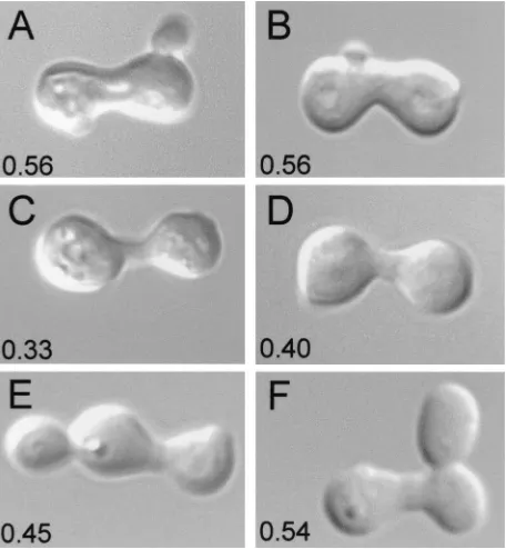
Figure 1 shows the morphogenetic responses of wild-type (WT)
andfig2⌬zygotes that result from mating cells on media
dif-fering only in osmolarity. Measurements of the width of the conjugation bridge in comparison to the mean parental width of zygotes previously showed that, in wild-type zygotes, this
ratio is approximately 0.52, whereas infig2⌬zygotes, the ratio
is⬃0.30 (15). Table 2 shows the mean values and standard
FIG. 1. Increased extracellular osmolarity suppresses the morpho-genetic defect offig2⌬cells. DIC images of representative wild-type andfig2⌬zygotes formed under the osmotic conditions indicated. The ratio of conjugation bridge/mean parental cell diameter is shown in the lower left portion of each image. Zygotes shown are from the following matings: YSE20⫻YSE21 (WT), YPAD (A); YSE20⫻YSE21 (WT), YPAD with 1.0 M sorbitol (B); YSE29⫻YSE30 (fig2⌬), YPAD (C); YSE29⫻YSE30 (fig2⌬), YPAD with 0.3 M sorbitol (D); YSE29⫻ YSE30 (fig2⌬), YPAD with 0.6 M sorbitol (E); YSE29⫻YSE30 (fig2⌬), YPAD with 1.0 M sorbitol (F).
on September 8, 2020 by guest
http://ec.asm.org/
error obtained by making this comparison directly from images of over 100 zygotes for each pair of strains and mating condi-tion. As is apparent from both Fig. 1 and Table 2, as osmotic conditions increase from 0.3 M sorbitol through 1.0 M sorbitol,
the defects offig2⌬mutants with respect to conjugation bridge
morphogenesis are progressively more suppressed to a mor-phology very near that of wild-type zygotes. When conditions are isosmotic to hyperosmotic, all cells mate at reduced
effi-ciencies (data not shown). Under these circumstances, fig2⌬
cells, despite forming zygotes with similar morphologies to the wild type, encounter other problems affecting their mating
success (described below). These data show that the fig2⌬
morphogenetic defects are suppressible by conditions that have previously been reported to compensate for defects in cell wall biogenesis or structure (26, 40, 48).
FIG2is also required for normal cell-cell fusion during
mat-ing. A number of different genes encoding cell polarity pro-teins and necessary for efficient mating cell fusion play roles in
both vegetative and mating cell morphogenesis (e.g., SPA2,
BNI1,KEL1, andPEA1) (reviewed in reference 63). Thus, the
defects in cell fusion seen infig2⌬matings might be a direct
result of morphogenesis defects. Alternatively, the mating cell
fusion defects might reflect a role ofFIG2in this process that
is independent of its morphogenesis functions. To address this question, we examined the fraction of cells in the different matings in the experiments just described that existed as cou-pled cells (prezygotes) or zygotes and noted any defects in cell
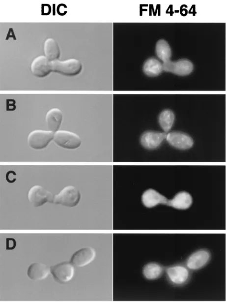
fusion processes. As Fig. 2 and Table 3 illustrate,fig2⌬ cells
mated on YPAD medium show an approximate twofold in-crease in the fraction of zygotes showing defects in cell fusion,
as previously reported (15). However, in contrast to the results obtained with respect to suppression of the narrow conjugation bridge phenotype, changes in external osmolarity failed to
res-cue defects of fig2⌬ mating cells in the cell fusion process.
These results suggest that the requirements forFIG2in mating
cell morphogenesis and cell fusion can be uncoupled by os-motic conditions and therefore are likely to be in some way distinct.
High temperature interferes with the osmotic suppression offig2⌬and cell fusion.Given that sorbitol suppressed defects
offig2⌬ cells in mating cell morphogenesis, and since Rho1
and Pkc1 participate in both cell integrity signaling and polar-ized cell growth, we further investigated whether elevated sig-naling by the Rho1/Pkc1 pathway might be responsible for
some of the phenotypes observed in fig2⌬ cells. We again
carried out the preceding experiments, except that cells were mated at an elevated temperature, 37°C, which has been shown to activate signaling through the Rho1/Pck1 pathway (23). Ta-ble 2 shows the results of these experiments in comparison to matings carried out at 30°C with respect to sorbitol suppression
of conjugation bridge morphogenesis defects present infig2⌬
zygotes. These data indicate that while elevated temperature
does not significantly enhance the baseline effect offig2⌬in the
FIG. 2. Suppression offig2⌬/fig2⌬zygote phenotypes by increased osmolarity acts specifically on morphogenetic and not cell-cell fusion defects. DIC and FM4-64-stained fluorescence images of zygotes formed in matings carried out in YPAD–1.0 M sorbitol medium. Mi-crographs are of A) YSE20⫻YSE21 (WT) (A) and (panels B, C, and D) YSE29⫻YSE30 (fig2⌬) zygotes display sorbitol-suppressed, nor-mal conjugation bridge morphogenesis, but remain defective in cell fusion.
TABLE 2. Conjugation bridge morphogenesis in WT andfig2⌬zygotes
Zygote
Bridge diam/parental cell width in YPAD with the following concn of sorbitola:
None 0.3 M 0.6 M 1.0 M
30°C
WT⫻WT 55.6⫾0.8 54.2⫾0.9 53.7⫾0.9 54.4⫾1.0 fig2⌬ ⫻fig2⌬ 34.6⫾0.6 38.9⫾0.8 45.5⫾0.8 50.6⫾0.8 37°C
WT⫻WT 58.5⫾1.6 56.4⫾1.6
fig2⌬ ⫻fig2⌬ 35.3⫾0.9 45.1⫾0.8
aValues represent ratios and associated standard errors of conjugation bridge
diameter to mean parental cell widths fornⱖ95 zygotes, measured as described in Materials and Methods.
TABLE 3. Cell fusion defects in WT andfig2⌬/fig2⌬zygotes
Zygote
% of cells with defectsa
30°C 37°C
YPAD YPAD–1 Msorbitol YPAD YPAD–1 Msorbitol
WT⫻WT 5.3 4.0 11.3 12.0
fig2⌬ ⫻fig2⌬ 12.3 10.6 23.8 20.0
aValues represent percentages ofnⱖ150 paired conjugating cells (prezygotes
and zygotes) displaying partially or completely unfused cells as visualized by FM4-64 and DAPI staining (15). The mating mixtures assayed were the same as those presented in Table 2.
on September 8, 2020 by guest
http://ec.asm.org/
absence of osmotic support, high temperature does reduce the
extent to which sorbitol suppresses the effects of fig2⌬ on
conjugation bridge morphogenesis (Table 2). We also exam-ined cell fusion in these experiments and observed a twofold increase in cell fusion defects when cells of all genotypes are mated at 37°C (Table 3).
Conditions of elevated temperature and polarized growth during mating activate the Rho1/Pkc1 pathway (4, 23, 47, 65).
Our data indicate that loss ofFIG2 also increases signaling
through this pathway during mating (described below). One interpretation of these results is that the combination of these
three conditions (mating, high temperature, and loss ofFIG2)
produces a level of signaling in this pathway that can no longer be suppressed by the osmotic quenching of pathway signaling that can occur under vegetative conditions (23).
FIG2is required for the maintenance of cell integrity during mating.A number of gene products have been shown to be required for maintenance of the viability of cells during
mat-ing, including the MID2 gene product, which functions as a
sensor upstream of the Rho1/Pkc1 pathway (24, 41, 45). A potential role for Fig2p in organizing components of the mat-ing cell wall would predict that the absence of the protein during mating might lead to diminished integrity of cells due to
defects in wall organization. We therefore tested whetherfig2⌬
strains were less viable than wild-type cells after the onset of mating cell morphogenesis. Two types of assays were employed to quantify viability under different conditions to test mating
cell function. We first tested whether treatment ofMATacells
with levels of␣-factor mating pheromone that elicit mating cell
morphogenesis, and which have previously been shown to re-veal defects in cell integrity for other mutants, would rere-veal
viability differences between wild-type and fig2⌬ cells under
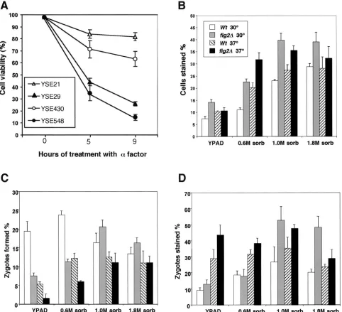
such conditions. The fraction of cells staining with the dye methylene blue, which is excluded from viable cells, was exam-ined immediately following pheromone treatment at the times indicated in Fig. 3A. Cells from two different strain back-grounds were examined, and the results of both show that,
relative to wild-type cells,fig2⌬cells are markedly reduced in
viability following extended treatment with morphogenesis-in-ducing levels of mating pheromone. This reduction in viability
was not accentuated by removal of Ca2⫹from the medium by
addition of EGTA, nor was it suppressed by addition of Ca2⫹
to levels up to 100 mM (data not shown). Thus, the effect of
fig2⌬on cell viability does not appear to occur through
block-ing of the essential transport of Ca2⫹ions into mating cells (21,
35).
We next sought to determine whetherfig2⌬cells displayed
reduced viability under actual mating conditions and whether zygotes or nonmating cells displayed differential sensitivities.
For these studies,MATaandMAT␣cells were mated on filters
for a period of 4 h, after which, the fraction of methylene blue-stained cells was determined by microscopy. Figure 3B, C, and D show the results of these studies, which indicate that
fig2⌬cells are significantly less viable than wild-type cells in
mating mixtures under nearly every condition tested. Figure 3B and D show that as osmolarity levels of 0.6 M sorbitol and
above are encountered,fig2⌬cells can be seen to show a sharp
reduction in viability relative to wild-type cells. Interestingly, when zygote formation was quantified in these experiments (Fig. 3C), we found that there was a significant decrease in the
rate of zygote formation offig2⌬cells under hypoosmotic
mat-ing conditions, but this inhibition was alleviated by elevated osmolarity. However, while elevated osmolarity leads to in-creased zygote formation in such cells, a large fraction of such
fig2⌬ zygotes are inviable (Fig. 3D). In each of the assays
described above, analyses of the responses of vegetative cells to elevated osmolarity conditions showed no difference in
viabil-ity or growth rate of fig2⌬ cells in comparison to wild-type
strains, consistent with the induced expression and known roles
ofFIG2in mating. In summary, the effects offig2⌬mutations
on mating cell viability in both ␣-factor-treated cells and in
mating mixtures suggests a common defect in the maintenance of cell integrity that cannot be rescued by elevated levels of osmotic support.
Rho1/Pkc1 pathway activity is elevated infig2⌬cells under-going mating morphogenesis.Our data concerning
morpho-genesis, cell fusion, and cell integrity defects offig2⌬mutants
suggested the hypothesis that the Rho1/Pkc1 cell integrity pathway might be activated in such cells in a manner inappro-priate to its normal signaling functions in mating. The Rho1/ Pkc1 pathway has previously been shown to undergo activation in response to pheromone-induced morphogenesis (4, 47). To test our hypothesis, we first examined whether Mpk1p/Slt2p, the MAPK that lies downstream of Pkc1p in its signaling
path-way, was activated to higher levels in fig2⌬cells undergoing
mating cell morphogenesis than it normally is in wild-type cells.
Wild-type andfig2⌬MATacells were treated with
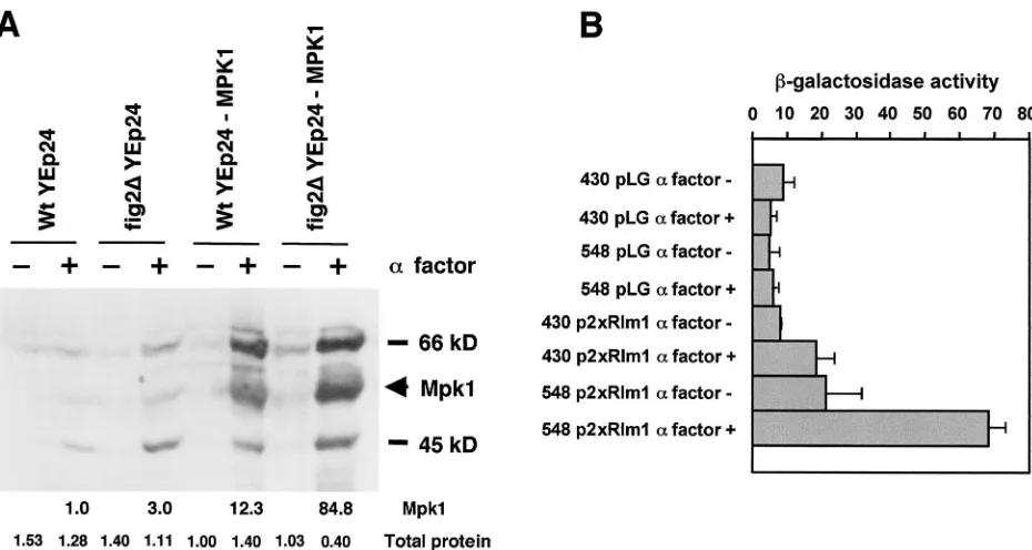
morphogen-esis-inducing levels of mating pheromone for 4 h, after which, the cells were harvested by centrifugation and lysates were prepared. Protein extracts were electrophoresed on SDS-poly-acrylamide gel electrophoresis (PAGE), and were semidry transferred to nylon membrane and then incubated with anti-p42/44 MAPK antibodies that preferentially recognize the phosphotyrosine form of MAPKs. This antibody has been used by others successfully to detect the MAPK Mpk1p/Slt2p and is specific to the activated, tyrosine-phosphorylated form of the MAPK (S. Vidan and M. Snyder, personal communication). As shown in Fig. 4 A, by detecting both the endogenous Mpk1p/Slt2p and the protein present in cells, where it is en-coded endogenously and on a high-copy plasmid,
YEp24-MPK1/SLT2 (12), we observed increased levels of activated
Mpk1p/Slt2p infig2⌬cells relative to wild-type cells. Scanning
densitometry of the films and normalization of signals to lane loading of total protein indicates that Mpk1p/Slt2p is found at
3.5- to 24.0-fold the levels in mating pheromone-treatedfig2⌬
cells relative to wild-type cells. These data therefore support the hypothesis that the absence of Fig2p from the mating cell wall leads to a further increase in Rho1/Pkc1 pathway activity. This level of Mpk1p activation may be inappropriately high for mating cells and thereby cause the morphogenesis, cell fusion, and zygotic spindle alignment defects (see below) associated
withfig2⌬cells.
We used a second approach to independently assess Mpk1p/
Slt2p activation levels infig2⌬cells in comparison to wild-type
cells. Plasmids carrying either acyc1-lacZreporter gene
lack-ing UAS sequences alone, pLG, or with two upstream Rlm1
binding sites, p2⫻Rlm1, were transformed into the strains we
had examined for Mpk1p activation. Rlm1p is a DNA binding transcriptional activator that lies directly downstream of Mpk1p,
and its activity is directly controlled by the Pkc1⬎⬎Mpk1
on September 8, 2020 by guest
http://ec.asm.org/
naling pathway. This reporter plasmid has previously been shown to be induced 6.5- to 16.5-fold after 4 h under different conditions of cell wall stress (U. S. Jung and D. Levin, personal
communication). MATa wild-type and fig2⌬ strains carrying
these plasmids were grown in liquid culture, and cells were
treated with morphogenesis-inducing levels of␣-factor
phero-mone for 4 h. Following pherophero-mone treatment, cells were
assayed for-galactosidase activity as described in Materials
and Methods. As Fig. 4B shows, wild-type andfig2⌬ strains
carrying the 2⫻Rlm1 reporter both show an ␣
-factor-depen-dent increase in activity. Infig2⌬ cells, however, there is an
elevation of basal signaling in the absence of␣-factor (⬃
2.2-fold) and a marked elevation under conditions of ␣-factor
induction (⬃3.6-fold). These data are in close agreement with
the relative increases we found in pathway signaling as mea-sured by immunoblot comparison of endogenous Mpk1p/Slt2p activation (3.5-fold). In summary, these experiments assessing Mpk1p and Rlm1p activity levels show that signaling through the Rho1/Pkc1 cell integrity pathway occurs at significantly
elevated levels infig2⌬cells relative to those in wild-type cells
during mating.
Zygotic spindle alignment requires FIG2 function. Initial
studies offig2⌬ ⫻fig2⌬matings noted an unusual appearance
and possible mislocalization of the nucleus in zygotes formed in these crosses, as visualized by DAPI staining (15). We sought to further investigate whether nuclear migration is
al-FIG. 3. Absence ofFIG2results in increased inviability under mating conditions. A) Viability in the presence of␣-factor.MATaWT(YSE20 and YSE430) andfig2⌬(YSE29 and YSE548) strains were treated with pheromone for the indicated times and assayed for viability by methylene blue exclusion. (B) Mating cells from bilateral crosses ofWt(YSE21⫻YSE22) andfig2⌬(YSE29⫻YSE30) strains were assayed for viability by methylene blue exclusion immediately following mating for 4 h on solid media under the conditions indicated. Values represent percentages from over 800 cells counted. (C) Zygote formation rates for the crosses indicated in panel B. (D) Viability of zygotes in mating mixtures shown in panel B.
on September 8, 2020 by guest
http://ec.asm.org/
tered in such cells and whether this phenotype is influenced by osmotic conditions. To quantify nuclear migration in zygotes,
we transformed MATa and MAT␣ wild-type and fig2⌬ cells
with a centromere-based plasmid carrying a Stu2::GFP fusion gene (61). The encoded Stu2::GFP fusion protein localized to spindles and spindle pole bodies in wild-type cells as previously reported. Spindle localization was seen as a bar of staining as shown in Fig. 5A to D. In zygotes that had initiated a bud, we quantified the alignment of the spindle axis with respect to the mother (zygote)-bud axis as described in Materials and Meth-ods. Table 4 shows that spindles are aligned with the
zygote-bud axis approximately 18% less well infig2⌬zygotes than in
wild-type zygotes. Because we found that osmotic support sup-pressed morphogenesis defects in such zygotes, we tested whether a similar suppression was possible for the spindle alignment defect. Table 4 shows that conditions that can com-pensate for cell wall stress and reduce Pkc1 pathway signaling (1.0 M sorbitol) also suppress 61% of the difference in spindle
alignment fidelity seen between wild-type and fig2⌬ cells.
These data also show that the process of spindle alignment in
fig2⌬/fig2⌬zygotes is more sensitive to cold than it is in
wild-type zygotes. These data, obtained with strains from this inde-pendent S288C background (YSE548 and YSE549), agree with
previous findings thatfig2⌬ ⫻fig2⌬matings are markedly cold
sensitive in comparison to the wild type (15). To further con-firm our observations concerning spindle alignment and nu-clear positioning, we also scored by DIC or fluorescence mi-croscopy of DAPI-stained cells the locations of single, fused zygotic nuclei in zygotes that had initiated a bud that had grown to medium to mature size. Zygotes were divided into the
following three classes: I, nucleus present in either parent of the zygote; II, nucleus present in the bridge of the zygote near the zygote-bud neck; and III, nucleus present completely in the bud. The distributions of these classes for wild-type zygotes
were I ⫽ 2.0%, II⫽ 93%, and III⫽ 5% (n ⫽ 204 zygotes
scored), and the distributions infig2⌬/fig2⌬zygotes were I ⫽
7.6%, II⫽85.7%, and III⫽6.7% (n⫽207 zygotes scored).
Collectively, these data indicate that spindle positioning by astral microtubule dynamics in zygotic divisions is impaired in
fig2⌬/fig2⌬zygotes. These defects and the morphogenesis
de-fects infig2⌬cells and zygotes may therefore occur through a
common mechanism.
Infig2⌬/fig2⌬zygotes, we also noted another difference in
Stu2::GFP localization relative to wild-type cells, such that
4.3% of all zygotes examined infig2⌬ ⫻fig2⌬mating mixtures
contained patterns of Stu2::GFP localization to a small num-ber of distinct foci, as shown in Fig. 5E and F, in comparison
to 1.1% observed in matings of wild-type cells (nⱖ280). These
foci occurred in clusters in cells and were not located near presumptive sites of spindle poles. Cells displaying these foci of Stu2::GFP were rarely observed in wild-type mating mixtures, and this localization was not strictly correlated with the stage of cells in mating with respect to their being unbudded, polarized, or a zygote, although the frequency of foci was somewhat higher in zygotes relative to the other classes. When cells were mated on media containing 1 M sorbitol, foci were observed in
only 1.3% offig2⌬/fig2⌬zygotes and 0.6% of wild-type zygotes
(n ⱖ 150). These data indicate that the mislocalization of
Stu2p infig2⌬ zygotes is also substantially suppressed by
in-creases in extracellular osmolarity.
FIG. 4. Activation levels of the Rho1/Pkc1 cell integrity pathway are greater infig2⌬cells than in the wild type. (A) Anti-MAPK immunoblot detecting activation of Mpk1p following pheromone treatment in YSE430 (WT) and YSE548 (fig2⌬) cells carrying the plasmid YEp24 or the same cells each carrying the plasmid YEp24-MPK1/SLT2. Numbers below the lanes indicate the relative intensity of Mpk1 in each lane and relative total protein levels determined as described in Materials and Methods. (B) Activity levels of the Pkc1 pathway assayed by a 2⫻Rlm1 binding site-dependent-galactosidase reporter, p2⫻Rlm1 in YSE430 (WT) and YSE548 (fig2⌬) cells following mating pheromone treatment. pLG is the parentcyc1-lacZ-containing vector, which contains no Rlm1 binding sites.
on September 8, 2020 by guest
http://ec.asm.org/
Enhanced agglutination of fig2⌬ cells during mating de-pends on agglutinins.Cells lackingFIG2show increased ag-glutination in bilateral matings under liquid conditions (15),
and when FIG2 is ectopically expressed in vegetative cells,
cell-cell adherence is modified (18). To help understand the basis of the enhanced agglutination observed in matings
in-volvingfig2⌬cells, we further investigated its mechanism.
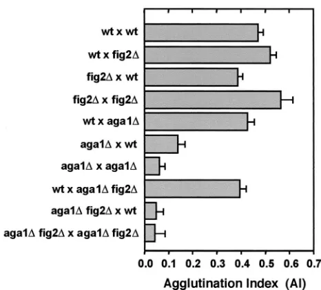
Fig-ure 6 shows again that agglutination is most strongly enhanced
when both mating partners are missing FIG2. However, in
unilateral matings in which only one partner isfig2⌬, increased
agglutination occurs only in the context in which thefig2⌬cells
are theMAT␣partner. This is evident by the strong
aggluti-nation ofwt⫻fig2⌬matings in comparison tofig2⌬ ⫻wtand
bilateral crosses ofwtandfig2⌬strains alone (Fig. 6). Thus, the
mechanism ofFIG2modulation of agglutinin activity is largely
␣ partner specific, although its absence from both partners
does lead to maximally increased agglutination. This suggests an additional interaction with a non-cell-type-specific compo-nent of the process. Similar results concerning this mechanism have also recently been found by others (C. Jue and P. Lipke, personal communication)
FIG2andAGA1encode GPI-anchored proteins that share
sequence homology in regions containing WCPL domains and
a CX4C domain (53; M. Zhang and S. Erdman, unpublished
data). These genes have also recently been found to be collec-tively important for some aspect of efficient mating that is in-dependent of the agglutinin Aga2p (18). To determine whether
the mechanism of enhanced agglutination induced by fig2⌬
strains operated through the agglutinins Aga1p, Aga2p, and
Ag␣1p, we tested the ability offig2⌬to enhance the
aggluti-nation of cells carryingaga1⌬mutations. As shown in Fig. 6,
the effect offig2⌬is largely dependent on interactions between
the agglutinins, as evidenced by strongly reduced agglutination
inMATaaga1⌬fig2⌬double mutants.
Increased osmolarity fails to reduce enhanced agglutination due to loss ofFIG2.Because sorbitol was effective in
suppress-ing other defects associated with loss ofFIG2, we sought to
determine whether osmotic support could compensate for the
agglutination effects observed in the absence ofFIG2. To test
this, we compared the agglutination behavior offig2⌬mutants
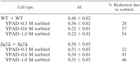
to that of wild-type cells mated under different osmotic condi-tions. As Table 5 shows, under conditions of elevated extracel-lular osmolarity, while cells of every genotype tested aggluti-nate much less, particularly at sorbitol concentrations of 0.6 M and above, there is no specific elimination of the enhanced
agglutination offig2⌬ cells relative to wild-type cells. These
data demonstrate that the effects offig2⌬on agglutination are
FIG. 5. Reduced zygotic spindle alignment fidelity and mislocaliza-tion of the spindle/microtubule-associated protein Stu2p infig2⌬/fig2⌬ zygotes. (A) Merged DIC-GFP fluorescence images of Stu2::GFP fu-sion proteins decorating a wild-type aligned spindle. (B, C, and D) GFP fluorescence images of wild type aligned (B), andfig2⌬/fig2⌬ unaligned (C and D), zygotic spindles. (E and F) GFP fluorescence images of Stu2::GFP fusion proteins incorrectly mislocalized to dis-crete foci infig2⌬/fig2⌬zygotes.
FIG. 6. Enhanced agglutination due to loss of Fig2p is dependent upon the mating agglutinins. Agglutination indices are shown for bi-lateral matings of wild type andfig2⌬cells, as well as reciprocal uni-lateral crosses involving wild-type,aga1⌬andaga1⌬fig2⌬strains.
TABLE 4. Zygotic spindle alignment in WT andfig2⌬zygotes
Zygote
% Aligneda
30°C 16°C
YPAD YPAD YPAD–1M sorbitol
WT/WT 89.4 92.0 83.2 (0.93)
fig2⌬/fig2⌬ 71.4 (0.79) 82.7 (0.89) 61.2 (0.86)
aValues represent percentages based onnⱖ150 zygotes examined; numbers
in parentheses indicate the fraction of WT spindle alignment fidelity occurring at 30°C represented by the given value.
on September 8, 2020 by guest
http://ec.asm.org/
independent of the osmolarity of the medium. Thus, the
phe-notypic effects offig2⌬mating cells with regard to
morphogen-esis, cell integrity, and spindle positioning are evidently sepa-rable from those involving agglutination.
DISCUSSION
In the present study, we describe new roles forFIG2in the
maintenance of mating cell integrity and zygotic spindle align-ment and further examine the nature of different phenotypic
effects offig2⌬mutations in the yeast mating process. These
studies suggest a complex interplay between pheromone
sig-naling and its downstream targets, includingFIG2and
signal-ing pathways maintainsignal-ing cell integrity. These pathways are likely to be related through both dependent relationships and feedback loops that are active under different physiological conditions.
Cell wall organization through Fig2p underlies several mat-ing processes.FIG2is one of many genes whose induced ex-pression occurs by activated mating pathway signaling and which serve to differentiate cells, enabling them to efficiently carry out the mating process (15, 31, 56). Studies thus far have
described both negative and positive roles forFIG2in
medi-ating cell adhesion processes (15, 18; C. Jue and P. Lipke, personal communication). One apparent positive role for Fig2p occurs in the absence of another adhesin, Aga1p, and is
independent of Aga2p, the functional portion of theMATacell
agglutinin molecule (18; S. Erdman, unpublished observa-tions). Currently, it remains unclear what this Aga1p-related and agglutination-independent role may be, although it has
been observed that few fig2⌬ aga1⌬ cells are found to be
mating in mating mixtures (18; S. Erdman, unpublished
obser-vations). Additional roles forFIG2in cell-cell fusion and
spa-tial control of morphogenesis during mating have been de-scribed previously (15). Here, we have identified additional
positive functions for FIG2 in mediating survival of mating
cells and in aiding the fidelity of the alignment process that zygotic spindles undergo.
The involvement of Fig2p in the multiple processes of cell adhesion, cell-cell fusion, cell morphogenesis, cell integrity maintenance, and zygotic spindle alignment prior to anaphase suggests that some of these defects may be secondary effects
due to the loss ofFIG2. Because Fig2p is located outside of the
cell, it follows that the morphogenesis and zygotic spindle-positioning functions of Fig2p must be indirect and therefore involve transmembrane signaling. Of the remaining processes that require Fig2p, regulation of agglutination and mainte-nance of cell integrity, the effects on agglutination might also be indirect due to general alterations in the structure of the cell wall caused by the absence of Fig2p. The absence of Fig2p might alter mating cell wall organization, making the
aggluti-nins more accessible, or loss ofFIG2might reduce competition
for anchor attachment machinery that could arise among the
GPI-anchored adhesin proteins Aga1p, Ag␣ 1p, and Fig2p
during their biogenesis. These aspects of thefig2⌬phenotype
involving cell adhesion control remain to be addressed.
Our results demonstrate thatfig2⌬cells are less viable
rel-ative to wild-type cells under conditions promoting mating cell morphogenesis and in response to prolonged mating pathway activation. Because Fig2p is a cell wall resident protein, it seems logical that a major and primary role for Fig2p in cells is to help modulate and organize cell wall structure in the region of the mating projection where it is localized. Fig2p may function directly to organize other cell wall resident manno-proteins or the cell wall (glucans and chitin) itself. This type of organizational role might be critical to the maintenance of cell wall structure and therefore cell integrity during the rapid cell wall remodeling events accompanying projection growth and cell-cell fusion during mating. These requirements would be expected to be even more important to mating cells that expe-rience changes in their environment in regard to external os-molarity. Such a function for Fig2p is consistent with several observations. Ultrastructural studies of the mating projection have demonstrated the thinned and differentiated nature of the cell wall in the region of the tip of the projection (28). Studies demonstrating chitin synthase induction by pheromone and chitin localization to polarized growth sites in the region of the mating projection are also consistent with the need to maintain integrity in this region of the cell (1, 5, 50). It has also
been observed that mutations such asmpk1⌬and swi4tsthat
lead to fragile cells cause lysis at this site during morphogenesis
(26, 29, 40). Thus, we posit that a key roleFIG2plays is in the
maintenance of mating cell integrity.
Control of Rho1/Pkc1 cell integrity signaling is essential to normal mating.A regulatory role of the Rho1/Pkc1 pathway governing cell-cell fusion has been previously described, and our data further support this hypothesis (42). The frequency of
cell fusion defects observed in the absence ofFIG2(twofold) is
relatively close to that reported to occur under conditions of
excess Rho1/Pkc1 activity due to a dominant active
PKC1-R398Pallele (eightfold; shown in row 2 in Table 6 in reference 42). The magnitudes of these effects are consistent with loss of
FIG2creating a partial activation of this cell fusion blockade
due to elevated Rho1/Pkc1 activity. We found thatfig2⌬
de-fects in cell fusion were not suppressible by 1 M sorbitol,
whereas Philips and Herskowitz (42) found that
PKC1-R398P-mediated defects were partially osmoremedial. One interpre-tation of these different results is that cell fusion functions
requiring FIG2 represent part of the remaining fraction of
osmolyte-insensitive cell fusion defects they observed. Under both of these conditions, elevated Rho1/Pkc1p activity may still remain, preventing or significantly delaying some prezygotes from completing mating cell fusion. We note that this effect of
TABLE 5. Effects of osmolarity on the enhanced agglutination offig2⌬cellsa
Cell type AI % Reduction dueto sorbitol
WT⫻WT 0.48⫾0.02
YPAD–0.3 M sorbitol 0.36⫾0.02 24 YPAD–0.6 M sorbitol 0.21⫾0.01 57 YPAD–1.0 M sorbitol 0.22⫾0.01 54 fig2⌬ ⫻fig2⌬ 0.58⫾0.03
YPAD–0.3 M sorbitol 0.51⫾0.03 12 YPAD–0.6 M sorbitol 0.34⫾0.01 41 YPAD–1.0 M sorbitol 0.31⫾0.03 46
aValues represent Al measurements from three independent experiments as
described in Materials and Methods for WT (YSE21⫻YSE22) andfig2⌬
(YSE29⫻YSE30) strains.
on September 8, 2020 by guest
http://ec.asm.org/
Fig2p on cell fusion does not preclude the possibility that Fig2p might also function more directly during cell fusion to facilitate cell-cell contact and adhesion.
The Rho1/Pkc1 cell integrity pathway has recently been
shown to act in G2to help coordinate a morphogenesis
check-point that aids damaged, shocked, or defective cells in accu-rately completing bud growth and the cell cycle (19). In addi-tion, its activities in response to defective polarized growth and cell wall assembly are well established (reviewed in references 17, 43, and 62). Studies have clearly demonstrated roles for pathway components in polarized growth of mating projections and the maintenance of mating cell integrity, placing them in good position to act as regulators as well (12, 13, 29, 30). In further support of this function we found evidence that Mpk1p/Slt2p, the MAPK controlled by Rho1/Pkc1 signaling is
more abundant and active in fig2⌬ cells exposed to mating
pheromone than in wild-type cells.
Mating processes dependent on Fig2p have distinct require-ments for its function.We observed that defects in some of the
processes dependent upon FIG2 are remedial in fig2⌬ cells
when extracellular osmotic support is provided, while for other functions, this relationship does not hold. Mating cell morpho-genesis and zygotic spindle alignment can be rescued by ele-vated osmolarity, while cell-cell fusion, agglutination control,
and cell integrity differences infig2⌬cells remain unaffected by
changes in osmolarity. Further studies involvingmid2⌬fig2⌬
double mutant strains have provided additional evidence that
the phenotypes arising due to the absence ofFIG2are distinct.
MID2encodes a sensor for Rho1p that functions during
mat-ing (24, 41); deletion of this gene would be expected to sup-press aberrant signaling to Rho1p caused by the absence of Fig2p. Our analysis of zygotic spindle positioning in zygotes
formed in bilateral matings of mid2⌬ fig2⌬ double mutant
strains shows that the frequency of misaligned spindles is
in-distinguishable from that in wild-type cells (⬃11%,n ⫽ 150
zygotes). At the same time, mid2⌬ fig2⌬ cells still possess
agglutination phenotypes characteristic offig2⌬strains, again
showing the separable nature of fig2-dependent effects.
Col-lectively, these data suggest that the roles of Fig2p in these processes are distinct activities of the protein. Alternatively, differential requirements may exist for the level of Fig2p ac-tivity in these processes, which, for example, can be rescued for such processes as morphogenesis, but remain insufficient for others that require greater levels of activity, such as cell fusion. Continued study of Fig2p and its roles in these processes should help elucidate the nature of these requirements.
We believe that severalFIG2-dependent mating processes
that are responsive to Rho1/Pkc1 signaling are likely to be related in a manner shown in the model presented in Fig. 7. In
the absence ofFIG2, mating cell wall assembly is aberrant, and
Rho1/Pkc1 signaling is elevated, as shown in the lower half of the model. This elevation in Rho1/Pkc1 signaling normally reduces the rate of cell wall erosion and cell fusion, instead maintaining increased assembly of the wall through glucan synthase effectors of Rho1p, which are also likely to be acti-vated under these conditions (32, 44). These delays in cell fusion may then translate into prolonged projection morpho-genesis and delays in subsequent lateral expansion of the con-jugation bridge, a process that occurs shortly following cell
fusion in bothS. cerevisiaeandSchizosaccharomyces pombe(8,
16). Such changes in the normal program of polarized cell growth during mating would be expected to produce the nar-row mating projection and conjugation bridge associated with
the absence ofFIG2from mating cells and zygotes in certain
strain backgrounds. Consistent with such a model, elevated temperature conditions, which activate the Rho1/Pkc1 path-way, were also found to increase cell fusion defects and to reduce the efficacy of sorbitol suppression of conjugation bridge morphogenesis defects occurring in the absence of
FIG2. Osmotic support strongly reduces signaling through the
Rho1/Pkc1 pathway (23) and suppresses defects in conjugation bridge morphogenesis and in spindle positioning (this study). Under such conditions, it may be that a checkpoint blocking normal conjugation bridge morphogenesis and expansion is relieved without removing the defect due to absence of Fig2p, and hence the fusion defects remain under these conditions. Alternatively, conjugation bridge morphogenesis and spindle positioning may be independently affected by high Rho1/Pkc1 activity levels in a manner that, unlike the mechanism control-ling cell fusion, can be effectively relieved by increased osmo-larity.
One question that remains is why cells lackingFIG2are not
osmoremedial for their defects in cell integrity during mating.
It is likely that infig2⌬cells, a reduction in signaling through
Rho1/Pkc1 via sorbitol reduces cell wall assembly by glucan synthase effectors of Rho1p. Because such cells are expected to
remain defective for absentFIG2-mediated cell wall assembly
or organization functions, it may not be surprising that they still possess reduced integrity relative to wild-type cells. Studies by other groups have also failed to observe sorbitol suppression of viability defects induced by mating morphogenesis in the
FIG. 7. Model ofFIG2function in mating morphogenesis, cell fu-sion, and cell integrity. In wild-type cells, Fig2p is deposited in the cell wall during and throughout mating cell morphogenesis. Polarized growth activates Rho1/Pkc1 signaling, increasing cell wall assembly in the projection area and delaying cell fusion until a partner is sensed. Once activated Rho1/Pkc1 levels fall and cell fusion begins, lateral expansion of the conjugation bridge and subsequent spindle position-ing occurs. Infig2⌬cells, Fig2p is absent from the mating cell wall, leading to its disorganization or perhaps increased pressure in the region of the projection where Fig2p is normally present. This change is signaled, leading to more elevated Rho1/Pkc1 activity. This higher level of activity normally feeds back to enhance cell wall biogenesis, partially compensating for absence of Fig2p. Elevated activity none-theless leads to delays in cell fusion and spindle position and may delay lateral morphogenesis of the conjugation bridge. Conditions of higher temperature or sorbitol support enhance or suppress these pheno-types, respectively, through their effects on Rho1/Pkc1 signaling.
on September 8, 2020 by guest
http://ec.asm.org/
mid2andspc110mutants (24, 41, 57). Perhaps the continuous polarized growth targeting involved in mating projection mor-phogenesis presents a potential cell integrity challenge that cannot be rescued in the time frame for mating or of such studies. Other groups have noted a significant level of cell death associated with passage through the mating process in both budding and fission yeasts (8), confirming general obser-vations that this process is inherently dangerous for fungal cells (31, 42, 63).
Adhesins perform diverse cellular roles as pathway target genes. It is interesting that different adhesins, Fig2p, Aga1p,
and Sag1/Ag␣1p and Flo11p represent the products of
term-inal differentiation genes of the mating and pseudohyphal/ invasive growth pathways, respectively. These genes modulate cell-cell adhesion, and these adhesion differences relative to vegetative cells, along with specific morphogenetic roles of Fig2p, help regulate growth forms. Here we have shown that
FIG2is required to maintain cell integrity during mating.
In-terestingly, another adhesin-related geneSED1, which encodes
a protein predicted to contain two WCPL domains similar to those found in Fig2p and Aga1p (53), has recently been iden-tified as a target of Rho1/Pkc1 pathway activation by genome-wide expression array analysis (22). Taken together, these re-sults suggest that some adhesins such as Fig2p, Flo11p, and perhaps Sed1p may have evolved to assume additional or in-dependent functions, such as cell wall organization, during differentiated phases of the fungal life cycle.
Our studies also further highlight the necessity of the Rho1/ Pkc1 pathway and cell wall resident proteins in properly reg-ulating polarized morphogenesis. The activity of this pathway is evidently carefully controlled by feedback loops that appear to help maintain the normal targeting of polarized growth and integrity of cells during mating. The function of Fig2p, the product of a terminal target gene of the mating MAPK signal-ing pathway influences the extent to which this pathway stim-ulates activation of the Rho1/Pkc1 cell integrity MAPK path-way during mating. We look forward to the identification and further study of adhesins in other fungi and eukaryotes that may play similarly diverse roles in cellular differentiation and polarized cell growth processes.
ACKNOWLEDGMENTS
We thank S. Vidan and M. Snyder, T. Huffaker, and D. Levin for plasmids and S. Vidan for advice concerning assays of Mpk1 activation. J. Belote and N. Mackin provided comments on the manuscript. We also acknowledge C. Jue and P. Lipke for communication of results prior to publication.
D. Bennett was supported by funds from the Ruth Meyer Scholars fund of Syracuse University.
REFERENCES
1.Appeltauer, U., and T. Achstetter.1989. Hormone-induced expression of the
CHS1gene fromSaccharomyces cerevisiae. Eur. J. Biochem.181:243–247. 2.Baudin, A., O. Ozier-Kalogeropoulos, A. Denouel, F. Lacroute, and C.
Cul-lin.1993. A simple and efficient method for direct gene deletion in Saccha-romyces cerevisiae. Nucleic Acids Res.21:3329–3330.
3.Bloom, K.2000. It’s a kar9ochore to capture microtubules. Nat. Cell Biol. 2:E96–E98.
4.Buehrer, B. M., and B. Errede.1997. Coordination of the mating and cell integrity mitogen-activated protein kinase pathways inSaccharomyces cerevi-siae. Mol. Cell. Biol.17:6517–6525.
5.Bulawa, C. E., M. Slater, E. Cabib, J. Au-Young, A. Sburlati, W. J. Adair, and P. W. Robbins.1986. TheS. cerevisiaestructural gene for chitin synthase is not required for chitin synthesisin vivo. Cell46:213–225.
6.Bussey, H.1996. Cell shape determination: a pivotal role for Rho. Science 272:224–225.
7.Butty, A. C., P. M. Pryciak, L. S. Huang, I. Herskowitz, and M. Peter.1998. The role of Far1p in linking the heterotrimeric G protein to polarity estab-lishment proteins during yeast mating. Science282:1511–1516.
8.Calleja, G. B., B. Y. Yoo, and B. F. Johnson.1977. Fusion and erosion of cell walls during conjugation in the fission yeast (Schizosaccharomyces pombe). J. Cell Sci.25:139–155.
9.Caro, L. H., H. Tettelin, J. H. Vossen, A. F. Ram, H. van den Ende, and F. M. Klis.1997. In silicio identification of glycosyl-phosphatidylinositol-anchored plasma-membrane and cell wall proteins ofSaccharomyces cerevisiae. Yeast 13:1477–1489.
10.Chant, J.1999. Cell polarity in yeast. Annu. Rev. Cell Dev. Biol.15:365–391. 11.Chen, D., B. Yang, and T. Kuo.1992. One-step transformation of yeast in
stationary phase. Curr. Genet.21:83–84.
12.Costigan, C., D. Kolodrubetz, and M. Snyder.1994.NHP6AandNHP6B, which encode HMG1-like proteins, are candidates for downstream compo-nents of the yeastSLT2mitogen-activated protein kinase pathway. Mol. Cell. Biol.14:2391–2403.
13.Costigan, C., and M. Snyder.1994.SLK 1, a yeast homolog of MAP kinase activators, has a RAS/cAMP-independent role in nutrient sensing. Mol. Gen. Genet.243:286–296.
14.Delley, P. A., and M. Hall.1999. Cell wall stress depolarizes cell growth via hyperactivation ofRHO1. J. Cell Biol.147:163–174.
15.Erdman, S. E., L. Lin, M. Malczynski, and M. Snyder.1998. Pheromone-regulated genes required for yeast mating differentiation. J. Cell Biol.140: 461–483.
16.Gammie, A. E., V. Brizzio, and M. D. Rose.1998. Distinct morphological phenotypes of cell fusion mutants. Mol. Biol. Cell9:1395–1410.
17.Gulli, M. P., and M. Peter.2001. Temporal and spatial regulation of Rho-type guanine-nucleotide exchange factors: the yeast perspective. Genes Dev. 15:365–379.
18.Guo, B., C. A. Styles, Q. Feng, and G. R. Fink.2000. ASaccharomycesgene family involved in invasive growth, cell-cell adhesion, and mating. Proc. Natl. Acad. Sci. USA97:12158–12163.
19.Harrison, J., E. Bardes, Y. Ohya, and D. Lew.2001. A role for the Pkc1p/ Mpk1p kinase cascade in the morphogenesis checkpoint. Nat. Cell Biol. 3:417–420.
20.Hostetter, M. K.1994. Adhesins and ligands involved in the interaction of
Candidaspp. with epithelial and endothelial surfaces. Clin. Microbiol. Rev. 7:29–42.
21.Iida, H., H. Nakamura, T. Ono, M. Okumura, and Y. Anraku.1994.MID1, a novelSaccharomyces cerevisiaegene encoding a plasma membrane protein, is required for Ca2⫹influx and mating. Mol. Cell. Biol.14:8259–8271.
22.Jung, U. S., and D. E. Levin.1999. Genome-wide analysis of gene expression regulated by the yeast cell wall integrity signalling pathway. Mol. Microbiol. 34:1049–1057.
23.Kamada, Y., H. Qadota, C. Python, Y. Anraku, Y. Ohya, and D. Levin.1996. Activation of yeast protein kinase C by Rho1 GTPase. J. Biol. Chem.271: 9193–9196.
24.Ketela, T., R. Green, and H. Bussey.1999.Saccharomyces cerevisiaeMid2p is a potential cell wall stress sensor and upstream activator of thePKC1-MPK1
cell integrity pathway. J. Bacteriol.181:3330–3340.
25.Klis, F. M.1994. Cell wall assembly in yeast. Yeast10:851–869.
26.Levin, D. E., and E. Bartlett-Heubusch.1992. Mutants in theS. cerevisiae PKC1gene display a cell cycle-specific osmotic stability defect. J. Cell Biol. 116:1221–1229.
27.Lipke, P. N., and J. Kurjan.1992. Sexual agglutination in budding yeasts: structure, function, and regulation of adhesion glycoproteins. Microbiol. Rev.56:180–194.
28.Lipke, P. N., A. Taylor, and C. E. Ballou.1976. Morphogenic effects of
␣-factor onSaccharomyces cerevisiaeacells. J. Bacteriol.127:610–618. 29.Madden, K., Y. Sheu, K. Baetz, B. Andrews, and M. Snyder.1997. SBF cell
cycle regulator as a target of the yeast PKC-MAP kinase pathway. Science 275:1781–1784.
30.Manning, B. D., R. Padmanabha, and M. Snyder. 1997. The Rho-GEF Rom2p localizes to sites of polarized cell growth and participates in cytoskel-etal functions inSaccharomyces cerevisiae. Mol. Biol. Cell8:1829–1844. 31.Marsh, L., and M. D. Rose.1997. The pathway of cell and nuclear fusion
during mating inS. cerevisiae, p. 827–888.InJ. R. Pringle, J. R. Broach, and E. W. Jones (ed.), The molecular and cellular biology of the yeast Saccha-romyces, vol. 3. Cold Spring Harbor Laboratory Press, Cold Spring Harbor, N.Y.
32.Mazur, P., N. Morin, W. Baginsky, M. El-Sherbeini, J. A. Clemas, J. B. Nielsen, and F. Foor. 1995. Differential expression and function of two homologous subunits of yeast 1,3--D-glucan synthase. Mol. Cell. Biol.15: 5671–5681.
33.Miller, R., D. Matheos, and M. Rose.1999. The cortical localization of the microtubule orientation protein, Kar9p, is dependent upon actin and pro-teins required for polarization. J. Cell Biol.144:963–975.
34.Miller, R., and M. Rose.1998. Kar9p is a novel cortical protein required for cytoplasmic microtubule orientation in yeast. J. Cell Biol.140:377–390.