THE EXPRESSION AND FUNCTION OF
INTEGRINS IN MALIGNANT
ORAL EPITHELIUM
by
JUDITH JONES
DEPARTMENT OF ORAL PATHOLOGY EASTMAN DENTAL INSTITUTE
This report is submitted in fulfilment of the requirements for the degree of
PhD
of the University of London
ProQuest Number: 10018651
All rights reserved
INFORMATION TO ALL USERS
The quality of this reproduction is dependent upon the quality of the copy submitted. In the unlikely event that the author did not send a complete manuscript and there are missing pages, these will be noted. Also, if material had to be removed,
a note will indicate the deletion.
uest.
ProQuest 10018651
Published by ProQuest LLC(2016). Copyright of the Dissertation is held by the Author. All rights reserved.
This work is protected against unauthorized copying under Title 17, United States Code. Microform Edition © ProQuest LLC.
ProQuest LLC
789 East Eisenhower Parkway P.O. Box 1346
Abstract
ABSTRACT
Integrins are cell surface molecules involved in cell-cell and cell-extracellular
matrix interactions. They consist of non-covalently linked a and B glycoprotein chains,
and play an important role in regulating cellular interactions during growth, development,
differentiation and the immune response as well as tumour cell invasion and metastasis.
Altered integrin expression has been reported in various epithelial tumours as well as
epithelial cell lines derived from them. However, the effect that altered integrin
expression has on the behaviour of epithelial cells is not fully understood. The purpose
of this study was to investigate the role of integrins in modulating the behaviour of
kératinocytes. Epithelial cell lines derived from oral squamous cell carcinomas (SCCs)
were used along with frozen sections of oral squamous cell carcinomas.
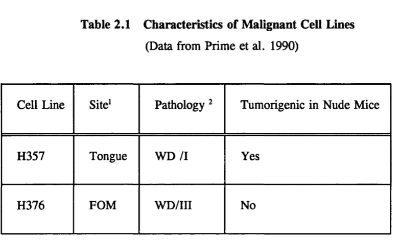
s e e cell lines H357 and H376 that were naturally deficient in the ocy and B4
integrin subunits respectively were "repaired" by transfection of the cell line with the
missing subunit along with a drug resistance gene. Empty vector controls were also
made. Positive clones were sorted by FACS, until after 4 sorts stable integrin expression
was observed in 7 ay and 5 B4 clones. B5 was the subunit found to be associated with
the «V subunit. Introduction of the integrin subunits had no consistent effect on the growth
rate of the clones on tissue culture plastic. However, anchorage-independent growth was
reduced in the ay expressing clones compared with the negative controls. Neither positive
transfectants nor parental cells caused tumour formation in nude mice. The capacity for
terminal differentiation, as measured by involucrin production, a precursor of the comified
envelope of kératinocytes, was increased in the ay clones, but not in the B4 clones.
Expression of the &od af^^ integrins in the cell lines enabled them to adhere to their
respective ligands, vitronectin and laminin. Expression of « 6 8 4 reduced the motility of the
H376 cells. Staining of frozen sections of oral squamous cell carcinomas showed a
reduction in «yBs and ay in poorly differentiated tumours compared with moderately and
well differentiated tumours. Bg was only expressed in squamous cell carcinomas whereas
B3 was not expressed in any of the specimens examined.
Contents
CONTENTS
Abstract 1
Contents 2
Table of Figures 9
Table of Tables 13
Chapter 1 INTRODUCTION
1 Overview 15
1.1 Integrins 15
1.1.1 Integrin Classification 16
1.1.2 Integrin Structure 17
1.1.3 Ligand Binding and Integrin Adhesion 19
1.1.4 Integrin Signaling 21
1.1.5 Regulation of Integrin Function 22
1.2 Normal O ral Mucosa 24
1.2.1 Stratified Squamous Epithelium 25
1.2.1.1 Culture of Kératinocytes 28
1.2.2 Basement Membrane 28
1.2.3 Connective Tissue 30
1.2.4 Extracellular Matrix Proteins 30
1.2.4.1 Fibronectin 30
1.2.4.2 Collagen 30
1.2.4.3 Laminins 31
1.2.4.4 Vitronectin 32
1.2.5 Cell-Extracellular Matrix and Cell-Cell Adhesion
in Epithelium 33
1.2.5.1 Cell-Extracellular Matrix Adhesion 33
Contents
1.3 Malignant Oral Mucosa 36
1.3.1 Oral Cancer 36
1.3.2 Characteristics of Malignant Cells 37
1.3.3 Characteristics of Malignant Tumours: Invasion and Metastasis 39
1.3.4 Changes in Extracellular Matrices in Malignancy 39
1.3.4.1 Basement Membrane Changes 39
1.3.4.2 Interstitial Stroma Changes 40
1.3.4.3 Morphology of Squamous Cell Carcinomas 40
1.4 Integrin Expression and Function in Skin and
Normal Oral Mucosa 41
1.4.1 Integrin Expression in Epidermis 41
1.4.2 Integrin Expression in Oral Epithelium 42
1.4.3 Integrin Expression in Cultured kératinocytes 43
1.4.4 Integrin Functions in Kératinocytes 43
1.4.4.1 Adhesion 43
1.4.4.2 Stratification 43
1.4.4.3 Commitment to Terminal Differentiation 43
1.4.4.4 Stem Cells 44
1.5 Integrins in Malignant Epithelium 44
1.5.1 In Vivo 44
1.5.2 In Vitro 46
1 . 6 Summary and Aims 48
1 .6 . 1 Aims 49
Chapter 2 MATERIALS AND METHODS
2.1 Cell Culture 51
2 .1 . 1 Routine Cell Culture 51
2 .1.1 . 1 Normal Keratinocyte Subculture 51
2 .1.1 . 2 Preparation of 3T3 Feeder Cells 52
2.1.1.3 Culture of Malignant Kerainocyte Cell Lines 52
2.1.1.4 Freezing Down Cell Stocks 53
Contents
2.1.2 Transfection of Integrin Subunits into the Cell Lines 54
2.1.3 Growth and Selection of the «y and B4 Positive Clones 55
2.2 Flow Cytometry 55
2.2.1 Fluorescent Activated Cell Scanning 55
2.2.2 Fluorescent Activated Cell Sorting 55
2.3 Growth Assays 56
2.3.1 Growth Rate on Tissue Culture Plastic 56
2.3.2 Growth in Suspension 56
2.3.3 Growth on Cellagen Inserts 56
2.3.4 Growth of Cells on De-epidermised Dermis (DED) 57
2.4. Protein Analyses 58
2.4.1 Preparation of Cell Lysates 58
2.4.1.1 Protein Extraction From Kératinocytes for
Western Blotting of Involucrin 58
2.4.1.2 Extraction of Labelled Protein
from Kératinocytes 59
2.4.2 Quantification of Protein Content of Cell Lysates 59
2.4.2.1 Bradford Assay for Protein Content 59
2.4.2.2 Quantification of ^^S Sulphate into Labelled
Protein Lysates by Tri-Chloro Acetic
Acid (TCA) Precipitation 59
2.4.3 Polyacrylamide Gel Electrophoresis (PAGE) of Proteins 60
2.4.4 Immunoprécipitations 60
2.4.4.1 Preparation of Sepharose-A and
Sepharose-G Beads 61
2.4.4.2 Immunoprécipitation 61
2.4.5 Coomassie Blue Staining 61
2.4.6 Western Blotting 62
2.4.6.1 Restaining Western Blots 64
2.5 RNA Isolation and Northern Blotting 64
2.5.1 RNA Extraction 64
2.5.2 Agarose Mini-Gel Electrophoresis 64
Contents
2.5.4 Preparation of Probes 6 6
2.5.4.1 Transformation of Competent bacteria 6 6
2.5.5 Hybridisation of Northern Blots 6 8
2.6 Preparation of Extracellular M atrix Proteins 68
2.6.1 Vitronectin 6 8
2.6.2 Laminin 5/Kalinin 69
2.6.2.1 Rouselle Method 69
2.6 .2.2 Trypsin Method 70
2.6 .2.3 Burgeson Method 70
2.7 Adhesion Assays 71
2.7.1 ^‘Cr Labelling 71
2.7.2 Adhesion Assays 71
2.8 Motility Assays 72
2.9 Tumour Formation in Nude Mice 72
2.10 Immunocytochemical Staining 73
2.10.1 Sections of Cultured Cells 73
2.10.2 Staining of Cells to Show Stable Anchoring
Contacts (SAC)s and Focal Contacts 74
2.10.3 Staining of Sections of Oral Squamous Cell
Carcinomas and Normal Oral Mucosa 74
2.10.3.1 Selection of Tissue 74
2.10.3.2 Staining of Sections 75
2.10.3.3 Assessment of Sections 76
2.10.3.4 Haematoxylin and Eosin Staining 76
Chapter 3 ay TRANSFECTION INTO H357 CELLS
3.0 Introduction 77
Contents
3.2 Selection of «y Positive Clones 80
3.3 ay Expression in the Selected Clones 80
3.3.1 tty Expression Demonstrated by Flow Cytometry 80
3.2.3 tty mRNA Expression 81
3.4 Effect of tty Transfection on Oi and II4 Integrin Levels 81
3.5 6 Partner Associated with the Transfected ay Integrin Subunit 82
3.5.1 Flow Cytometry 82
3.5.2 Northern Blotting 82
3.6 Adhesiveness of ay Transfectants 82
3.6.1 Preparation of Vitronectin 83
3.6.2 Adhesion to Vitronectin 83
3.6.3 Blocking Adhesion with Antibodies 84
3.7 Growth of ay Transfectants 85
3.7.1 Growth on Tissue Culture Plastic 85
3.7.2 Growth in Suspension 85
3.8 Differentiation of ay Transfectants 8 6
3.9 Formation of Epithelial Layers on Cellagen Membranes
and DEDs 8 6
3.9.1 Morphology of Epithelial Sheets 87
3.9.2 Staining of Cell Layers for Involucrin and Integrins 87
3.10 Tumour Formation in Nude Mice 8 8
Contents
C hapter 4 EXPRESSION OF 8 3 AND INTEGRINS IN
NORMAL ORAL MUCOSA AND SQUAMOUS CELL CARCINOMAS
4.0. Introduction 117
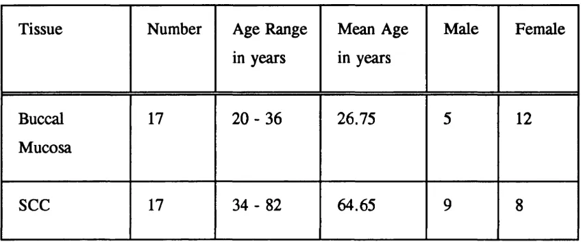
4.1 Normal Buccal Mucosa 118
4.2 Squamous Cell Carcinomas 119
4.3 Discussion 120
Chapter 5 B4 TRANSFECTION INTO H376 CELLS
5.0 Introduction 127
5.1 Transfection of H376 Cells with the B4 Integrin Subunit 128
5.2 Cloning and Selection of B4 Positive Clones 128
5.3 DNA Fingerprinting 128
5.4 B4 Expression in the Selected Clones 129
5.5 Effect of B4 Transfection on the Expression of O ther
Integrin Subunits 129
5.6 Immunoprécipitation 130
5.7 Immunofluorescence Staining of Integrins and Vinculin 130
5.8 Adhesiveness of B4 Transfectants 131
5.8.1 Adhesion to Laminin 1 132
5.8.2 Adhesion to Laminin 5 (Kalinin) 132
5.9 Motility of B4 Transfectants 135
5.10 Growth of B4 Transfectants 136
5.10.1 Growth on Tissue Culture Plastic 136
Contents
5.11 Differentiation of B4 Transfectants 137
5.12 Discussion 138
C hapter 6 DISCUSSION
6.1 Experimental Approach 167
6.2 Production of Clones 167
6.3 Anchorage-Independent Growth 168
6.4 Differentiation 168
6.5 Motility 169
6 . 6 Relevance to Squamous Cell Carcinomas In Vivo 169
6.7 Conclusion 171
APPENDIX 1 Media, Solutions and Buffers 172
APPENDIX 2 Antibodies and Probes 186
APPENDIX 3 Suppliers 189
APPENDIX 4 Tables of Results from Chapters 3 and 5 190
ACKNOWLEDGEMENTS 201
REFERENCES 202
Figure List
LIST OF FIGURES
Figure 1.1
Figure 1.2
Figure 1.3
Figure 1.4
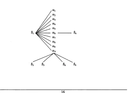
Integrin Subunit Associations 16
Schematic Structure of an Integrin 18
Diagram Showing the Arrangement of the Histological
Layers of Kératinocytes in Human Epidermis 27
Diagram Showing the Arrangement of the Histological
Layers of Kératinocytes in Human Oral Mucosa 27
Figure 2.1
Figure 2.2
Figure 2.3
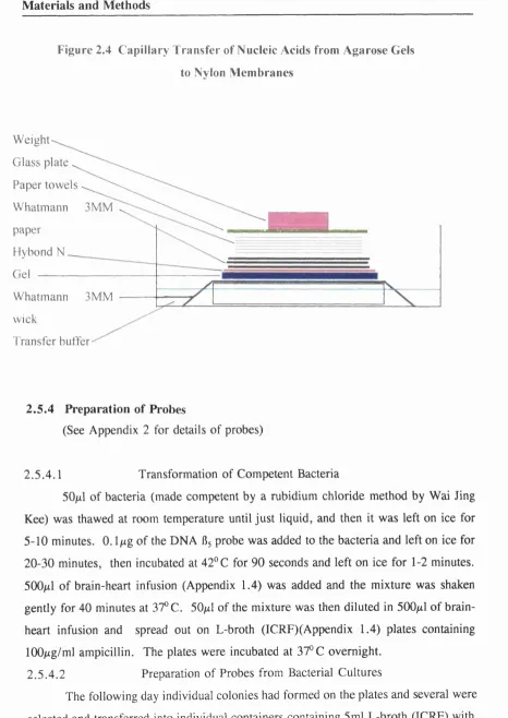
Figure 2.4
Growth of Cells on Cellagen Inserts 57
Growth of Cells on De-Epidermised Dermis 58
Western Blotting 63
Capillary Transfer of Nucleic Acids from Agarose
Gels to Nylon Membranes 6 6
Figure 3.1
Figure 3.2
Figure 3.3
Figure 3.4
Figure 3.5
Figure 3.6
Figure 3.7
Integrin Expression of H357 Parental Cell Line 91
Sort Profiles for Clones VI - V7 92-98
«V Expression of Clones (VI - V7, 1A4, PRC,
1C6 and 1D3) and H357 Parental Cell Line 99
Flow Cytometry Profiles of V3 and H357 Parental
Cell Line with Anti-Integrin Antibodies 1(X)
Integrin Expression of Clones (VI - V7, 1A4, PRC,
1D3 and 1C6) and H357 Parental Cell Line 101
Flow Cytometry Profiles of V2, V7 and 1A4 Clones
with Anti-Integrin Antibodies 102
Northern Blot of Clones V2, V4, V6 , V7 and
Figure List
Figure 3.8
Figure 3.9
Figure 3.10
Figure 3.11
Figure 3.12
Figure 3.13
Figure 3.14
Figure 3.15
Figure 3.16
Figure 3.17
Figure 3.18
Figure 3.19
Figure 3.20
Figure 3.21
SDS-Polyacrylamide Gel Electrophoresis of Fractions
of Vitronectin Purified from Plasma 104
Adhesion Assay on Increasing Concentrations
of Vitronectin 105
Adhesion Assay on Increasing Concentrations
of Vitronectin 105
Adhesion Assay on Increasing Concentrations
of Vitronectin 106
Adhesion Assay on Vitronectin in the Presence
of Blocking Antibodies 107
Growth Curves for Clones (VI - V7) and H357
Parental Cell Line 108
Photographs of Colonies Formed in Suspension 109
Photographs of Colonies Formed in Suspension 110
Quantitative Data from Growth in Suspension Assays 111
Quantitative Data from Growth in Suspension Assays 112
Detection of Involucrin By Western Blotting 113
Growth of Cells from Clones VI and V6 on
De-Epidermised Dermis 114
Growth of Cells from Clones V4, V5, 1A4 and
H357 Parental Cell line on Cellagen Membranes 115
Involucrin and Integrin Expression by
Cultures on Cellagen Membranes 116
Figure 4.1
Figure 4.2
Figure 4.3
Figure 4.4
Expression of a^, (XyBg, 8 3 and ilg Integrins in
Normal Oral Mucosa
Expression of ay Integrin in Oral SCCs
Expression of Integrin in Oral SCCs
Expression of 8 3 and 8 ^ Integrins in Oral SCCs
123
124
125
Figure List
Figure 5.1
Figure 5.2
Figure 5.3
Figure 5.4
Figure 5.5
Figure 5.6
Figure 5.7
Figure 5.8
Figure 5.9
Figure 5.10
Figure 5.11
Figure 5.12
Figure 5.13
Figure 5.14
Figure 5.15
Figure 5.16
Figure 5.17
Figure 5.18
Figure 5.19
Integrin Expression by H376 Parental Cell Line 141
Sort Profiles for Clones 142-146
DNA Fingerprinting 147
8 4 Integrin Expression in the Clones B3, B5 - B8,
1A2 and H376 Parental Cell Line 148
Flow Cytometry Profiles of Clones B3, B6 and 1A2
with Anti-Integrin Antibodies 149
Flow Cytometry Profiles for Clone B6 and H367
Parental Cell Line with Anti-Integrin Antibodies 150
Expression of Integrins on Clones B5, B6 , 1A3 and
H376 Parental Cell line 151
Immunoprécipitation of oc^, and 8 4 152
Immunofluorescence Staining of Clone B3, H376
Parental Cells and normal kératinocytes 153
Immunofluorescence Staining of Clones B3, B6 , H376
Parental Cells and normal kératinocytes 154
Adhesion Assay on Laminin 1 155
Adhesion Assay on Laminin 5 156
Adhesion Assay on Laminin 5 in the Presence
of Blocking Antibodies 157
Adhesion Assay on Laminin 5 in the Presence
of Blocking Antibodies 157
SDS-Polyacylamide Gel Electrophoresis of Protein
Collected from Keratinocyte Cultures 158
Adheison Assay on Laminin 5 in the Presence
of Blocking Antibodies 159
Traces of Movement of Cells During a Motility Assay
on Clones B3, B6 , 1A2 and H376 Parental Cell Line 160
Quantified Data from the Motility Assay 161
Growth Rate of Clones (B3, B5 - B8, 1A3, 1B5,
Figure List
Figure 5.20
Figure 5.21
Figure 5.22
Figure 5.23
1C2) and H376 Parental Cell Line 162
Quantified Data from Growth in Suspension of Clones
B3, 1A3, 1C2 and H376 Parental Cell Line 163
Quantified Data from Growth in Suspension of Clones
B3, B5-B8, 1A2, 1C2 and H376 Parental Cell Line 164
Quantified Data from Growth in Suspension of Clones
B5, 1A3, 1C2 and H376 Parental Cell Line 165
Table List
LIST OF TABLES
Table 1.1 Ligands of Keratinocyte Integrins 35
Table 2.1
Table 2.2
Characteristics of Malignant Cell Lines
Tissue Used for Immunohistrochemistry
53
75
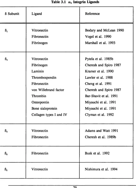
Table 3.1 «V Integrin Ligands 79
Table 4.1 Expression of «y and «yfis in SCCs 122
Table 5.1
Table 5.2
Data from Motility Assay on Clones and H376
Parental Cell Line
Clones Included in Suspension Assays
136
137
Appendix 2 Table of Integrin Antibodies Used
Table of Secondary Antibodies Used
Table of Other Antibodies Used
Table of Northern Blotting Probes
186
187
187
188
Appendix 4 Table of Data
Table of Data
Table of Data
Table of Data
Table of Data
Table of Data
Table of Data
Table of Data
Table of Data
from Figure 3.1 and 3.5
from Figure 3.9
from Figure 3.10
from Figure 3.11a
from Figure 3.11b
from Figure 3.12
from Figure 3.13a-d
from Figure 3.16a
from Figure 3.17a
Table List
Table of Data from Figure 5.1 196
Table of Data from Figure 5.4 196
Table of Data from Figure 5.7 197
Table of Data from Figure 5.13 197
Table of Data from Figure 5.14a and b 198
Table of Data from Figure 5.15 198
Table of Data from Figure 5.16 199
Table of Data from Figure 5.18 199
Introduction
Chapter 1
INTRODUCTION
1.0 Overview
Neoplastic transformation results from loss of normal controls over the growth
and differentiation of cells (Weinberg 1991). A characteristic of malignant cells is that
their growth involves invasion into surrounding tissues and metastasis to distant sites.
The process of tumour growth and metastasis involves a number of complex coordinated
mechanisms. Enlargement of the primary tumour is accompanied by growth of new
blood vessels. Invasion of the tumour into the surrounding tissue and into blood and
lymphatic vessels is then followed by release of cells from the primary mass. These
cells must survive in the circulation, arrest at a secondary site and then leave the blood
or lymphatic vessel and migrate into the adjacent tissues. Following this the cells must
grow and produce a tumour at the secondary site. To accomplish these steps, a
tumour cell must change its ability to interact and adhere to cells and extracellular
matrix proteins. Cell adhesion molecules are found on the surface of all cells and
enable them to interact and adhere with other cells and the extracellular matrix.
Therefore, the roles of these molecules in malignant disease is the subject of great
interest. Integrins are one of the families of cell surface adhesion molecules involved
in such interactions and therefore modulation of integrin expression and function may
be important in mediating the behaviour of malignant cells.
1.1 Integrins
Integrins are a family of cell surface receptors involved in cell-cell and cell-
extracellular matrix (ECM) interactions (Hynes 1992, Ruoslahti 1991, Albelda and Buck
1990). Each integrin is composed of an a and a 15 subunit which are glycoprotein
chains. Integrins serve as a link between the extracellular matrix and the cytoskeleton
of the cell (Chen et al. 1985, Burridge et al. 1988).
Introduction
1.1.1 Integrin Classification
The integrins are subdivided into families based on the B chains. There are 8
B and 15 a subunits. Some a subunits are capable of combining with more than one
B subunit, whereas others can only combine with one B subunit, ay is unique as it
potentially associates with 5 B subunits. Ligand binding specificity depends, to a large
extent on heterodimer composition, but the same integrin in different cell types can
show different binding properties (Elices and Hemler 1989, Kirchofer et al. 1990).
Most integrins can combine with more than one ligand and one ligand can combine with
more than one integrin. However, different integrins that bind to the same ligand may
not necessarily serve the same function within the cell. Individual cells can vary their
adhesive properties by selective expression of integrins and further versatility is
achieved by cells modulating the binding properties of integrins. Figure 1.1 shows the
relationships of the integrin subfamilies. The B2 integrins which are expressed on
leucocytes and cells of the immune system, have not been included.
Introduction
1.1.2 Integrin Structure
Each integrin subunit consists of a short cytoplasmic domain of about 50 amino
acids (except B4, which has a tail of about 1(XX) amino acids (Hogervorst et al. 1990)),
a hydrophobic transmembrane domain and an extracellular domain. The a subunits
vary in size between 120-180 kDa and the B subunits between 90-110 kDa. The
extracellular domains of a and B subunits are greater than lOOkDa and 75 kDa
respectively, and they combine non-covalently to form aB heterodimers. The
transmembrane and cytoplasmic domains are not needed for aB interactions as truncated
integrins that lack transmembrane and cytoplasmic domains can be expressed and do
form functional aB dimers (Dana et al. 1991, Bodary et al. 1991). Electron
microscope images of purified integrins (Nermut et al. 1988), cross linking experiments
(Smith and Cheresh 1988, D ’Souza et al. 1988) and detailed mutational analysis of
ligand-binding domains suggest an association of the aminoterminal globular domains
of the a and B subunits to form the extracellular ligand binding regions of the receptor.
Some integrins have an extra domain of 180 - 200 amino acids between the last
calcium binding site and the amino terminus (Hemler 1988) known as the I (inserted)
domain, which plays a role in ligand binding (Haas and Plow 1994).
The B subunits have a 40% homology in their amino acid sequences (Hemler
1990), with specific structural features being conserved over a wide variety of species
including mammals, birds, amphibians, insects and fungi (Marcantoni and Hynes 1988).
Bi - B3 and B5 - By subunits have very similar cytoplasmic domains, with three amino
acid clusters that are highly conserved (reviewed by Sastry and Horwitz 1993). The
high degree of conservation of these clusters suggests a fundamental structural and
functional role for them. However, variant sequences have been reported for the B
subunit cytoplasmic domains of Bj, B3 and B4, and two variants of B^, Bib and Bjs, have
been reported (Balzac et al. 1993, Languino and Ruoslahti 1992, Altruda et al. 1990).
B4 has a unique cytoplasmic domain as it is very large (see above) and is associated with
the intermediate filament network rather than the actin cytoskeleton with which Bj
integrins interact (Sonnenberg et al. 1991, Stepp et al. 1990). Three variant forms of
B4 exist, B4A, B4B, and B4C (Tamura et al. 1990), which probably arise from alternative
Introduction
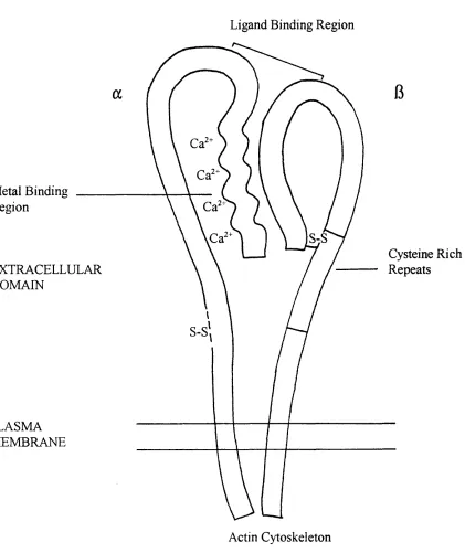
Figure 1.2 Schematic Structure of an Integrin
Ligand Binding Region
Metal Binding Region
S-S
Cysteine Rich Repeats EXTRACELLULAR
DOMAIN
S-S
PLASMA MEMBRANE
Introduction
splicing of 6 4 mRNA. The B4A is the most commonly found variant (Suzuki and Naitoh,
1990).
The a subunits exhibit more sequence heterogeneity than the B subunits, but
their cytoplasmic domains are highly conserved among different species, for example,
the «V subunit is 83% identical in amino acid sequence between chick and human
(Bossy and Reichardt 1990). In contrast to the B subunits, the different cytoplasmic
domains show little homology , but one domain, the GFFKR motif, adjacent to the
transmembrane domain is conserved in all a subunits. O’Toole et al. (1994) have
shown that this region is important for maintaining integrins in a default low affinity
state.
The current view is that the a and B amino terminal domains form a ligand
binding head on each integrin. Two stalks each made up of one of the subunits connect
the heads to the membrane spanning segment and thus to the cytoplasmic domains.
The cytoplasmic domains interact with cytoskeletal proteins and other cytoplasmic
components (Horwitz et al. 1986, Burridge et al. 1988, Otey et al. 1990). Therefore
a link between the cytoskeleton of the cell and the extracellular matrix exists through
the integrin molecule. The schematic structure of an integrin is shown in Figure 1.2.
1.1.3 Ligand Binding and Integrin Adhesion
A similar mechanism of ligand binding occurs in different integrins (Humphries
1992). Many integrins recognize a specific amino acid sequence, the "RGD" sequence
(arginine-glycine-aspartate) which is present in a wide variety of matrix proteins
including fibronectin, vitronectin, collagen, laminin, von Willebrand factor, fibrinogen,
osteopontin and thrombospondin. Some integrins recognize one or more of these
ligands in the context of the RGD sequence, whereas others are more restricted in their
ligand binding specificity. For example, «v^ 3 recognizes all the listed matrix
molecules, a^Bg binds vitronectin and binds fibronectin, yet they can all be
inhibited with the same RGD peptides. Therefore, it is possible that alternative regions
within the ligand contribute to the specificity of these interactions. The specificity may
also be determined by the unique conformation that the RGD sequence may adopt in
Introduction
different proteins (Pierscbacher and Ruoslahti 1987). Experiments involving integrins
and the RGD sequences in fibrinogen have implied that integrin-mediated ligand binding
depends on secondary and tertiary structural requirements as well as primary sequence
recognition. However, not all integrins recognize RGD even though they bind to
proteins that contain RGD sequences. Some integrins of the Bi, Bg and B7 families bind
other cell adhesion molecules, such as ICAM-1 and VCAM-1, for example, a^Bi is
capable of binding with fibronectin and VCAM-1 (Elices et al. 1990).
Integrins require divalent cations to bind their ligands (Smith and Cheresh 1991),
and in some cases the ligand affinity of some integrins is affected by divalent cations.
For example Mn^"^ enhances the binding of CX5B1 to fibronectin (Gailit and Ruoslahti
1988).
Integrins provide a mechanism for cell adhesion and localize to structures such
as focal adhesions and adhesion plaques when the cells are adherent to the appropriate
ligand. Adhesion often results in changes in cell shape and involves reorganization of
cytoskeletal proteins and alignment of stress fibres with focal adhesions through integrin
association with the actin cytoskeleton. Integrins provide a link between the
extracellular matrix and the cytoskeleton via talin (Horwitz et al. 1986), vinculin (Chen
et al. 1985), a actinin (Otey et al. 1990) and actin (Burridge et al. 1988). The role of
these adhesion structures in epithelial cells is discussed in Section 1.2.5.
There is more evidence for interactions of the B subunits with cytoskeletal
proteins than for a. subunits, and current data point to the integrin B subunit
cytoplasmic domain as the structure that targets integrins to focal adhesions. La
Flamme et al. (1992) showed that the Bj and not the « 5 cytoplasmic domain directs
host protein to focal adhesions. In addition, truncations of the « 5 cytoplasmic domain
do not influence the targeting of a^Bi to focal adhesions in the presence of fibronectin
(Bauer et al. 1993). A combination of deletions and single amino acid substitutions has
identified putative sequences in the cytoplasmic domain required for the localization of
Bi integrin subunits to focal contacts (Reska et al. 1992). Bi and B3 integrins both
localize in focal adhesions and have very similar cytoplasmic domains. Localization to
Introduction
localizes to focal adhesions on fibronectin substrates, whereas on vitronectin the
and not the receptor localizes (Singer et al. 1988, Path et al. 1989). It is thought
that the « 5 cytoplasmic domain interferes with the ability of the Bi cytoplasmic domain
to interact with the cytoskeleton unless the receptor is ligated. Hence the suggestion
that the a cytoplasmic domain inhibits localization of unoccupied integrins. However,
this is not the only determining factor as cells plated on vitronectin have ay^ 3 but not
ttyBs localised in focal contacts even though both react with the RGD sequence site on
vitronectin (Wayner et al. 1991). On vitronectin, there is co-localisation of «yBg, but
not cKyBg with the cytoplasmic proteins talin, a actinin, tensin and actin (Lewis et al.
1996).
1.1.4 Integrin Signaling
Integrins function not only as structural receptors that link the ECM with the
cytoskeleton, but also as signal transducers. They are able to transmit signals from the
exterior of the cell into the interior, the so called "outside-in" signaling, as well as the
"inside-out" signaling (Hynes 1992), discussed in regulation of integrin function.
Section 1.1.5.
Ligation of integrins has been shown to activate a number of intracellular
signaling events or secondary messengers resulting in a wide variety of cellular
responses including differentiation, proliferation, differential gene expression,
cytoskeletal assembly and migration. Integrin induced signals include Ca^^ influx, H^
exchange, protein tyrosine and non-tyrosine phosphorylation, alterations in
phosphoinositide metabolism, activation of mitogen-activated protein (MAP) kinases,
changes in gene expression and growth stimulation (reviewd by Hynes 1992, Juliano
and Haskill 1993, Parsons et al. 1994, Clark and Brugge 1995).
Integrin cytoplasmic domains do not possess kinase or phosphatase activities,
and so must interact with initiators of signaling cascades. Current models postulate
that integrin cytoplasmic tails bind to signaling molecules, such as one of the kinases,
which then initiates the signaling cascade after integrin ligation (Yamada and Miyamoto
1995). Focal adhesions are areas where integrin-mediated signal transduction can occur
Introduction
since they consist of regulatory proteins such as tyrosine kinases as well as the
structural proteins. Focal adhesion kinase (FAK), a tyrosine protein kinase binds to the
Bi integrin cytoplasmic domain (Schaller and Parsons 1994) and it co-clusters with fij
integrins that are aggregated by non-inhibitory anti-integrin antibodies (Miyamoto et
al. 1995a). «v^ 3 and integrins are able to induce co-localisation of FAK (Lewis
et al. 1996). Akiyama et al. (1994) report that Bg and B5 as well as Bi can trigger
phosphorylation of FAK after clustering. However, Lewis et al. (1996) report
phosphorylation only in expressing cells, stimulation with phorbol esters being
necessary for phosphorylation of FAK in the expressing cells. The signal
transduction pathways that are activated after clustering and activation of kinases, may
resemble other established pathways (Clark and Brugge 1995).
Aggregation of integrin receptors, even in the absence of ligand occupancy, is
sufficient to bring about a transmembrane accumulation of at least 2 0 signal transduction
molecules (Miyamoto et al. 1995b). However, tyrosine kinase-mediated
phosphorylation and actin cytoskeletal integrity, as well as integrin occupancy and
aggregation, are required for the accumulation of F-actin, paxillin and filamin
cytoskeletal elements. Talin, a-actinin and vinculin require both integrin aggregation
and occupancy, but not tyrosine phoshorylation for membrane accumulation (Miyamoto
et al. 1995b). It appears therefore, that these integrin-based signaling pathways and
cytoskeletal organising systems are highly sophisticated, with each element having
specific requirements for membrane accumulation and specific functions.
1.1.5 Regulation of Integrin Function
As mentioned above cells are able to alter their adhesive properties by selective
expression of integrins, but further versatility is introduced by the ability of cells to
modulate the binding properties of integrins. Integrin function and affinity for their
ligands can be controlled by intracellular or extracellular events.
Integrins can be functionally active or inactive (Adams and Watt 1990), and
both subunits can contribute to the regulation of integrin activity. In the so called
Introduction
within the cell. An example of inside-out signaling is seen in platelets where the aiJlg
integrin on resting circulating platelets is in an inactive state and does not bind any of
its soluble ligand. However, following platelet activation a conformational change
occurs and the integrin becomes an active receptor (Keiffer and Phillips 1990,
Phillips et al. 1991). Changes in the affinity of integrins for their extracellular ligands
in respones to cytoplasmic signals have also been observed in integrins (Faull et al.
1994).
The cytoplasmic tails of both integrin subunits play a role in regulating integrin
function. The a subunit tail is essential for maintaining integrins in their active state
and is required for the upregulation of integrin function by signals from within the cell
(Kassner and Hemler 1993, Kawaguchi and Hemler 1993). The Bi subunit tail is
required for adhesive function and for the migration of integrins into focal adhesions
(La Flamme et al. 1992), (see Section 1.1.3.) These functions are mediated by separate
regions of the cytoplasmic tail (Reszka et al. 1992, Hemler et al. 1994). There are
several phosphorylation sites on the B% tail and correlative evidence suggests that
changes in phosphorylation are associated with changes in the activity of Bj integrins
(Hemler et al. 1994).
Integrins interact with their ligands in a cation-dependent fashion and integrins
are known to be differentially affected by divalent cations (Albelda and Buck 1990,
Ruoslahti 1991, Hynes 1992). This provides another mechanism for integrin regulation.
For example, in the presence of Mg^'^, Ca^^ inhibited the ability of integrin ayBi to
bind RGD containing peptides or its ligand fibronectin, while Ca^^ enhanced the binding
of integrin a^Bg to RGD containing peptides or vitronectin (Kirchhofer et al. 1991).
Each oc chain contains three repeats of a cation binding motif, which appear to be
directly involved in ligand binding interactions (D’Souza et al. 1994, Haas and Plow
1994).
It is possible to activate or inhibit ligand binding with monoclonal antibodies
against the Bi subunit (Akiyama et al. 1989, Kovach et al. 1992). It is thought that
these antibodies cause conformational changes in the binding site which affect the ability
to bind ligand. Some antibodies only recognise the Bj subunit in the active state.
Introduction
probably by binding to a region that is exposed in the active configuration, supporting
this hypothesis (Kovach et al. 1995). This is therefore another possible means of
controlling integrin function.
The availability of recognized sequences on the extracellular matrix protein also
provides a means of control over integrin function, for example the RGD sequence on
the laminin A chain is not exposed and the two RGD sequences on the a fibronectin
chain are not readily detectable (Cheresh et al. 1989a, Aumailly et al. 1990). These
sites are exposed by proteolytic digestion of the ligand. This finding suggests that in
vivo matrix-integrin interaction might be partly regulated by proteolytic cleavage, as
occurs in acute inflammation or in areas of invasive growth of neoplastic cells.
The plasma membrane may play a role in the control of integrin function as it
has been shown that the phospholipid composition of the surrounding plasma membrane
influences the receptor affinity (Conforti et al. 1990) and may well explain the presence
of specific matrix-adhesion domains on the plasma membrane.
1.2 Oral Mucosa
The oral cavity is lined by mucosa that consists of a superficial layer of stratified
squamous epithelium and an underlying lamina propria, separated by a basement
membrane.
Oral epithelium has several distinct patterns of maturation in different regions
of the oral cavity. Keratinised epithelium is found in the masticatory mucosa of the
hard palate and in parts of the dorsum of the tongue and gingiva. Keratinised
epithelium of the oral cavity has a similar structure to epidermis. In some areas, such
as in gingiva the epithelium keratinises but the nuclei are not lost and the epithelium
is termed parakeratinised. Non-keratinised epithelium forms the surface of the
distensible lining mucosa of the soft palate, ventral surface of the tongue, floor of
mouth, alveolar mucosa, vestibule, lips and cheek. In general it is thicker and more
permeable than keratinised epithelium.
Stratified squamous epithelium is composed of multiple layers of kératinocytes
Introduction
basement membrane. As such it provides a barrier to the outside world preventing the
entry of microorganisms and other noxious substances as well as preventing the loss of
body fluids. Also present within the epithelium are melanocytes, Merkel cells,
Langerhans cells and lymphocytes. The basal kératinocytes are attached to the basement
membrane which undulates as connective tissue papillae intedigitate with rete ridges.
1.2.1 Stratified Squamous Epithelium
There are three main phases in the life of a keratinocyte: 1) growth and
proliferation in the basal layers, 2) maturation and outward displacement more
superficially and 3) desquamation at the outer layer.
Basal kératinocytes are cuboidal cells attached to the underlying basement
membrane by hemidemosomes and to each other by desmosomes. Most cell divisions
occur within this layer and the cells are the least differentiated of the epithelial layers.
The cytoplasm contains the distinct network of keratin intermediate filaments which give
kératinocytes their name. The keratin filaments connect with the hemidesmosomes and
desmosomes (Staehelin 1974, Jones et al. 1989, Schwarz et al. 1990).
After dividing a cell may remain within the basal layer or move upward and
commence differentiation and maturation. There are two types of proliferating cells in
the basal layer: stem cells, which retain a high capacity for self-renewal throughout
adult life, and transit amplifying cells, which have a lower capcity for self-renewal and
a high probability of undergoing terminal differentiation after a few rounds of cell
division (Potten 1981, Hall and Watt 1989). The earliest event in the initiation of
keratinocyte terminal differentiation is irreversible withdrawal from the cell cycle,
which is associated with an increase in cell size, loss of proliferative capacity
(Barrandon and Green 1985) and expression of involucrin (Watt and Green 1981). A
cell committed to terminal differentiation will detach from the basement membrane,
following the inactivation and loss of integrins on the cell surface (Hotchin et al.
1993). In stratified cultures of kératinocytes, upward migration is a consequence, not
a cause of terminal differentiation (Watt and Green 1982) and occurs because
kératinocytes become less adherent to their substratum and to one another (Watt 1984),
Introduction
and this is also true in vivo (Iverson et al. 1968).
As cells leave the basal layer they become more flattened and take on a spinous
appearance. At a histological level the spines correspond to the insertion of bundles of
keratin filaments into neighbouring cells. This spinous or prickle cell layer is 4 or 5
cells thick, with the more superficial cells being larger, more flattened and containing
more keratin bundles than those adjacent to the basal cells.
Lying superficial to the spinous layer is the granular layer, so called due to the
accumulation of 0.5-5/im diameter keratohyaline granules in the kératinocytes (Holbrook
1994). Also present in the cells are membrane coating granules which fuse with the
plasma membrane and discharge their contents into the intercellular space, which play
a role in controlling the permeability of the epithelium. The cells again become larger
and more flattened before entering the most superficial layer.
Within keratinising epithelium, such as the epidermis and the hard palate,
gingiva and dorsum of the tongue in the mouth, the most superficial layer is the
comified layer and consists of fiattenend dead squames, the cytoplasm of which is full
of keratin and devoid of organelles. This process of kératinisation is accompanied by
the deposition of a tough comified envelope, made of crosslinked involucrin and loricrin
proteins just below the plasma membrane.
In epithelium that does not keratinise the cells become flatter and larger as they
progress towards the surface, but they do not lose their nuclei or form a comified layer,
and the cells that are shed from the surface contain a variety of organelles and
unbundled filaments.
The final step in the maturation process is desquamation and in order to maintain
a constant thickness of epithelium this must be equivalent to the rate of cell division in
the basal layer.
Schematic diagrams of epidermal and oral keratinocyte layers are shown in
Introduction
Figure 1.3 Diagram Showing the Arrangement of the Histological Layers of
Kératinocytes in Human Epidermis
Cornified L ay e r__
Granular Layer
Spinous Layer
Basal Layer
Basement Membrane
Figure 1.4 Diagram Showing the Arrangement of the Histological Layers of
Kératinocytes in Human Oral Mucosa
Superficial L ay e r___
Granular Layer
Prickle Cell Layer
Suprabasal Layer
Basal Layer
Basement Membrane
Introduction
1.2.1.1 Culture of Kératinocytes
It is possible to culture kératinocytes so that they form a stratified squamous
sheet which mimics growth in the epidermis (Watt 1994), and the oral cavity. The
cells can be grown using the Rheinwald and Green (1975) culture method, which
involves plating a single cell suspension on a feeder layer of growth arrested mouse 3T3
fibroblasts, in medium containing serum and other additives as described in Section 2.1.
The colonies formed in this method grow and coalesce to form a confluent sheet, 6 - 8
cell layers thick, in which desmosomes and adherens junctions and stable anchoring
contacts (SACs, see Section 1.2.5.1) are formed (Watt et al. 1984, Magee et al. 1987,
O’Keefe et al. 1987, Carter et al. 1990b), but hemidesmosomes are not (Holbrook and
Hemmings 1983). Basal cells in these cultures express integrins and cells above the
basal layer express differentiation markers such as involucrin (Watt and Green 1982).
This method provides a valuable tool for investigating the growth of normal
kératinocytes as well as the changes that can occur in the progression from normal to tumorigenic growth.
1.2.2 Basement Membrane
Basement membranes are cell associated extracellular matrices that may
completely surround groups of cells as in muscle or make contact with only one cell
surface as in stratified squamous epithelium. This description of basement membranes
will be limited to those that are present in epidermis and oral mucosa. Basement
membranes spatially orientate kératinocytes, organise their cytoskeletal networks, affect
their internal properties (Ingber et al. 1981), and are involved in controlling
keratinocyte differentiation in the epidermis (Adams and Watt 1990, Adams and Watt
1993). They also form selectively permeable barriers between tissue compartments and
provide immobilised ligands for cellular receptors. The proteins in the basement
membrane are produced by both fibroblasts and kératinocytes (Marinokovich et al.
1993).
Conventional electron microscopy of the basement membrane reveals 3 layers;
Introduction
reticular layer (Briggaman and Wheeler 1975). The lamina lucida is a 45nm thick
electronlucent layer that lies between the basal surface of the kératinocytes and the
lamina densa and is traversed by anchoring filaments running from the kératinocytes to
the lamina densa. The lamina densa is an electrondense layer, 55nm thick that contains
abundant collagen IV. The reticular layer is an electron lucent layer traversed by
anchoring fibrils of collagen VII connecting the dermis to the hemidesmosomes.
However, Rouselle et al. (1991) suggest that the lamina lucida may be an artefact due
to shrinking of the basal cells away from the basement membrane during preparation
of the sections, and that the lamina densa is the residue of the whole basement
membrane. Laminin 5 forms the anchoring filaments that are seen to run from the
hemidesmosomes into the basement membrane, and plays an important role in
maintaining the dermo-epithelial junction as demonstrated by the disease Herlitz’s
junctional epidermolysis bullosa in which individuals have mutations in the laminin 5
gene (Verrando et al. 1991, Aberdam et al. 1994, Pulkkinen et al. 1994).
Laminin 1 is the most abundant non collagenous protein in the basement
membrane, and is located in the lamina lucida. Laminin 1 binds to collagen type IV
and heparan sulphate proteoglycans so that the heparan sulphate and laminin are
embedded within a network of polymerized type IV collagen (Laurie et al. 1985).
Multiple interactions between the various collagen, heparan and laminin binding
domains are believed to orientate the three molecules within the network.
Nidogen/Entactin is a 150kDa glycoprotein present in epidermal basement membranes
(Yurchenco and O’Rear 1994). It is able to bind to other basement membrane proteins
and is thought to play a role in the assembly of the proteins into a three dimensional
network in vivo (Yurchenco and Schittney 1990, Yurchenco and O’Rear 1994). A small
amount of fibronectin is present in the basement membrane (Fleischmayer and Timpl
1984). Also present are proteoglycans, such as heparan sulphate, chondroitin sulphate
and hyaluronic acid, perlecan and veriscan (Caughman et al. 1987, Murdoch et al.
1994, Zimmerman et al. 1994, Van den Bom et al. 1994). These molecules are able
to bind growth factors and present them to receptors at the cell surface (Aviezer et al.
1994)
Introduction
1.2.3 Connective Tissue
Immediately below the basement membrane lies a specialised layer of dense
connective tissue known as the dermis in the skin and the lamina propria in the oral
mucosa. This layer imparts strength to the skin and mucosa. It consists of a dense
latticework of collagen and elastin embedded in a viscoelastic ground substance of
proteoglycans and glycoprotein. Other components include elastin, tenascin (Chiquet-
Ehrismann 1991) and fibronectin (Yamada 1989) and vitronectin associated with dermal
elastic fibres (Dahlback et al. 1989). The stroma also contains cells such as fibroblasts,
osteoblasts, chondroblasts, mast cells and leucocytes. Capillary loops and lymphatics,
nerves and nerve endings enter the lamina propria and dermis from the underlying
tissue.
1.2.4 Extracellular Matrix Proteins
1.2.4.1 Fibronectin
Fibronectin is a ubiquitous 540kDa glycoprotein found in plasma and serum and
widely distributed in tissues, it is involved in a wide variety of cellular interactions with
extracellular matrices. The molecule is composed of two identical subunits held
together by two disulphide bonds near the carboxytermini to produce a " V shaped
molecule. Each subunit contains domains that bind other fibronectin dimers, collagen
types I and IV, heparin and the cytokine transforming growth factor 13 (Fava and
McClure 1987, Hynes 1989, Potts and Campbell 1994). Molecules are made of three
general types of short amino acid sequences that are repeated many times.
1.2.4.2 Collagen
The fundamental structural unit of collagen is a 300nm long, 1.5nm diameter
tropocollagen molecule composed of three coiled polypeptide units or a domains. At
the amino and carboxy terminal of each chain are 16-25 non helical residues known as
globular domains. In the past decade about 20 different collagens have been identified,
plus numerous other non-structural proteins that contain at least one triple helix as a
Introduction
should be regarded as members of the collagen superfamily (Hulmes 1992). Collagens
type I, II and III are the most abundant types in the interstitial stroma and types IV and
VII are present in basement membranes. Integrins can bind to collagen type I via RGD
sequences (Ruoslahti and Piersbacher 1987) or via other peptides (Toda et al. 1987).
Type IV collagen is the integral collagen of basement membranes and forms a
covalently stabilised polymer network (Yurchenco 1994), it is abundant in the lamina
densa under stratified squamous epithelium (Briggaman et al. 1991, Mihara et al. 1992).
In epidermal basement membranes the molecule is a trimer consisting of [al(IV) ] 2
a2(IV) chains (Yurchenco and O’Rear 1994). It is a flexible molecule capable of
interacting with other collagen molecules at the carboxyl and amino terminals and by
lateral associations (Yurchenco and Furthmayer 1984, Yurchenco and Schittney 1990).
Type VII collagen forms the anchoring fibrils that traverse the reticular layer of
the basement membrane and connect the type IV collagen of the basement membrane
to anchoring plaques composed of type IV and VII collagen (Burgeson 1993).
Becker et al. (1986) report that type V collagen is present in the lamina propria,
but not within epithelial or vascular basement membranes, and type VI collagen is more
prominent in the upper parts of connective tissue papillae.
1.2.4.3 Laminins
Laminins are glycoproteins present in basement membranes and are thought to
be important for cell binding and adhesion. They support the attachment of numerous
cell types and form complexes with other extracellular matrix components: type IV
collagen, heparan sulphate proteoglycan, heparan and nidogen. Recently laminins have
been reclassified and numbered and the subunits termed a, B and 7 (Burgeson et al.
1994). Laminin 1 consists of a l , B1 and 7 I, arranged in a cross shape (Yurchenco
and Schittney 1990). Laminin 2, consisting of «2, B2 and 7 I subunits, was formerly
known as merosin and is present at a low level in the epidermal basement membrane
(Solberg et al. 1992).
Laminin 5 was previously known as kalinin or epiligrin and was first found in
epidermal basement membranes (Verrando et al. 1988, Carter et al. 1991, Rouselle et
Introduction
al. 1991). This variant of laminin is composed of «3, 133 and 7 2 subunits. It exhibits
a complex pattern of bands by SDS-PAGE that reflects post-synthetic proteolytic
processing (Marinokovich et al. 1993, Vailly et al. 1994).
Laminin 6 (K laminin) is also present in the epidermal basement membrane
(Marinokovich et al. 1992). Its distribution coincides with that of laminin 5 and it is
composed of 61 and 7 I probably associated with an «3 subunit. Preliminary evidence
suggests that laminin 5 and 6 are linked as a disulphide bonded complex that binds to
cells via domains in laminin 5 and is connected to type IV collagen in the lamina densa
via nidogen bound to the 7 I subunit of laminin 6 (Marinokovich et al. 1992,
Marinokovich 1993).
1.2.4.4 Vitronectin
Vitronectin is a glycoprotein found in the circulation and some tissues. It was
first identified as a cell attachment factor ("serum spreading factor") and later named
vitronectin to denote its binding to glass (Hayman et al. 1983). It is identical to the
complement component, "S-protein" (Suzuki et al. 1985, Tomasini and Mosher 1986).
The vitronectin molecule is divided into several domains: the first region of 44
residues is known as the somatomedin B region, as it is identical to the plasma peptide
somatomedin B (Barnes et al. 1984), and it is likely that vitronectin is the precursor
of somatomedin B. The second structural region is a connecting strand which begins
with an RGD motif that has cell attachment activity. The next region contains the
hemopexin repeat domains, and the final region is the heparin binding domain.
Plasma vitronectin is predominantly derived from the liver, but platelets
(Preissner et al. 1989), megakaryocytes (Kanz et al. 1988) and monocytes/macrophages
(Hetland et al. 1989) also contain an immunologically identical protein. It is
synthesized and processed as a 75-80 kDa single chain polypeptide in hepatocytes
(Barnes and Reing 1985), but a 2-chain form (65kDa and lOkDa) is also found in the
circulation. Approximately 50% of vitronectin isolated from pooled plasma is in the
2-chain form. However the exact proportion of single to double chain vitronectin
Introduction
known. Vitronectin is present in plasma at concentrations of 200-400/ig/ml, making up
0.2-0.5% of the protein content. Vitronectin has a number of activities that may be
important in haemostasis and thrombosis as well as playing a role in cell adhesion.
Vitronectin has been demonstrated by immunofluorescence studies to be
deposited in loose connective tissue (Hayman et al. 1983), and to be associated with
dermal elastic fibres in skin, in an age-dependent manner (Dahlback et al. 1986,
Dalback et al. 1989): there is virtually no vitronectin in the skin of 0-10 year olds, but
it increases at puberty and then plateaus in adulthood. Vitronectin is also known to
associate with collagen types I-VI (Gebb et al. 1986). Vitronectin is absent from
basement membranes, except diseased renal tubular basement membrane (Falk et al.
1987). Hinter et al. (1989) reported the binding of vitronectin to kératinocytes in skin
sections and to keratin filament aggregates after incubation of tissue sections with serum
to isolated vitronectin. The in vivo relevance of this association is unclear as no
immunochemical co-localization of vitronectin with kératinocytes could be detected in
skin sections incubated with saline alone.
Owing to its multidomain structure with a repertoire of various ligand binding
sites and the possibility of maintaining different conformations, vitronectin appears to
be a versatile adhesive component, allowing for multiple interactions at interfaces or
pericellular sites.
1.2.5 Cell-Extracellular Matrix and Cell-Cell Adhesion in Epithelium
1.2.5.1 Cell-Extracellular Matrix Adhesion
Hemidesmosomes, SACs, focal adhesions and integrins provide a means
whereby epithelial cells can attach to the basement membrane.
Basal kératinocytes adhere to the basement membrane zone through
hemidesmosomes (Staehelin 1974) which are sites for keratin filament insertion at the
basal plasma membrane (Staehelin 1974, Jones et al. 1989). Hemidesmosome
adhesion is partly mediated by (Carter et al. 1990b, Stepp et al. 1990), which
adheres to laminin 1 and 5 in the basement membrane. The cytoplasmic region of the
8 4 integrin subunit interacts with the cytoskeletal element of hemidesmosomes, and is
Introduction
reponsible for assembly into hemidesmosomes (Spinardi et al. 1993).
Hemidesmosomes are not found in standard keratinocyte cultures, and kératinocytes
adhere to the substrate via multiple types of adherent structures including focal
adhesions (FAs; Burridge et al. 1988). FAs are the closest contacts of the cells with
the extracellular adhesive ligands (Burridge et al. 1988) and they contain integrin
receptors (Hynes 1987, Buck and Horwitz 1987, Carter et al. 1990a, Chen and Singer
1982, Ruoslahti 1988). Structures called stable anchoring contacts (SACs) are found
on the basal surface of cultured kératinocytes which may represent precursors of
hemidesmosomes, since they contain and 230kD hemidesmosomal plaque protein,
bullous pemphigoid antigen (BPA) (Carter et al. 1990b, Marchisio et al. 1990).
SACs are distinct from FAs but similar to hemidesmosomes in skin: they are
relatively stable to detergent/urea extraction (Carter et al. 1990b), and occur only in
nonmigrating cells. BPA colocalises with a^B^in SACs, but does not colocalise to FAs,
which are associated with the actin cytoskeleton instead of keratin filaments.
Differences in structure, sites and composition of these two adhesion structures suggests
distinct roles. FAs are thought to act as mediators of cell spreading, attachment and
migration through their association with actin-containing stress fibres and their ligands,
whereas SACs play a role in anchoring of cells (Carter et al. 1990b). Adhesion to
extracellular matrix at FAs is mediated by integrins, which are linked to actin
microfilament bundles by intermediate proteins such as talin, vinculin and a actinin
(Otey et al. 1990, Johnson and Craig 1995). FAs are also areas of signal transduction
see Section 1.1.4. This complex of stress fibres, integrin and ligand plays a major
role in dynamic processes including adhesion , spreading and migration (Strauss et al.
1989, Bretscher 1989, Zeiske et al. 1989). Integrins, talin, vinculin and focal adhesion
kinase are localised at the basal surface of basal kératinocytes in the epidermis,
suggesting that focal contacts may exist in vivo (Kaiser et al. 1993, Kubler and Watt
1993, Gates et al. 1994).
Introduction
Table 1.1 Ligands of Keratinocyte Integrins
Integrin Ligand Reference
Collagen type I
Collagen type IV
Laminin 1
Wayner et al. 1987
Adams and Watt 1991
Languino et al. 1989
« 3 6 1 Laminin 1
Laminin 5
Carter et al. 1990a
Carter et al. 1991
M l Fibronectin Adams and Watt 1991
« 0 6 4 Laminin 1
Laminin 5
Niessen et al. 1994
Niessen et al. 1994
Vitronectin Adams and Watt 1991
Q f y f i ô Fibronectin Busk et al. 1992
1.2.5.2 Cell-Cell Adhesion
Within epithelium the major cell-cell junctions are desmosomes and adherens
junctions . Desmosomes are disc like structures at which the plasma membrane of two
cells attach to each other. The junctions have a very dense cytoplasmic plaque of 14-
20nm in thickness where keratin filaments insert (Schwarz et al. 1990). The major
Introduction
transmembrane components of desmosomes are desmogleins and desmocollins, members
of two subgroups of the cadherin supergene family (Buxton and Magee 1992). In the
plaque these transmembrane proteins are associated, directly or indirectly, with the
plaque proteins plakoglobin and desmoplakin. Adherens junctions anchor bundles of
actin microfilaments and contain cadherins, such as E-cadherin, as their transmembrane
glycoproteins (Hirano et al. 1987, Kaiser et al. 1993). The plaques of adherens
junctions contain a-, fi- and 7 - catenin (cadherin associated cytoplasmic proteins),
plakoglobin, radixin, vinculin , and a-actinin (Tsukita et al. 1990).
The cadherins found in desmosomes and adherens junctions have distinct
cytoplasmic structures and associate with different plaque proteins (Amagai 1995). E-
cadherin is present in all cell layers in the epidermis, whereas P-cadherin is only
expressed in the basal layers (Nose and Takeichi 1986, Shimoyama et al. 1989).
Cadherin adhesion is homophilic and calcium dependent (Takeichi 1991) and appears
to play a role in the regulation of integrin expression during keratinocyte differentiation
(Hodivala and Watt 1994).
Expression of « 2 8 1 and « 3 8 1 integrins on the lateral and apical surface of
kératinocytes has lead to the proposal that these integrins may be involved in cell-cell
adhesion (Marchisio et al. 1990, Carter et al. 1990a, Laijava et al. 1990, Symington
et al. 1993). However, a recent report by Weitzman et al. (1995) states that cell-cell
adhesion mediated by integrins is not a widespread phenomenon and « 3 expression may
actually cause diminished cell-cell adhesion. Carter et al. (1990a) found that serum or
calcium-induced aggregation of kératinocytes in culture was associated with a relocation
of «2 ^ 1 « 3 8 1 from focal adhesions to areas of cell-cell contact and suggested that this
may play a role in causing kératinocytes to leave the basement membrane and hence
enter the differentiating zones of the epithelium.
1.3. Malignant Oral Mucosa
1.3.1 Oral Cancer
The term "oral cancer" includes cancer of the lip ((9* International Classification
Introduction
(ICD9 143-145)(Parkin 1993). Although the term oral cancer encompasses all tumours
in the afore mentioned sites, the commonest type of oral cancer is squamous cell
carcinoma of the covering epithelium, accounting for 95% of cases.
World-wide, the incidence of oral cancer varies widely. Generally the highest
rates are found in the developing world where oral cancer and cancer of the pharynx
together are the third commonest cancers. The highest rates are in India and Sri Lanka
where oral cancer is the most common cancer. However, there are isolated pockets of
high incidence in the developed world, for example the Bas-Rhin region in France. In
England and Wales there are over 20(X) cases of oral cancer a year (Office of
Population Censuses and Surveys, 1994). Increases in mortality from cancer of the
oral cavity and pharynx over the past 20-30 years have also been reported from almost
all EC countries (Johnson 1990) and over 50% die of their disease within 5 years
(OPCS 1994). The poor prognosis is due to the fact that over 60% of patients
presenting with oral cancer have tumours greater than 2cm in diameter, and prognosis
is known to be worse for larger lesions (Platz et al. 1986). Stell and McCormick
(1985) reported an increase in the presentation of late stage disease since 1960. In
addition, more aggressive treatment including combination therapy may locally control
the cancer, yet second primary tumours and deaths from distant métastasés appear to
be increasing (Carr and Langdon 1989).
1.3.2 Characteristics of Malignant Cells
Cancer is a multi-step process which is initiated by carcinogen-induced damage
in certain cells. If undetected by the cells’ DNA repair mechanism, the cell may
develop a selective growth advantage and form a tumour. Statistical analysis of age-
incidence curves for human neoplasms have suggested that 6-7 mutation-like events are
needed for the production of carcinomas (Faber 1980). Different stages involve genetic
alterations in both oncogenes and tumour suppressor genes.
Alterations in oncogenes and tumour suppressor genes have been reported in oral
cancer. In the Western world over expression of erbB-l, ras gene family and the c-
myc oncogene have been reported (Field 1992), and over expression of the c-myc gene