OncoTargets and Therapy 2016:9 2265–2271
OncoTargets and Therapy
Dove
press
submit your manuscript | www.dovepress.com 2265
O r i g i n a l r e s e a r c h
open access to scientific and medical research
Open access Full Text article
smad3 and phospho-smad3 are potential markers
of invasive nonfunctioning pituitary adenomas
chunhui liu1,2
Zhenye li1–3
Dan Wu4
chuzhong li1–3
Yazhuo Zhang1–3
1Beijing neurosurgical institute, capital Medical University, 2Beijing institute for Brain Disorders, Brain Tumor center, 3Department of neurosurgery, Beijing Tiantan hospital, capital Medical University, 4Department of neurology, Beijing renhe hospital, Beijing, People’s republic of china
Background: Transforming growth factor-β (TGF-β) signaling plays important roles in tumor development. Nevertheless, the roles of TGF-β/Smad signaling in nonfunctioning pituitary adenomas (NFPAs) have not been fully studied.
Methods: Tumor samples were obtained from patients who had NFPAs and underwent endo-scopic transsphenoidal surgery or craniotomy at Beijing Tiantan Hospital from March 2008 to December 2012. Immunohistochemistry was performed to determine the expression of Smad transducer proteins in NFPAs. Ki-67 was evaluated as an indicator of the proliferative activity of NFPAs.
Results: A total of 161 patients with NFPAs were identified; 59 (36.6%) had invasive NFPAs and 102 (63.4%) had noninvasive NFPAs. Protein levels of Smad3 and phospho-Smad3 (p-Smad3) were significantly lower in patients with invasive NFPAs than in patients with non-invasive NFPAs(P,0.05 and P,0.01, respectively). The Ki-67 index was markedly greater in invasive NFPAs than in noninvasive NFPAs (P,0.05) and was significant correlated with p-Smad3 levels (P,0.05, r=−0.702).
Conclusion: A low level of Smad3 and p-Smad3 proteins was associated with the invasion of NFPAs.
Keywords: nonfunctioning pituitary adenoma, invasion, transforming growth factor-β, Smad, Ki-67
Introduction
Pituitary adenomas (PAs), with a prevalence of 80–90/100,000 people,1,2 comprise ~15%
of intracranial tumors. Majority of PAs secrete pituitary hormones, resulting in elevated blood hormone levels and clinical syndromes. Nonfunctioning pituitary adenomas (NFPAs) constitute ~30% of PAs and do not cause clinical hormone hypersecretion.3
Most PAs are histologically benign. However, as hormonal inactivity leads to delayed diagnosis, a part of NFPAs becomes invasive.4 Invasive NFPAs often invade cavernous
sinus; hence, curative radical surgery becomes difficult. In addition, there are no effec-tive drugs for NFPAs and radiotherapy remains controversial. Therefore, NFPAs are a therapeutic challenge.
The effects of transforming growth factor-β (TGF-β) signaling are diverse in dif-ferent tumor periods and tumor types.5–7 TGF-β ligands carry out biological effects
by binding to TGF-β receptor complexes (TGF-β RI/II) and activating them. Smads are TGF-β signal transducer proteins. Activated TGF-β receptor complexes can phos-phorylate Smad2 and Smad3, which associate with Smad4 to form a complex to get into the nucleus and regulate transcriptional activity. Smad7 is an inhibitory factor that restrains the phosphorylation of Smad2 and Smad3.7–9
correspondence: Yazhuo Zhang Beijing neurosurgical institute, capital Medical University, 6 Tiantan Xili, Dongcheng District, Beijing 100050, People’s republic of china Tel +86 10 6709 6763 Fax +86 10 6705 7391 email [email protected]
Journal name: OncoTargets and Therapy Article Designation: Original Research Year: 2016
Volume: 9
Running head verso: Liu et al
Running head recto: Smad3 and phospho-Smad3 DOI: http://dx.doi.org/10.2147/OTT.S99699
OncoTargets and Therapy downloaded from https://www.dovepress.com/ by 118.70.13.36 on 26-Aug-2020
For personal use only.
Number of times this article has been viewed
This article was published in the following Dove Press journal: OncoTargets and Therapy
Dovepress
liu et al
It has been suggested that TGF-β/Smad signaling may be associated with the development and invasion of NFPAs in our previous work.10 However, the validity of this conclusion
remains uncertain due to a small sample size and potential selection bias. Therefore, we built a PA database at Beijing Tiantan Hospital, which includes information on all patients with PAs. In this study, with the use of the database, we aimed to investigate whether TGF-β/Smad signaling is associated with the invasion of NFPAs and evaluate the proliferative activity (Ki-67) in NFPAs.
Materials and methods
Patients and specimens
From March 2008 to December 2012, patients with NFPAs who underwent endoscopic transsphenoidal surgery or craniotomy at Beijing Tiantan Hospital were included in this study. Patients with previous radiation therapy or recurrence were excluded. NFPA was diagnosed according to clinical feature, hormonal levels, magnetic resonance imaging, histopathological examination, and immunohistochemical staining for all anterior pituitary hormones. Invasive PAs were defined as Knosp classification grade IV.11 NFPA
sam-ples were obtained from patients, and immunohistochemistry was performed to evaluate protein levels. The study protocol was approved by the Ethics Committee of Beijing Tiantan Hospital, and written informed consent was obtained from all the patients.
Tissue microarray
Formalin-fixed, paraffin-embedded tissue blocks were sliced, and hematoxylin–eosin-stained slides were produced. Three core biopsies with a 2.0 mm diameter were selected from the paraffin-embedded tissues. The cores were transferred to tissue microarray (TMA) using the Minicore tissue-arraying instrument (Mitogen, Alphelys, France). Samples were ran-domly ordered and anonymized on the TMA slides. TMAs were cut into 4 µm sections using a serial microtome.
immunohistochemistry
All the TMA slides were evaluated in advance using a hematoxylin–eosin staining to assess tumor content and quality. The TMAs were placed in a Leica BOND-III arrayer (Leica Microsystems, Wetzlar, Germany), which is a fully automated, random, and continuous access slide staining system that processes immunohistochemistry tests simultaneously. Bond™ Polymer Refine Detec-tion (Leica Microsystems) was used for the detecDetec-tion of
primary antibodies. Immunostainings were standardized using appropriate positive and negative controls for each antibody. All the TMAs were stained in the same run for each antibody to avoid interassay variability. The slides were digitally scanned, and protein expression was examined using Aperio AT2 (Leica Microsystems). The following antibodies were used: Ki-67 (1:100; Abcam, Cambridge, UK), Smad2 (1:100; Abcam), phospho-Smad2 (p-Smad2; Ser465/467) (1:100; Abcam), Smad3 (1:100; Abcam), p-Smad3 (Ser423/425) (1:100; Abcam), and Smad4 (1:100; Abcam).
The sections were examined and scored by two patholo-gists who were unaware of clinical records of patients. For Smad2, Smad3, and Smad4, the cytoplasmic staining pattern was considered to be the positive expression. For p-Smad2 and p-Smad3, the nuclear staining pattern was considered to be the positive expression. Five fields at 200×
magnification were randomly selected. A semiquantitative system was used to evaluate the staining scores. The pro-portion of positive cells was scored as follows: 0 (,5%), 1 (6%–25%), 2 (26%–50%), 3 (51%–75%), and 4 (.75%). Staining intensity was evaluated as follows: 0 (no staining), 1 (weak staining, light yellow), 2 (moderate staining, yel-lowish brown), and 3 (strong staining, brown). Sum score was determined by multiplying the positive proportion score by intensity score: negative (0), weakly positive (1–4), mod-erately positive (5–8), and strongly positive (9–12). In this study, the expression was considered low expression when the score was less than nine, and high expression when the score was $9.12
statistical analysis
SPSS software (version 16.0; SPSS Inc., Chicago, IL, USA) was used for statistical analysis. Data were expressed as mean ± standard deviation. Differences between the groups were determined by one-way analysis of variance or inde-pendent two-sample Student’s t-test. Correlation analysis was assessed by Spearman’s rank correlation. P,0.05 was considered statistically significant.
Results
Patient characteristics
A total of 161 patients with NFPA who underwent endo-scopic transsphenoidal surgery or craniotomy were included in this study. These patients had a medium age of 52 years; 98 (60.9%) were men and 63 (39.1%) were women. Of the 161 patients, 59 (36.6%) had invasive NFPAs and
OncoTargets and Therapy downloaded from https://www.dovepress.com/ by 118.70.13.36 on 26-Aug-2020
Dovepress smad3 and phospho-smad3
102 (63.4%) had noninvasive NFPAs (Table 1). Visual deficit and hypopituitarism were more common in patients with invasive NFPAs than in patients with noninvasive NFPAs, and tumor size was larger in patients with invasive NFPAs than in patients with noninvasive NFPAs.
expression of smad2 and p-smad2 in
nFPas
To determine the expression of Smad2 and p-Smad2 in invasive and noninvasive NFPAs, we evaluated the protein levels of Smad2 and p-Smad2 by immunohistochemistry. We found that there was no significant difference in the Smad2 or p-Smad2 protein levels between invasive and noninvasive NFPAs (Figure 1). Smad2 was highly expressed in 33.9% (20 of 59 cases) of invasive NFPAs and 29.4% (30 of 102 cases) of noninvasive NFPAs (P=0.553). p-Smad2
was highly expressed in 39.9% (23 of 59 cases) of invasive NFPAs and 33.3% (34 of 102 cases) of noninvasive NFPAs (P=0.470).
expression of smad3 and p-smad3 in
nFPas
Next, we sought to investigate the protein levels of Smad3 and p-Smad3 by immunohistochemistry in invasive and noninvasive NFPAs. Surprisingly, we found that the protein levels of Smad3 and p-Smad3 were significantly lower in invasive NFPAs than in noninvasive NFPAs (Figure 2). Smad3 was highly expressed in 15.3% (nine of 59 cases) of invasive NFPAs and 31.4% (32 of 102 cases) of noninvasive NFPAs (P,0.05). p-Smad3 was highly expressed in 10.2% (six of 59 cases) of invasive NFPAs and 40.2% (41 of 102) of noninvasive NFPAs (P,0.01).
expression of smad4 in nFPas
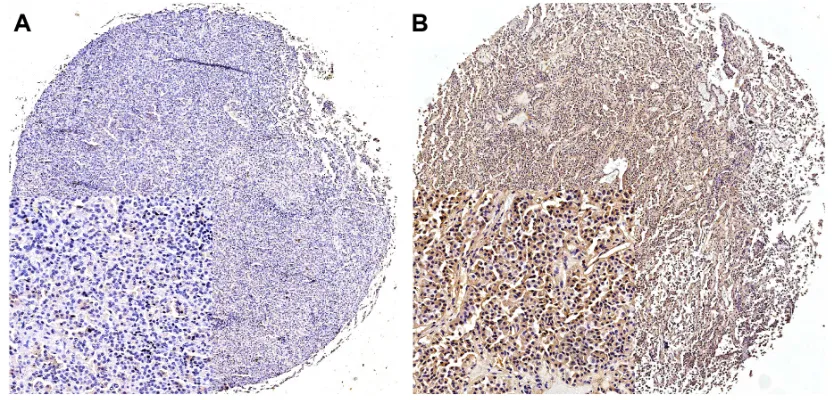
We then investigated the protein levels of Smad4 by immu-nohistochemistry in invasive and noninvasive NFPAs. We found that there was no significant difference in the Smad4 protein levels between invasive NFPAs and nonin-vasive NFPAs (Figure 3). Smad4 was highly expressed in 37.3% (22 of 59 cases) of invasive NFPAs and 36.3% (37 of 102 cases) of noninvasive NFPAs (P=0.898).
expression of Ki-67 in nFPas
Ki-67 can serve as an indicator of proliferative activity in NFPAs. We thus evaluated the levels of Ki-67 by immunohis-tochemistry in invasive and noninvasive NFPAs. We found
Table 1 characteristics of patients Characteristic Invasive NFPAs
(N=59) mean ± SD
Noninvasive NFPAs (N=102) mean ± SD age, (years), no (%) 49.0±11.5 51.8±10.7
sex
Male, no (%) 37 (62.7) 61 (59.8)
symptom, no (%)
headache 35 (59.3) 72 (70.6)
Visual deficit 28 (47.5) 36 (35.3)
hypopituitarism 20 (33.9) 17 (16.7)
none 2 (3.4) 15 (14.7)
Tumor size (cm), no (%)
,3 8 (13.6) 79 (77.5)
$3 51 (86.4) 3 (22.5)
Abbreviations: nFPas, nonfunctioning pituitary adenomas; sD, standard deviation.
Figure 1 (Continued)
OncoTargets and Therapy downloaded from https://www.dovepress.com/ by 118.70.13.36 on 26-Aug-2020
Dovepress
liu et al
Figure 2 smad3 and p-smad3 expression in nFPas.
Notes: (A) low expression of smad3 in invasive nFPas, (B) high expression of smad3 in noninvasive nFPas, (C) low expression of p-smad3 in invasive nFPas, (D) high expression of p-smad3 in noninvasive nFPas; (A–D) magnification: 200×.
Abbreviation: nFPas, nonfunctioning pituitary adenomas. Figure 1 smad2 and p-smad2 expression in nFPas.
Notes: (A) low expression of smad2, (B) high expression of smad2, (C) low expression p-smad2, (D) high expression p-smad2; (A–D) magnification: 200×. Abbreviation: nFPas, nonfunctioning pituitary adenomas.
OncoTargets and Therapy downloaded from https://www.dovepress.com/ by 118.70.13.36 on 26-Aug-2020
Dovepress smad3 and phospho-smad3
Figure 3 smad4 expression in nFPas.
Notes: (A) low expression and (B) high expression; (A, B) magnification: 200×. Abbreviation: nFPas, nonfunctioning pituitary adenomas.
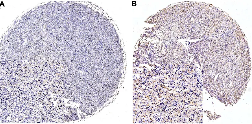
Figure 4 Ki-67 expression in nFPas.
Notes: (A) low expression in noninvasive nFPas and (B) high expression in invasive nFPas; (A, B) magnification: 200×. Abbreviation: nFPas, nonfunctioning pituitary adenomas.
that the Ki-67 index was markedly greater in invasive NFPAs than in noninvasive NFPAs (P,0.05). Furthermore, Spear-man’s rank correlation analysis showed that the Ki-67 index was significantly correlated with p-Smad3 levels (P,0.05, r=−0.702), but not with the levels of Smad2, p-Smad2, Smad3, or Smad4 (Figure 4).
Discussion
In the present study, we investigated TGF-β/Smad signaling and its proliferative activity in a large number of invasive and noninvasive NFPAs. We found that a low level of Smad3
and p-Smad3 was associated with the invasion of NFPAs, and that proliferative activity was greater in invasive NFPAs than in noninvasive NFPAs.
Smad3 is a key factor in TGF-β/Smad3-mediated tumor suppressor effect. Wolfraim et al13 demonstrated that a
reduction in Smad3 expression contributes to promote T-cell leukemogenesis. Moreover, Sodir et al14 reported that Smad3
deficiency promotes tumorigenesis in distal colon. In addi-tion, it has been reported that Smad3 mutation in human cancer can functionally inactivate the Smad3 protein.15
In this study, we found that the expression of Smad3 and
OncoTargets and Therapy downloaded from https://www.dovepress.com/ by 118.70.13.36 on 26-Aug-2020
Dovepress
liu et al
p-Smad3 was decreased in invasive NFPAs as compared with noninvasive NFPAs. Furthermore, we found that the Ki-67 index, an indicator for the proliferative activity of tumor cells, was markedly greater in invasive NFPAs than in noninvasive NFPAs and was significant correlated with p-Smad3 levels. These data suggest that Smad3 may act as a tumor suppressor in NFPAs and that TGF-β signaling may contribute to the invasion of NFPAs.
Smad2 and Smad4 have been identified as tumor sup-pressors in various human tumors,16–19 including gastric,
esophagus, liver, and breast cancers. Liu et al20 reported
that Smad4 expression is reduced in ductal breast carcinoma as compared with surrounding uninvolved ductal breast epithelia, suggesting that Smad4 has a potential role in the development of breast carcinoma and is a potential prognos-tic marker of breast carcinoma.20 However, in the present
study, we found that there was no significant difference in Smad2 and Smad4 protein levels between invasive NFPAs and noninvasive NFPAs. The discrepancy on Smad2 and Smad4 between PAs and other tumors suggests that the roles of Smad2 and Smad4 in PAs may be different and need to be further investigated.
Conclusion
In summary, this study showed that the protein levels of Smad3 and p-Smad3 are decreased in invasive NFPAs. This decrease in Smad3 and p-Smad3 may be associated with the invasion of NFPAs, and Smad3 and p-Smad3 may be useful biomarkers for the diagnosis of invasive NFPAs. Patients who have NFPAs with low expression of Smad3 and p-Smad3 may benefit from aggressive treatment and close surveillance.
Acknowledgments
This work was supported by the National High Technology Research and Development Program of China (863 Program) (2014AA020610), the Research Special Fund for Public Welfare Industry of Health (201402008), and the National Natural Science Foundation of China (31200796).
Disclosure
The authors report no conflicts of interest in this work.
References
1. Daly AF, Rixhon M, Adam C, Dempegioti A, Tichomirowa MA, Beckers A. High prevalence of pituitary adenomas: a cross-sectional study in the province of Liege, Belgium. J Clin Endocrinol Metab.2006; 91(12):4769–4775.
2. Fernandez A, Karavitaki N, Wass JA. Prevalence of pituitary adenomas: a community-based, cross-sectional study in Banbury (Oxfordshire, UK). Clin Endocrinol (Oxf).2010;72(3):377–382.
3. Katznelson L, Alexander JM, Klibanski A. Clinical review 45: clini-cally nonfunctioning pituitary adenomas. J Clin Endocrinol Metab. 1993;76(5):1089–1094.
4. Colao A, Di Somma C, Pivonello R, Faggiano A, Lombardi G, Savastano S. Medical therapy for clinically non-functioning pituitary adenomas. Endocr Relat Cancer.2008;15(4):905–915.
5. Wakefield LM, Roberts AB. TGF-beta signaling: positive and negative effects on tumorigenesis. Curr Opin Genet Dev.2002;12(1):22–29. 6. Massague J. TGFbeta in cancer. Cell.2008;134(2):215–230. 7. Massague J. TGFbeta signalling in context. Nat Rev Mol Cell Biol.2012;
13(10):616–630.
8. Heldin CH, Miyazono K, ten Dijke P. TGF-beta signalling from cell membrane to nucleus through SMAD proteins. Nature.1997; 390(6659):465–471.
9. Nakao A, Afrakhte M, Moren A, et al. Identification of Smad7, a TGFbeta-inducible antagonist of TGF-beta signalling. Nature.1997; 389(6651):631–635.
10. Zhenye L, Chuzhong L, Youtu W, et al. The expression of TGF-beta1, Smad3, phospho-Smad3 and Smad7 is correlated with the development and invasion of nonfunctioning pituitary adenomas. J Transl Med. 2014;12:71.
11. Knosp E, Steiner E, Kitz K, Matula C. Pituitary adenomas with invasion of the cavernous sinus space: a magnetic resonance imaging classification compared with surgical findings. Neurosurgery.1993; 33(4):610–617; discussion 617–618.
12. Li ZY, Zhang XH, Chen Y, et al. Clinical significance of B7-H4 expres-sion in matched non-small cell lung cancer brain metastases and primary tumors. Onco Targets Ther.2013;6:869–875.
13. Wolfraim LA, Fernandez TM, Mamura M, et al. Loss of Smad3 in acute T-cell lymphoblastic leukemia. N Engl J Med.2004;351(6):552–559. 14. Sodir NM, Chen X, Park R, et al. Smad3 deficiency promotes tumori-genesis in the distal colon of ApcMin/+ mice. Cancer Res.2006;66(17): 8430–8438.
15. Ku JL, Park SH, Yoon KA, et al. Genetic alterations of the TGF-beta signaling pathway in colorectal cancer cell lines: a novel mutation in Smad3 associated with the inactivation of TGF-beta-induced transcrip-tional activation. Cancer Lett.2007;247(2):283–292.
16. Hahn SA, Schutte M, Hoque AT, et al. DPC4, a candidate tumor sup-pressor gene at human chromosome 18q21.1. Science.1996;271(5247): 350–353.
17. Wang LH, Kim SH, Lee JH, et al. Inactivation of SMAD4 tumor sup-pressor gene during gastric carcinoma progression. Clin Cancer Res. 2007;13(1):102–110.
18. Natsugoe S, Xiangming C, Matsumoto M, et al. Smad4 and transforming growth factor beta1 expression in patients with squamous cell carcinoma of the esophagus. Clin Cancer Res.2002;8(6):1838–1842.
19. Yakicier MC, Irmak MB, Romano A, Kew M, Ozturk M. Smad2 and Smad4 gene mutations in hepatocellular carcinoma. Oncogene.1999; 18(34):4879–4883.
20. Liu N-n, Xi Y, Callaghan MU, et al. SMAD4 is a potential prog-nostic marker in human breast carcinomas. Tumor Biol.2013;35(1): 641–650.
OncoTargets and Therapy downloaded from https://www.dovepress.com/ by 118.70.13.36 on 26-Aug-2020
OncoTargets and Therapy
Publish your work in this journal
Submit your manuscript here: http://www.dovepress.com/oncotargets-and-therapy-journal
OncoTargets and Therapy is an international, peer-reviewed, open access journal focusing on the pathological basis of all cancers, potential targets for therapy and treatment protocols employed to improve the management of cancer patients. The journal also focuses on the impact of management programs and new therapeutic agents and protocols on
patient perspectives such as quality of life, adherence and satisfaction. The manuscript management system is completely online and includes a very quick and fair peer-review system, which is all easy to use. Visit http://www.dovepress.com/testimonials.php to read real quotes from published authors.
Dovepress
Dove
press
smad3 and phospho-smad3
OncoTargets and Therapy downloaded from https://www.dovepress.com/ by 118.70.13.36 on 26-Aug-2020