Research report
Ingested placenta blocks the effect of morphine on gut transit in
Long–Evans rats
James W. Corpening*, Jean C. Doerr, Mark B. Kristal
Behavioral Neuroscience Program, Department of Psychology, Park Hall, University at Buffalo, Buffalo, NY 14260-4110, USA
Accepted 1 May 2004 Available online 15 June 2004
Abstract
Opioids produce antinociception, and ingested placenta or amniotic fluid modifies that antinociception. More specifically, ingested placenta enhances the antinociception produced by selective activation of centraln-opioid ory-opioid receptors but attenuates that produced
by activation of centralA-opioid receptors. Opioids also slow gut transit by acting on central or peripheralA-opioid receptors. Therefore, we hypothesized that ingested placenta would reverse the slowing of gut transit that is produced by morphine, a preferentialA-opioid-receptor agonist. Rats were injected with morphine either centrally or systemically and fed placenta, after which gastrointestinal transit was evaluated. We report here that ingested placenta reversed the slowing of gut transit produced by centrally administered morphine but did not affect the slowing of gut transit produced by systemically administered morphine. These results suggest another likely consequence of placentophagia at parturition in mammals—reversal of opioid-mediated, pregnancy-based disruption of gastrointestinal function—as well as an important consideration in opioid-based treatments for pain in humans—enhancement of desirable effects with attenuation of adverse effects.
D2004 Elsevier B.V. All rights reserved.
Theme:Endocrine and autonomic regulation
Topic:Gastrointestinal and urogenital regulation
Keywords:Opioid; Mu; Constipation; Placenta; Gut transit; Morphine
1. Introduction
Placenta or amniotic fluid, when ingested, enhances the antinociception produced by endogenous and exogenous opioids[17 – 19], including the endogenous opioid-mediated antinociception that is characteristic of late pregnancy
[13,21] and that is produced by vaginocervical stimulation
[9,16,20]. The mechanism of action for this enhancement of opioid processes has been elucidated by demonstrations that enhancement is blocked by gastric vagotomy[27]and does not rely on digestion of placenta[25], that ingested amniotic fluid enhances the central nervous system (CNS) action, but not the peripheral nervous system (PNS) action, of injected
morphine [8], and that ingested placenta enhances opioid antinociception mediated by n- or y-opioid receptors but actually attenuates that mediated byA-opioid receptors[7].
Another effect of central or systemic opioids is a slowing of gut transit [3,6,15], which is believed to be mediated by A-opioid receptor activity both centrally and peripherally [2,12,23,24,28]. This is believed to occur centrally by action in the dorsal vagal complex of the hindbrain [10], which is part of the vagovagal neuro-circuitry that influences gastrointestinal function [11], yet it is believed to occur peripherally by disruption of the role of local opioids in intestinal muscular contraction and relaxation [22,23].
Because ingested placenta has been shown to inhibit opioid processes that are mediated byA-opioid receptors, we hypothesized that ingested placenta would reverse the slow-ing of gut transit that is produced by administration of morphine, a preferentialA-opioid-receptor agonist.
0006-8993/$ - see front matterD2004 Elsevier B.V. All rights reserved. doi:10.1016/j.brainres.2004.05.006
* Corresponding author. c/o Dakota Therapeutics, 428 W. Delavan Ave., Buffalo, NY 14213-1413, USA. Tel.: 886-9341; fax: +1-716-886-9341.
E-mail address:[email protected] (J.W. Corpening).
2. Materials and methods
2.1. Subjects
Nulliparous, female Long-Evans rats were used, all of which were born and maintained in the Behavioral Neuro-science Research Facility of the University at Buffalo De-partment of Psychology. Rats were housed individually under a 14:10 light/dark cycle (lights on at 0500h EST) in 322020-cm plastic cages (Experiment 1) or in 24.51818-cm wire-mesh cages (Experiment 2) with water and food (Harlan Teklad Rodent Diet 8640) available ad lib. All rats exhibited at least two normal estrous cycles prior to testing, as determined by daily microscopic inspec-tion of vaginal epithelial cells obtained by vaginal lavage with physiological saline. All rats weighed 230 – 320 g and were 75 – 125 days of age at the time of testing. Rats in Experiment 2 underwent stereotaxic surgery and were allowed at least 2 weeks for postoperative recovery prior to testing. Food and water were removed from all cages 2 h prior to testing. All procedures were approved by the University at Buffalo Institutional Animal Care and Use Committee.
2.2. Materials
Morphine sulfate (Sigma) was dissolved in physiological saline vehicle prior to injection. A test meal, used to determine gastrointestinal transit rate, was composed of methyl cellulose (Sigma), deionized water, and green food color (McCormick), mixed 3.5 g:100 ml:1 ml, respectively, and allowed to stand for at least 1 h after mixing to reach maximum viscosity. A 4 ml:1 ml mix of Ketaset (ketamine hydrochloride, 100 mg/ml; Sigma) and Rompun (xylazine, 20 mg/ml; Sigma), respectively, was used as anesthetic for surgery.
2.3. Placenta collection and administration
Rat placenta was collected from time-bred pregnant female rats killed by CO2 asphyxiation on Day 21 of
pregnancy (presence of vaginal sperm indicates Day 1). The collection procedure in our laboratory is to remove placentas from a rat immediately after death and, between 20 and 30 min after death, freeze the placentas at 20j C
for later use [18]. Pregnant-rat liver, which was also col-lected during the placenta-collection procedure and treated similarly, was used as a control substance for placenta in all experiments, because it is similar in consistency and com-position to placenta and it has been shown not to exhibit an opioid-modifying effect[1]. Placenta or liver was presented to each rat in a 3725-mm Stender dish.
2.4. Procedural habituation
Rats were habituated to the testing procedure for 5 days prior to testing by daily orogastric intubation without
infusion. Rats used in Experiment 2 were further habituated each day to the handling they received during administration of drug. Because it was necessary that rats eat placenta and liver within 5 min during testing, all rats were also habit-uated to these substances under the following schedule: rats were each presented daily with 0.5 g of lean ground beef, which they typically eat promptly, until they ate the ground beef within 5 min on two consecutive days; then presented daily with 0.5 g of ground beef with placenta (0.25 g each) until they ate that within 5 min on 1 day; then presented daily with 0.5 g of placenta until they ate that within 5 min on two consecutive days; then presented daily with 0.5 g of liver until they ate that within 5 min on 1 day. In our laboratory, this habituation takes a mean of 7.54F2.85 days to complete.
2.5. Stereotaxic surgery
To prepare for i.c.v. administration of drug in Experiment 2, a Kopf stereotaxic apparatus was used to implant an indwelling, stainless steel, 22-ga, 6-mm-long guide cannula into each rat’s brain. Each rat was anesthetized with an injection (1.0 ml/kg i.p.) of the Ketaset-Rompun mix, described above, and had a guide cannula implanted with the ventral tip lying 0.5 mm above the right lateral ventricle of the brain (coordinates were AP = 0.0 mm from bregma, ML = 2.0 mm from the midsagittal sinus, DV = 2.8 mm from surface of the brain, with the incisor bar at + 3.0 mm). The guide cannula was secured to the skull with dental cement and three stainless steel anchor screws fastened to the skull. A stainless steel wire, cut to be flush with the ventral tip of the guide cannula, was kept in the cannula to maintain patency.
2.6. Drug administration
Anesthetic was injected with a 25-ga hypodermic needle and a 1-cm3plastic syringe. Morphine was injected either in a similar fashion (i.p.) or i.c.v. at a rate of 1.0Al/min through a stainless steel, 28-ga, 7-mm-long injection cannula inserted through an indwelling guide cannula. This arrange-ment allowed the injection cannula to extend into the lateral ventricle 1 mm beyond the ventral tip of the guide cannula. During i.p. injections, rats were held vertically in the experimenter’s hand; during i.c.v. injections, rats sat on the lap of the experimenter as the experimenter sat next to the infusion pump.
2.7. Gastrointestinal transit evaluation
Gastrointestinal transit was evaluated by monitoring the passage of a test meal through the small intestine. Test meals were administered by orogastric infusion, and rats were killed by CO2asphyxiation 15 min after infusion of the test
intestine was promptly clamped at the farthest-traveled portion of the infused test meal. The small intestine was then severed at both the duodenogastric and ileocecal junctions, removed intact from the diaphragm, and laid out taut for measurement. The length of the upper segment of small intestine, from its duodenogastric end to the farthest portion of the test meal, was measured, as was the entire small intestine. Gastrointestinal transit was determined by calculating the percentage of the small intestine that the test meal traveled. The experimenter measuring gastrointestinal transit was blind to the rat’s experimental condition at the time of measurement.
2.8. Experimental design and procedure
Experiment 1 was performed to determine whether ingested placenta can reverse the effect of systemically administered morphine on gastrointestinal transit. Six rats were randomly assigned to each of eight groups in a 42 design, Systemic Morphine DoseIngestate. Dur-ing testDur-ing, rats were injected with morphine sulfate at one of four doses (0.00, 0.05, 0.50, 3.00 mg/kg, i.p.). Ten minutes later they were presented with either pla-centa or liver (0.5 g), which they promptly ate, and 5 min later were infused orogastrically with a test meal. Infusion of the test meal lasted approximately 15 s. Gastrointestinal transit was evaluated 15 min after infu-sion of the meal.
Experiment 2 was performed to determine whether ingested placenta can reverse the effect of centrally admin-istered morphine on gastrointestinal transit. Six rats were randomly assigned to each of six groups in a 32 design, Central Morphine DoseIngestate. During testing, rats were presented with either placenta or liver (0.5 g), which they promptly ate. Five minutes later they were injected centrally with morphine sulfate in 0.5 Al physiological saline vehicle at one of three doses (0.0, 2.5, 20.0 Ag, i.c.v.), and 15 min later were infused orogastrically with a test meal. Gastrointestinal transit was evaluated 15 min after infusion of the meal. To verify cannula placement after transit was evaluated, each rat was injected with methyl blue dye (0.5 Al, i.c.v.) in the same fashion that morphine was administered. Each rat’s brain was then removed, frozen at
14j C, and sliced at 40 Am. Accurate placement and infusion was indicated by the presence of dye in the lateral ventricle. The experimenter who verified placement site was blind to the rat’s experimental condition at the time of verification.
2.9. Statistical analyses
Parametric analyses were performed when appropriate, and, when possible, analyses were performed on a personal computer using statistical software. The specific tests used are described below. All analyses were performed using
a= 0.05.
3. Results
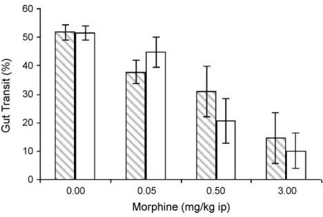
In Experiment 1, ingested placenta did not affect gut transit that had been slowed by systemically administered morphine.Fig. 1shows gastrointestinal transit as a function of morphine dose and ingestate for rats injected systemically with morphine. Six of the twelve rats (three liver-fed, three placenta-fed) that were injected with the highest dose of morphine showed 0% gastrointestinal transit, which likely contributed to the statistically significant difference between the highest and lowest group variances, F(7, 40) = 3.15, p= 0.01. Because of these extreme values, nonparametric statistical analyses were used. Independent Kruskal – Wallis one-way analyses revealed a significant effect of dose of morphine on gut transit in placenta-fed rats, H(3) = 14.98, p= 0.002, and in liver-fed rats,H(3) = 9.22,p= 0.026, such that a larger dose of morphine produced a greater slowing of gut transit. The same effect was seen in all (pooled) rats, H(3) = 22.97,p< 0.001. There was no difference, however, between the liver- and placenta-fed rats at any of the four doses of morphine, as revealed by Mann – WhitneyU-tests (p>0.39).
In Experiment 2, however, ingested placenta did reverse the slowing of gut transit that was produced by centrally administered morphine.Fig. 2shows gastrointestinal transit as a function of morphine dose and ingestate for rats injected centrally with morphine. The extreme variances seen in Experiment 1 were not seen in this experiment, so parametric statistical analyses were used. A two-way ANOVA revealed a significant effect of dose of morphine, F(2, 30) = 10.50, p< 0.001, such that a larger dose of morphine produced a greater slowing of gut transit, and a significant interaction between dose of morphine and inges-tate, F(2, 30) = 5.49, p= 0.009. Because of the problem interpreting main effects in the presence of an interaction, independent one-way ANOVAs were performed on the data from the liver- and placenta-fed rats. There was a significant effect of dose of morphine on gut transit in liver-fed rats,
F(2, 15) = 15.35, p< 0.001, such that a larger dose of morphine produced a greater slowing of gut transit, but there was no effect of dose of morphine in placenta-fed rats, F(2, 15) = 2.00,p= 0.17. This latter result was undoubtedly due to the fact that ingested placenta blocked the slowing of gut transit that was produced by 20 Ag morphine, t(10) = 3.72, p< 0.01. Of the rats injected with 20 Ag morphine, the mean percent transit exhibited by liver-fed rats was 8.79F8.79, whereas that exhibited by placenta-fed rats was 38.84F5.14. Specifically, five of the six liver-fed rats injected with 20 Ag morphine showed 0% gastrointes-tinal transit, but none of the six placenta-fed rats showed 0% transit. Ingested placenta blocked the transit-slowing effect of 20Ag morphine, such that gastrointestinal transit was not different from that of rats injected with either 2.5 Ag morphine or saline. There was no difference between the liver- and placenta-fed rats that were injected with either 2.5 Ag morphine or saline (p>0.54).
4. Discussion
The present research reveals that ingested placenta can reverse the slowing of gut transit that is produced by morphine acting centrally (Experiment 2). This same rever-sal was not seen, however, when morphine was injected systemically (Experiment 1). It is known that systemically administered morphine can inhibit gastrointestinal transit independent of a CNS morphine effect[14], and it is known that ingested amniotic fluid modifies the CNS, but not PNS, antinociceptive action of opioids[8], so it is reasonable to conclude that the lack of placental influence in Experiment 1 was due to the peripheral action of morphine. The results of Experiments 1 and 2 are consistent, then, with the results of prior research that focused on antinociception, in that they support the evidence that ingested placenta selectively modifies the CNS-based action of opioids, and that such modification is inhibitory when the opioid action is medi-ated byA-opioid receptors.
Most of the research in our laboratory has used an opioid-placenta order of presentation, similar to our protocol
in Experiment 1, which is the order that would occur at parturition. We were reminded at the start of Experiment 2, however, that a rat could disqualify herself from a trial if, after being injected with morphine, she did not eat placenta or liver within the required interval. Given the significant resources invested in each rat (i.e., CNS surgery and recovery), the time required to allow for metabolism of the morphine before non-eating rats could be retested, and the risk of damage during the interim to the rat and to the cannula stage mounted to the rat’s head, we chose to present placenta or liver 5 min before the morphine injection. Other research in our laboratory has shown that the order of presentation is irrelevant ([19], and personal observation), and this placenta-opioid order of presentation is permissible, given the duration of placental action [9].
As mentioned, ingested placenta or amniotic fluid enhan-ces the antinociception of late pregnancy [13,21] and of vaginocervical stimulation[9,6,20]. In addition to antinoci-ception, a slowed gut transit is characteristic of late preg-nancy [4]. Of particular importance, here, are the findings that pregnancy antinociception is mediated byn-opioid[26]
andy-opioid[5]receptors, that the opioid-based slowing of gut transit is mediated by A-opioid receptors [2,12,23, 24,28], and that ingested placenta enhances n- and y -receptor-mediated antinociception but inhibits A -receptor-mediated antinociception[7]. Therefore, as suggested by the present results, ingestion of placenta or amniotic fluid at parturition may enhance the antinociception of pregnancy as well as help restore normal gut transit after parturition, particularly when transit has been slowed by activity at A -opioid receptors in the CNS.
The present results confirm that ingested placenta inhib-its CNS A-receptor-mediated opioid processes, in this case the slowing of gastrointestinal transit produced by mor-phine. One of the widely studied side effects of opioid treatment for pain in humans is a slowing of gastrointestinal transit (constipation). Therefore, the present results, together with prior results suggesting that ingested placenta or amniotic fluid modifies, specifically, opioid function, should make clear that the mechanism of placental modification of opioid function has an important implication for human pain management. This is particularly true given the possibility that an understanding of the mechanism of placental action could conceivably contribute to the development of adjunct treatments for pain that enhance the desirable effects of CNS opioids while actually blocking the undesirable effects of those opioids.
References
[1] P. Abbott, A.C. Thompson, E.J. Ferguson, J.C. Doerr, J.A. Tarapacki, P.J. Kostyniak, J.A. Syracuse, D.M. Cartonia, M.B. Kristal, Placental opioid-enhancing factor (POEF): generalizability of effects, Physiol. Behav. 50 (1991) 933 – 940.
[2] J.C. Bornstein, H.L. Fields, Morphine presynaptically inhibits a gan-glionic cholinergic synapse, Neurosci. Lett. 15 (1979) 77 – 82. Fig. 2. Mean percent gastrointestinal transit (FS.E.M.) for rats that were
[3] L. Bueno, J. Fioramonti, Action of opiates on gastrointestinal func-tion, Bailliere’s Clin. Gastroenterol. 2 (1988) 123 – 139.
[4] F.-Y. Chang, S.-D. Lee, G.-H. Yeh, C.-C. Lu, P.-S. Wang, S.-W. Wang, Disturbed small intestinal motility in the late rat pregnancy, Gynecol. Obstet. Investig. 45 (1998) 221 – 224.
[5] M. Dawson-Basoa, A.R. Gintzler, Involvement of spinal cordyopiate receptors in the antinociception of gestation and its hormonal simu-lation, Brain Res. 757 (1997) 37 – 42.
[6] A. De Luca, I.M. Coupar, Insights into opioid action in the intestinal tract, Pharmacol. Ther. 69 (1996) 103 – 115.
[7] J.M. DiPirro, M.B. Kristal, Placenta ingestion by rats enhancesn- and y-opioid antinociception, but suppresses A-opioid antinociception, Brain Res. (2004) (in press).
[8] J.M. DiPirro, A.C. Thompson, M.B. Kristal, Amniotic-fluid ingestion enhances the central analgesic effect of morphine, Brain Res. Bull. 26 (1991) 851 – 855.
[9] J.C. Doerr, M.B. Kristal, Enhancement of opioid-mediated analgesia by ingestion of amniotic fluid: onset latency and duration, Physiol. Behav. 46 (1989) 913 – 915.
[10] S. Duan, N. Shimizu, A. Fukuda, T. Hori, Y. Oomura, Hyperpolariz-ing action of enkephalin on neurons in the dorsal motor nucleus of the vagus, in vitro, Brain Res. Bull. 25 (1990) 551 – 559.
[11] W.R. Ewart, D.L. Wingate, Central representation and opioid modu-lation of gastric mechanoreceptor activity in the rat, Am. J. Physiol. 244 (1983) G27 – G32.
[12] J.J. Galligan, T.F. Burks, Centrally mediated inhibition of small in-testinal transit and motility by morphine in the rat, J. Pharmacol. Exp. Ther. 226 (1983) 356 – 361.
[13] A.R. Gintzler, Endorphin-mediated increases in pain threshold during pregnancy, Science 210 (1980) 193 – 195.
[14] D.E. Gmerek, A. Cowan, J.H. Woods, Independent central and pe-ripheral mediation of morphine-induced inhibition of gastrointestinal transit in rats, J. Pharmacol. Exp. Ther. 236 (1986) 8 – 13.
[15] A.F. Green, Comparative effects of analgesics on pain threshold, respiratory frequency and gastrointestinal propulsion, Br. J. Pharma-col. 14 (1959) 26 – 34.
[16] B.R. Komisaruk, J. Wallman, Antinociceptive effects of vaginal stim-ulation in rats: neurophysiological and behavioral studies, Brain Res. 137 (1977) 85 – 107.
[17] M.B. Kristal, Enhancement of opioid-mediated analgesia: a solution to the enigma of placentophagia, Neurosci. Biobehav. Rev. 15 (1991) 425 – 435.
[18] M.B. Kristal, A.C. Thompson, H.L. Grishkat, Placenta ingestion enhances opiate analgesia in rats, Physiol. Behav. 35 (1985) 481 – 486.
[19] M.B. Kristal, A.C. Thompson, P. Abbott, Ingestion of amniotic fluid enhances opiate analgesia in rats, Physiol. Behav. 38 (1986) 809 – 815.
[20] M.B. Kristal, A.C. Thompson, S.B. Heller, B.R. Komisaruk, Placenta ingestion enhances analgesia produced by vaginal/cervical stimulation in rats, Physiol. Behav. 36 (1986) 1017 – 1020.
[21] M.B. Kristal, A.C. Thompson, P. Abbott, J.M. DiPirro, E.J. Ferguson, J.C. Doerr, Amniotic-fluid ingestion by parturient rats enhances preg-nancy-mediated analgesia, Life Sci. 46 (1990) 693 – 698.
[22] R.A. North, Effects of morphine on myenteric plexus neurones, Neu-ropharmacology 15 (1976) 719 – 721.
[23] W.D.M. Paton, The action of morphine and related substances on contraction and on acetylcholine output of coaxially stimulated guin-ea-pig ileum, Br. J. Pharmacol. Chemother. 11 (1957) 119 – 127. [24] O. Pol, L. Valle, P. Sa´nchez-Bla´zquez, J. Garzo´n, M. Puig, Antibodies
and antisense oligodeoxynucleotides toA-opioid receptors, selectively block the effects ofA-opioid agonists on intestinal transit and perme-ability in mice, Br. J. Pharmacol. 127 (1999) 397 – 404.
[25] T.M. Robinson, P. Abbott, M.B. Kristal, Blockade of digestion by famotidine pretreatment does not interfere with the opioid-enhancing effect of ingested amniotic fluid, Physiol. Behav. 57 (1995) 261 – 263. [26] H.W. Sander, R.M. Kream, A.R. Gintzler, Spinal dynorphin involve-ment in the analgesia of pregnancy: effects of intrathecal dynorphin antisera, Eur. J. Pharmacol. 159 (1989) 205 – 209.
[27] J.A. Tarapacki, A.C. Thompson, M.B. Kristal, Gastric vagotomy blocks opioid analgesia enhancement produced by placenta ingestion, Physiol. Behav. 52 (1992) 179 – 182.