Study of the structure and function of the sheath of tropically adapted bulls
and risk factors for preputial eversion and preputial prolapse
Lex Brian Turner BVSc, Grad Dip Biotech
A thesis submitted for the degree of Doctor of Philosophy at
The University of Queensland in 2013
Abstract
Preputial prolapse is an obvious condition affecting bulls from many breeds. Unfortunately, the losses in production and welfare concerns associated with preputial prolapse can remain undetected for long periods of time in the extensive beef areas of northern Australia where the bulls are not inspected regularly. Thus, there is a critical need to identify the structural factors predisposing to preputial prolapse in young bulls so that they can be culled early.
Despite there being no firm scientific evidence of an association between preputial eversion and preputial prolapse, it seems logical that the increased exposure of the sensitive prepuce as a consequence of preputial eversion may increase the risk of bulls developing preputial
pathology, in particular preputial prolapse. This may be particularly relevant in Bos indicus bulls as they have a more pendulous sheath and thus eversion of the prepuce may be associated with a greater risk of injury to the prepuce compared to that in Bos taurus bulls. Further, studies of preputial eversion in Bos taurus bulls have concluded that there is an association between polledness and increased prevalence and severity (length of everted prepuce and duration of eversion) of preputial eversion due primarily to the absence or poor development of the caudal preputial muscles. No similar definitive work in Bos indicus bulls has been conducted and thus anatomical studies reported in this thesis were conducted to determine if a similar association occurred in Bos indicus bulls.
A survey of a sample of large beef breeding herds in northern Australia found that preputial prolapse is a significant problem in Bos indicus and Bos indicus derived bulls and affected both young and older bulls. The importance of preputial prolapse confirmed the value of further research into the causes of this problem. A series of anatomical studies confirmed that preputial eversion in Bos indicus derived bulls was not more prevalent in polled bulls than horned bulls and was not associated with deficiency of the caudal preputial muscles as was established in
observed in young polled bulls that had poorly developed or absent caudal preputial muscles. It was concluded that deficiency of the caudal preputial muscles in polled Bos indicus derived bulls may predispose to preputial prolapse at an early age, but no predisposing anatomical factors were found for horned Bos indicus derived bulls. In these studies, preputial eversion and preputial prolapse were found in horned Bos indicus derived bulls that did not have any
preputial muscle deficiency and it was noted that preputial eversion was not related to the length of the prepuce.
Further studies confirmed that preputial eversion was linearly and consistently associated with position of the glans penis within the sheath in Bos indicus derived bulls, and movement of the glans penis towards the preputial orifice consistently resulted in preputial eversion in these bulls. A method to objectively measure the relationship between movement of the glans penis within the sheath and preputial eversion was developed. Studies in humans have linked function of some abdominal muscles to function of the pelvic organs. This relationship was investigated in Bos indicus derived bulls to determine whether the function of specific
abdominal muscles affected position of the penis in the sheath. Using the method developed to objectively measure the relationship between penis movement and preputial eversion, the abdominal muscles that potentially were associated with movement of the glans penis or preputial eversion were examined but no significant relationships were observed.
In the anatomical study of Bos indicus derived bulls not affected with preputial prolapse a more pendulous sheath was associated with increased prevalence of preputial eversion. This
relationship was confirmed for horned and polled bulls in the penis movement studies. Bos
indicus derived bulls with more pendulous sheaths evert their prepuces more than bulls with
less pendulous sheaths thus increasing the risk of damage to the prepuce either from the
environment, other bulls, or from them inadvertently stepping on the everted prepuce when they get to their feet. Culling Bos indicus derived bulls with more pendulous sheaths should reduce the incidence of preputial eversion and possibly preputial prolapse. The anatomical study of
Bos indicus derived bulls that did not have preputial prolapse demonstrates that there are herds
Declaration by author
This thesis is composed of my original work, and contains no material previously published or written by another person except where due reference has been made in the text. I have clearly stated the contribution by others to jointly-authored works that I have included in my thesis.
I have clearly stated the contribution of others to my thesis as a whole, including statistical assistance, survey design, data analysis, significant technical procedures, professional editorial advice, and any other original research work used or reported in my thesis. The content of my thesis is the result of work I have carried out since the commencement of my research higher degree candidature and does not include a substantial part of work that has been submitted to qualify for the award of any other degree or diploma in any university or other tertiary institution. I have clearly stated which parts of my thesis, if any, have been submitted to qualify for another award.
I acknowledge that an electronic copy of my thesis must be lodged with the University Library and, subject to the General Award Rules of The University of Queensland, immediately made available for research and study in accordance with the Copyright Act 1968.
Publications during candidature
Bullpower. Final report to Meat & Livestock Australia (2005) pp. 116-135
Publications included in this thesis
Contributions by Others to the Thesis
Professor McGowan contributed to the early direction and design for chapters 3, 4 and 5 and the overall focus of the thesis. Sue Markwell contributed to the concept of abdominal muscle involvement in pelvic organ functions in chapter 7.
Departmental biometricians (particularly Vivienne Doogan, Gary Blight and David Mayer) contributed to the statistical analysis and interpretation for chapters 3, 4, 5, 6 and 7.
Biometricians were involved in the planning stages of all the studies to ensure data collected could be successfully analysed. Some of the more complicated regression analyses were performed by these professional biometricians. All data analyses and interpretation were completed in consultation with these biometricians. Dr Dianne Vankan provided professional editorial input into two of the chapters.
Statement of parts of the thesis submitted to qualify for the award of another degree
Acknowledgements
The author wishes to thank Mick Sullivan (DAFF) and the company staff involved in the survey of bull wastage in Northern Australia for their time, enthusiasm and input.
The author would also like to thank the management and staff at the South Burnett Meat Works Co-operative at Murgon for their generous help and for the use of their facilities; the University of Queensland School of Veterinary Science for the use of equipment; Anthony Coates for his help in coordinating the livestock as well as completing the on-farm scoring and the Santa Gertrudis Breeders’ (Australia) Association for funding part of the research.
The author would like to thank the management and staff, in particular Dr. Boris Dobrenov, at the AMH abattoir at Dinmore for their generous help and encouragement; all the producers who provided background information on the prolapsed bulls and the biometricians and members of the Bullpower group for their assistance.
The author would like to thank Sue Markwell, Dallas Baker and staff at the University of
Queensland Veterinary Science Farm for their generous assistance and ideas; Trevor Griffiths and staff of Yackatoon Grazing Company for their assistance and for providing access to bulls. Also thanks to Professor Keith Entwistle, Dr Scott Norman, Dr Dick Holroyd, Dr Wynne Collins, Dr Dianne Vankan and Dr Vivienne Perry, who have helped with the thesis and to all other UQ and DAFF staff who have assisted with this work. A special thanks to Professor Michael
Keywords
bull, bos indicus derived, preputial eversion, preputial prolapse, survey, anatomy, abdominal muscles, penis movement, bos indicus
Australian and New Zealand Standard Research Classifications (ANZSRC)
ANZSRC code: 070702 Veterinary Anatomy and Physiology 60%, ANZSRC code: 070709 Veterinary Pathology 10%,
ANZSRC code: 070799 Veterinary Sciences not elsewhere classified 30%
Fields of Research (FoR) Classification
Table of Contents
Study of the structure and function of the sheath of tropically adapted bulls
and risk factors for preputial eversion and preputial prolapse... i
Lex Brian Turner ... i
BVSc, Grad Dip Biotech ... i
Table of Contents ... x
List of figures ... xiii
List of tables...xv
Abbreviations ... xviii
Chapter 1. ... 1
Introduction... 1
Chapter 2. ... 4
Literature review: Factors affecting preputial function in bulls... 4
Introduction ... 4
Influence of bull fertility on overall herd fertility... 4
Importance of Bos indicus and Bos indicus derived cattle in tropical/subtropical regions of the world ... 6
Extent of bull fertility problems due to preputial problems... 7
Historical perspective of the importance of sheath conformation. ... 9
Bull external reproductive anatomy and function ... 10
Anatomical Development ... 12
Musculature ... 13
Vasculature to the sheath and prepuce ... 19
Innervation to the external reproduction organs... 20
Methods of defining the conformation of external reproductive structures in the bull ... 21
Sequence of preputial abnormalities ... 24
Factors predisposing to preputial prolapse ... 26
Structures predisposing to preputial pathology ... 31
Treatment and prevention of preputial problems ... 42
Chapter 3. ... 44
Causes of bull wastage from northern Australian beef cattle herds... 44
Introduction ... 44
Objectives ... 44
Materials and methods ... 44
Results... 47
Discussion ... 63
Chapter 4. ... 70
A quantitative anatomical study of the sheath and prepuce of Santa Gertrudis bulls relative to the occurrence of preputial eversion... 70
Introduction ... 70
Objectives ... 70
Materials and methods ... 71
Results... 77
Discussion ... 82
Chapter 5. ... 87
A study of the anatomy of the external genitalia of Santa Gertrudis bulls with chronic preputial prolapse... 87
Introduction ... 87
Objectives ... 87
Materials and methods ... 88
Results... 90
Discussion ... 94
Chapter 6. ... 99
Relationship between the position of the penis and preputial eversion in Bos indicus derived bulls... 99
Introduction ... 99
Objectives ... 99
Results... 102
Discussion ... 108
Chapter 7. ... 112
A study of the involvement of abdominal muscles in the position of the penis and prepuce within the sheath of Bos indicus derived bulls... 112
Introduction ... 112
Objective... 113
Materials and Methods ... 114
Results... 121
Discussion ... 132
Chapter 8. ... 136
General discussion and conclusions... 136
Bibliography ... 142
Appendices ... 156
Appendix 1. Examples of sheath scores... 156
Appendix 2. Examples of caudal preputial muscle network scores... 159
Appendix 3. Examples of preputial eversion... 165
List of figures
Page
Figure 3.1; Percentage of culled bulls of all ages culled for preputial prolapse on
property E over three years...60
Figure 3.2; Numbers by age of bulls culled with preputial prolapse from property E over three years...60
Figure 4.1; BREEDPLAN sheath scoring system. ...72
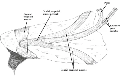
Figure 4.2; Diagram of the dissection indicating basic muscle positions. ...73
Figure 6.1; Relationship between length of preputial eversion and position of the tip of the glans penis relative to the preputial orifice for Bull 1 on two different days. ...103
Figure 6.2; Individual regression lines for the relationship between the length of preputial eversion and position of the tip of the glans penis relative to the preputial orifice for all bulls in the study. ...105
Figure 6.3; Relationship between length of preputial eversion and position of the tip of the glans penis relative to the preputial orifice for bulls of different horn status...106
Figure 6.4; Relationship between length of preputial eversion and position of the tip of the glans penis relative to the preputial orifice for the different age groups of the bulls. ...107
Figure 6.5; Relationship between length of preputial eversion when the glans penis is at the preputial orifice and vertical distance from the ventral abdominal wall to the preputial orifice. ...108
Figure 7.1; Ultrasound measuring positions (Adapted from McKiernan and Sundstrom 2000)...115
Figure 7.2; Position of measurement of the multifidus lumborum muscle. ...116
Figure 7.3; Position of measurement of the obliquus internus abdominis muscle...117
Figure 7.5; Position of backline measurements of the bulls. (The sites marked on the bull are indicators of the level of the site of the appropriate
vertebral processes and are not on the actual site of measurement)...119 Figure 7.6; Regression of length of preputial eversion against position of the tip of
List of tables
Page
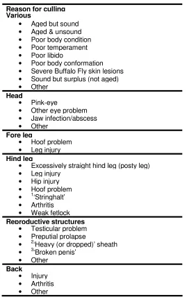
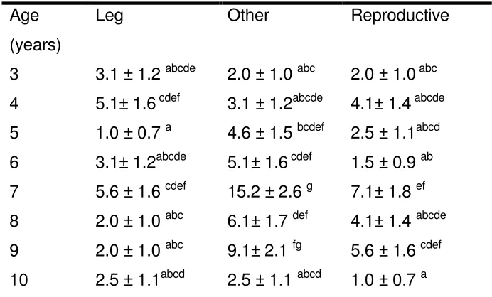
Table 3.1; List of possible reasons for culling bulls on the selected beef cattle
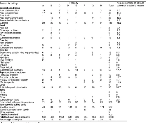
properties...46 Table 3.2; Summary of number of bulls culled by culling reason in 1998 on eight
commercial beef cattle properties in north west Queensland and the
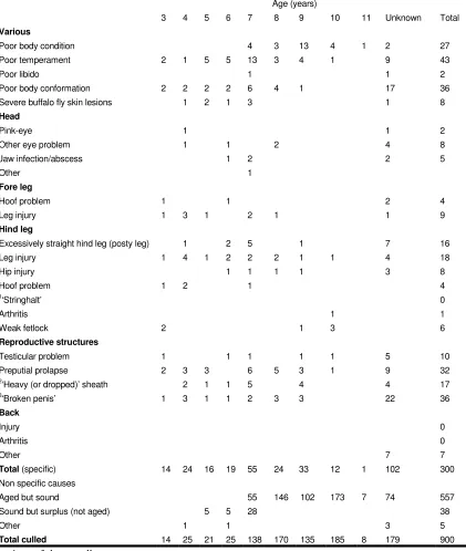
Barkly Tableland...49 Table 3.3. Age profile of bulls culled in 1998 for various abnormalities across
eight northern beef cattle properties. ...51 Table 3.4. Age structure of the bull population on each cattle property...52 Table 3.5; Age distribution of bulls culled with preputial prolapse from each
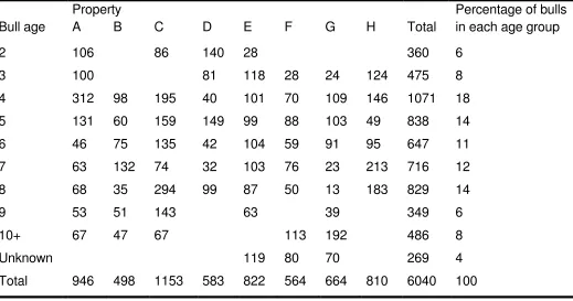
property during one year (1998)...54 Table 3.6; Summary of prevalence of preputial prolapse by breed and property. ...56 Table 3.7; Total numbers of bulls by age on all eight properties over a three year
period 1998-2000. ...57 Table 3.8; Total bull numbers on property A over the three years 1998-2000. ...58 Table 3.9; Age structure of all bulls (Brahmans) culled from property E over three
years. ...59 Table 3.10; Predicted frequency and SE of bulls culled for leg, reproductive and
other reasons in relation to age at culling. ...61 Table 3.11; Significance of age on culling for reproductive problems, leg problems
and preputial prolapse...62 Table 3.12; Frequency of different causes of reproductive problems. ...63 Table 4.1; Numbers of animals by horn status for classifications of sheath size,
preputial eversion and the caudal preputial muscle network. ...77 Table 4.2; Relationship between horn status, size of the caudal preputial muscle,
Table 4.3; Relationship between sheath length and preputial eversion. ...79 Table 4.4; Relationship between occurrence of preputial eversion and preputial
measurements...80 Table 4.5; Relationship between the size of the caudal preputial muscle network
and the occurrence of preputial eversion. ...80 Table 4.6; Mean, SD and normal range of various measurements taken in 40
Santa Gertrudis bulls aged 2-3 years...81 Table 5.1; Summary of the mean ( ± SD) preputial, penis and sheath
measurements from 32 Santa Gertrudis bulls affected with chronic
preputial prolapse...91 Table 5.2; Frequency of bull age and caudal preputial muscle network score
groups by horn status for bulls affected with chronic preputial
prolapse...92 Table 5.3; Comparison of means between preputial, penis and sheath
measurement from horned and polled bulls affected with chronic
preputial prolapse...93 Table 5.4; Summary of significant correlations between hot standard carcase
weight and penis and penis muscle measurements. ...94 Table 6.1; Relationship between length of preputial eversion and position of the
tip of the glans penis relative to position of the preputial orifice for individual bulls, and physical characteristics of the bulls that exhibited
eversion. ...104 Table 7.1; Individual bull data for weight, age, horn status, fat measurements,
sheath measurements and tail lift in relation to urination...122 Table 7.2;. Abdominal muscle thickness measurements (mm) on individual bulls...123 Table 7.3; Individual back and rump measurements and data from the correlation
between penis movement and preputial eversion. ...124 Table 7.4; Pearson correlations indicating significant relationships between
sheath depth, abdominal muscles thickness measurements, backline
Table 7.5; Probability-values from analyses of variance for horn status and ‘tail lift to urinate’ factors and sheath, abdominal muscle thickness
measurements, backline measurements and preputial eversion data. ...127 Table 7.6; Relationship between horn status and obliquus internus abdominis
thickness measurements. ...128 Table 7.7; Correlation coefficients between weight, age, and fat depth, and
sheath, abdominal muscle thickness measurements, backline
measurements and preputial eversion figures. ...129 Table 7.8; All significant relationships identified from simple correlation and
ANOVA analyses, factors identified through regression analyses that need to be accounted for in determining significance and significant relationships after accounting for these factors using multiple linear
regression analysis...130 Table 7.9; Correlations (with probability values) between fat depth measurements
Abbreviations
AI – artificial insemination g – gram
H - horns Kg - kilogram M – missing data mm – millimetre mth – month n – number
NS – not significant
P – probability (statistical figure) P – polled (bull)
R – correlation coefficient
R2 – coefficient of determination S – scurred
Chapter 1. Introduction
Preputial prolapse in bulls is a very obvious clinical abnormality. The welfare implications of sensitive preputial tissue continuously exposed and injured are evident to the general community as well as the cattle producer. The associated losses in production are also well understood by the cattle producers as bulls with untreated preputial prolapse cannot usually mate successfully with cows.
Affected bulls are usually culled when preputial prolapse is detected but losses in production have usually already occurred by this stage and the losses can be quite large under some management conditions (Wolfe et al. 1983). In the more extensive production areas the welfare of an affected bull can be compromised for some time before the condition is detected. If bulls that were more susceptible to preputial prolapse were able to be identified and removed from the breeding herds, welfare issues and production losses could be reduced.
To establish the causes, and the significance of each of the causes, of preputial prolapse in
Bos indicus and Bos indicus derived bulls, a longitudinal study is needed. To be effective, the longitudinal study would have to objectively measure the possible contributing factors to
preputial prolapse on a large number of Bos indicus and Bos indicus derived bulls and observe them over a long period of time (years) for development of preputial prolapse.
From the literature it was obvious that there was some confusion regarding the predisposing causes of preputial prolapse in Bos indicus and Bos indicus derived bulls so studies
determining the possible main predisposing causes were needed before a longitudinal study could be efficiently conducted. Much of the confusion in the literature was due to differences between Bos indicus and Bos taurus bulls in their susceptibility and mechanisms for
derived bulls and if these can be objectively measured, bulls could be removed from the breeding population before pathology develops.
The importance of preputial prolapse in these bulls in the extensive areas of northern Australia needs to be accurately determined. A survey involving large numbers of extensively managed
Bos indicus and Bos indicus derived bulls is to be conducted to determine the prevalence of
preputial prolapse in the beef industry in northern Australia. The survey results would allow cattle producers to balance their selection processes with genuine estimates of what proportion of Bos indicus and Bos indicus derived bulls are likely to develop preputial prolapse each year and what emphasis should be placed on selection against anatomical structures that may predispose bulls to preputial prolapse.
The anatomy of bulls affected with preputial prolapse and bulls not affected with preputial prolapse could be compared to determine the importance of the anatomy of reproductive structures in the development of preputial prolapse. Two studies are needed. An anatomical study is to be conducted on Bos indicus derived bulls that are not affected with preputial
prolapse to describe the anatomy of the external reproductive organs and associated structures of unaffected bulls. This study is to provide normal bull baseline measurements to compare with anatomical measurements of Bos indicus derived bulls affected with preputial prolapse. A companion study of the anatomy of reproductive organs and associated structures of Bos indicus derived bulls affected with preputial prolapse is to be conducted to identify anatomical factors that may predispose bulls to preputial prolapse.
Although not confirmed as a major cause of preputial prolapse in Bos indicus and Bos indicus
The causes of preputial eversion in Bos indicus and Bos indicus derived bulls are not well described in the literature and are often assumed to be similar to the causes of preputial eversion identified in Bos taurus bulls. Other, less obvious causes, may be affecting preputial eversion. Research in humans identified some abdominal muscles as important in the pelvic area functions such as micturition, defaecation and parturition (Getty 1975). Despite the difference in stance between humans and cattle, the literature stated that abdominal muscle function is similar between these species (Lansman and Robertson 1992). An ultrasound study is to be conducted to determine the association of the identified abdominal muscles with
preputial eversion in bulls utilising the newly developed technique for quantifying preputial eversion and penis position in bulls. If measurements of abdominal muscles could determine which bulls evert their prepuces more than others, these objective measurements could be utilised in initial bull selection to avoid bulls that are likely to excessively evert their prepuces, and may reduce the likelihood of the development of preputial problems in the selected bulls.
In summary, the objectives of this thesis are to:
• determine the importance of preputial prolapse to the Northern Australian beef industry;
• find anatomical differences in the external reproductive organs between normal Bos indicus derived bulls and Bos indicus derived bulls with preputial prolapse;
• confirm the relationship of the position of the penis in the sheath to preputial eversion;
Chapter 2.
Literature review: Factors affecting preputial function in bulls
Introduction
This thesis is specifically concerned with preputial function in Bos indicus and Bos indicus
derived bulls but the conditions of the prepuce are general and affect other genotypes. This review includes preputial function in these other groups for comparison. More work has been recorded in Bos taurus bulls in some aspects of preputial function and preputial problems. This work has been covered to give some direction to the studies in the Bos indicus and Bos indicus
derived bulls. Specific information on preputial function in Bos indicus bulls is limited and much of the information that is available is not recent.
Influence of bull fertility on overall herd fertility.
Reduced bull fertility affects overall cattle herd fertility (Benesch and Wright 1950). Factors affecting bull fertility may be congenital or acquired and may be temporary or permanent. Permanent loss of fertility is obviously a source of revenue loss but even temporary infertility may result in great loss due to wasted breeding time in any cattle industry and also a loss in milk yield in the dairy industry (Benesch and Wright 1950). Reported importance of the bull to herd fertility varied in the literature from bulls being responsible for 72% of herd fertility
Economic estimates of the importance of bull fertility are often presented in general terms and copulation failure has been described as economically devastating to cattle producers (Wolfe et al. 1983). Others described the loss of use of bulls as representing a major source of concern to breeders (Bellenger 1971). With the introduction of artificial insemination (AI), some authors noted that bull fertility has become even more important (Santamarina and Reece 1957). AI allows greater use of selected sires and reduced fertility of these sires would result in greater potential loss to the industry.
Causes of bull infertility can be subdivided into a number of categories, and analysis of records from Southern Africa suggests 76% are due to functional unsoundness (Smit 1994). Part of this functional unsoundness includes preputial problems in bulls. These are a common cause of impotence (Wolfe et al. 1983; Wolfe 1986) and occur frequently in the bovine (Frank 1959). This was reiterated by authors who stated that pathological changes in the genital system of the male in cattle are very common (Hungerford 1990) and that laceration complicated by prolapse of the prepuce is commonly encountered (Walker and Vaughan 1980).
Preputial prolapse may lead to mating difficulties (Lagos and Fitzhugh 1970) and it has been stated that the subsequent inability to copulate due to prolapse has forced the slaughter of many bulls (Walker 1966).
An accurate and repeatable method of predicting bull fertility could have an important economic impact on beef production. The ability to consistently select herd sires of high fertility could result in more cows calving early in the calving season, and thus producing more kilograms of calf weaned in beef herds (Smith et al. 1981).
Importance of Bos indicus and Bos indicus derived cattle in tropical/subtropical regions of the world
Bos indicus breeds play a dominant role in beef production in tropical and sub-tropical parts of the world (Swanepoel and Hoogenboezem 1993) because they are adapted to hot climates. To promote heat loss it has been suggested that the surface of skin is increased by greatly
developed dewlaps and, in many of these breeds, an excessively pendulous sheath (Hofmeyr 1987). To test the theory that excess skin may help the Bos indicus breeds cope with hot weather, some researchers removed the dewlap from a Bos indicus bull and found that it did not change his ability to resist high temperatures (USDA 1956). Despite being contrary to current thinking, in that study this led to the conclusion that the outsize Bos indicus dewlap has no influence on heat tolerance. Despite this finding in one bull, selection against excess skin (including the sheath) could be detrimental to the survivability of the cattle in some harsh environments if the excess skin is, in fact, important for survival in hot conditions.
The importance of utilising livestock breeds adapted to specific environments needs to be emphasised. This is particularly true for tropical environments where, in the absence of resources for substantial improvements of the production environment, the most viable and widely available option is the utilisation of adapted animal genetic resources. Bos indicus are essentially beef animals that survive under poor grazing conditions and are able to respond to better nutrition or feedlot management. These breeds have a long productive life and do not appear to be unduly troubled by ticks and other ectoparasites and exhibit considerable
tolerance to infectious keratoconjunctivitis (pink-eye), and squamous cell carcinoma of the eye. In addition these breeds are considerably tolerant of heat and gastrointestinal nematodes and have a relatively low incidence of calving problems. The international popularity of Bos indicus
Extent of bull fertility problems due to preputial problems
Preputial problems are one of the most common abnormal conditions affecting the ability of the male bovine to copulate (Walker 1970; Walker and Hull 1984; Memon et al. 1988), and are frequently observed in some breeds of cattle (Walker 1970). Preputial problems include traumatic injury to the prepuce that is most common under range breeding conditions (Walker and Vaughan 1980). Pearson (1972) has reported that even minor lesions of the prepuce may render a bull incapable of natural service and are frequently the cause of insurance disputes. Preputial prolapse adversely affects penis function because protrusion of the penis depends on free movement of the preputial mucosa and any fibrotic lesion in this tissue may seriously impede the ability of the penis to extend (Arthur et al. 1982).
Data obtained from some Australian abattoir studies has not confirmed how prevalent the preputial problems may be in Bos indicus derived breeds, and the breeds involved in these abattoir studies may partially explain the lack of agreement between studies. Pathological conditions of the prepuce were detected in 0.8% of 504 bulls surveyed at three Western Australian abattoirs over an 18 month period (Turnbull 1977). The mean age of the bulls (with histories provided) was 4.4 years. The survey included mostly Bos taurus breeds including Hereford (30%), Shorthorn (16%), Friesian (14.3%) and Angus (13.9%) bulls and the conditions detected were three abscesses and one diverticulum, which presented as a pocket in the
prepuce. Of 80 bulls culled through a NSW abattoir, none were culled for preputial problems. All were over 30 months old and the breeds were again mostly Bos taurus and included Hereford (32.5%), Poll Hereford (20%), Shorthorn (13.8%), Angus (15%), Santa Gertrudis (3.7%) and Devon (1.3%) (Bellenger 1971).
age and older but no supporting data were provided. As less than 2% of the bulls in the data set had Bos indicus content this only reflected the situation in the local Bos taurus breeds (Carroll
et al. 1963).
Similar results were seen by Chenoweth and Osborne (1978) and Chenoweth (1978) who examined 702, 16-31 month old bulls from three different Queensland properties. Prolapse of the prepuce was seen in 0.8% of the bulls. The breeds examined included Bos indicus and Bos
indicus derived (26%), Africander and Africander derived (26%) and Hereford or Hereford
derived (48%). The percentage found in the study may be a reflection of the age of the bulls examined and it was noted that total preputial abnormalities (including ulceration and abscess) was 9.5%.
In contrast there is a view that the economic importance of preputial problems is appreciable, considering the high cost of breeding bulls, the age at which the condition occurs, and the losses caused by the temporary cessation of service. For example, out of 368 bulls presented at a Colombian University clinic for abnormalities of the genital tract, 15.2% presented with preputial inflammation (Amaya Posada 1979). Without presenting supporting data, Amstutz (1981) stated that injuries to the prepuce were the most common lesions seen at the Purdue University Veterinary teaching clinic. This was contrasted by the fact that bull soundness examination records of two Southern Queensland veterinary practices showed that only 11 bulls (0.37%) with preputial problems were found from the total of 2996 bulls examined (Bertram et al. 1993). This may not be surprising as preputial problems are generally readily detected by producers and the bulls would not usually be submitted for a breeding examination. Thus the reported prevalence of preputial abnormalities is likely to be underestimated.
Vaughan (1980) found that deep to the epithelium of the visceral prepuce is a series of elastic lamellae that are continuous with those surrounding the penis. Lacerations or contusions that penetrate the epithelium and involve the elastic layers interfere with the normal unfolding action of the prepuce and thus prevent normal extension of the penis. Many authors suggest that normal preputial eversion does not interfere with copulation unless the everting portion becomes traumatised (Wheat 1951; Zemjanis 1970; Wolfe 1986).
Semen straw production from bulls affected with preputial problems may also be interrupted. Preputial eversion in bulls at AI centres may lead to an unhygienic situation, which may impact normal semen quality due to contamination with environmental pathogens (Klug et al. 1979). Semen production problems were seen by Shires and Evans (1978) in a six year old Poll Hereford bull but these were due to prolapse rather than eversion. In this bull, chronic preputial prolapse with phimosis resulted in intrapreputial ejaculation and grossly contaminated sperm. This sperm was unfit for freezing and storage. In a study in which prepuces of 60 Murrah buffalo were examined, (Rao et al. 1988) sheaths were classified as tight, medium, pendulous and pendulous with eversion. Classification criteria were not given but photos of an example of each classification were provided. Results showed that the classification of the sheath did not influence the semen quality. In this study, preputial eversion was not considered a problem for semen production in buffalos.
Historical perspective of the importance of sheath conformation.
Historically, in the beef industry, greater selection pressure has been placed on growth rate than on sheath conformation. To improve selection balance, Hoogenboezem and Swanepoel (1995) suggested that information on the relationship of growth rate and sheath area would be helpful. It has also been stated that there is a paucity of objective measurements describing the variation in sheath characteristics in tropically adapted breeds, particularly in relation to bull mating performance (Bertram et al. 1997).
area or its relationship to preweaning growth or body size, but most Zebu and derived breed associations discriminate against large and pendulous sheaths (Franke and Burnes 1985). Selective breeding against pendulous sheaths is meeting with some success in reducing the problem of sheath damage (Walker 1970). This is important because of the effects on mating ability, which are as significant as semen quality for fertility in the bull (Zemjanis 1970).
Bull external reproductive anatomy and function
Definitions
Although definitions by most authors are similar, there are some variations in descriptions, which cause confusion. A major problem in this field is that the prepuce is often called the sheath (Trotter and Lumb 1958). The two terms of sheath and prepuce are sometimes used interchangeably. One author who used the terms interchangeably defined the prepuce or sheath as a double invagination of skin which contains and covers the free portion of the penis when not erect and which covers the body of the penis behind the glans when the penis is erect (Roberts 1971). The terms, eversion and prolapse are also interchangeably used in the
literature.
Sheath
One definition is that the sheath (or external layer of the prepuce) extends from the scrotum to within five cm of the umbilicus where it is reflected ventrally and laterally forming the thick margin of the preputial orifice (Donaldson and Aubrey 1960). A more general definition has the sheath extending to the scrotum and the inguinal area (Walker and Hull 1984).
For this review the sheath is defined as the hair-covered skin appendage that supports and protects the penis along the ventral abdomen. It extends from the scrotum to the preputial orifice (Wolfe 1986).
Prepuce
acknowledged that differences in nomenclature do occur (Bellenger 1971). Other authors referred to the whole structure as the prepuce and the sheath as the external layer of the
prepuce. The internal layer or prepuce proper then extends backwards from the preputial orifice and turns forwards on the penis to the glans penis (Donaldson and Aubrey 1960; Getty 1975). Other researchers divided the prepuce into sections. After dissection of 10 penises from adult bulls one group divided the prepuce into penile and prepenile portions (Ashdown et al. 1968). They found the collagen fibres much denser in the penile than in the prepenile prepuce and the penile prepuce to be less extensible than the prepenile prepuce. Another author divided the prepuce up into the internal lamina (or lining membrane, parietal layer, or mucous membrane), and the visceral layer (Hofmeyr 1987). The internal lamina is covered by squamous epithelium and extends from the orifice to the fornix. Usually, with the penis at rest, this is two-thirds of the distance between the orifice and scrotum. At the fornix it is reflected onto the penis, forming the visceral layer.
Williams (1918) description of the prepuce is when the penis is in the resting position. The prepuce extends back from the mucosal attachment at the base of the glans and then forward to the position of the apex of the penis (when in resting position) on the parietal layer. The sheath extends from this position forward to the orifice. Although not easily identified in live bulls, this definition is based on the embryonic history of the two membranes. The preputial epithelium is delicate and easily abraded and the epithelium of the sheath is coarser and offers higher resistance. The preputial mucosa is even and smooth, and the sheath mucosa is thrown into wavy longitudinal rugae.
For this thesis, the prepuce has been defined as the hairless epithelium within the sheath that extends caudally from the preputial orifice, is reflected at the fornix, then extends cranially to attach at the base of the glans penis (Walker and Hull 1984).
Preputial eversion
Preputial prolapse
Preputial prolapse is a readily diagnosed condition but penile haematoma can be mistaken for prolapse of the prepuce, so careful examination is needed (Farquharson 1952). This confusion may occur because there is extensive swelling of the tissue along the sheath associated with rupture of the corpus cavernosum of the penis and the swelling may extend to the preputial orifice.
For this thesis preputial prolapse is defined as the protrusion of the prepuce with no tendency or ability to spontaneously retract it (Arthur 1964).
Anatomical Development
Large differences in rates of development of the penis and sheath are seen in bulls as demonstrated in a longitudinal study of five young bulls (three Jersey and two
Galloway/Ayrshire cross bulls; Ashdown 1960). At the age of four months, the penis in these bulls could not be exteriorised. Despite all five animals repeatedly mounting each other while quite young, the penis was not protruded until they were approaching sexual maturity at eight to nine months. A similar study of another five calves and ten foetuses found that the wall of the penis portion of the prepuce was adherent to the free end of the penis during foetal and early postnatal life (Ashdown 1960). In a more extensive macroscopic and microscopic examination of 103 penises from bull calves and young bulls it was found that in the first week of life no preputial cavity exists between the glans penis and the prepuce because it is filled with irregularly arranged epidermal cells (the ectodermal lamella; Abdel-Raouf 1960).
surfaces of penis and internal lamina. The frenulum, a fine band of connective tissue, connects the penis and prepuce ventrally where the lamella is incomplete (Hofmeyr 1987).
By cleavage of the lamella two stratified squamous epithelial layers develop, one lines the prepuce and the other covers the glans penis. It was found that the process of cleavage starts at the age of four weeks and is finished sometime after the age of 32 weeks (Abdel-Raouf 1960). A mechanism for this was presented as being due to male hormonal action in late foetal life. With this action the lamella is split into keratinised epithelial layers of penis and prepuce (Hofmeyr 1987). Variation on the age of commencement of separation was found with others stating that at about four months the separation commences and is usually complete at nine months (Hofmeyr 1987). In breeds that show early sexually maturity, this process is concluded much sooner. The explanation of the timing of this separation was simplified by stating that keratinisation separated the penis from the prepuce in later life and proceeded in the ectodermal lamella (Ashdown 1960).
Parts of this development are comparable to other species and one group found in their work on 110 rams that the age at which the prepuce became completely separated from the glans penis was variable and associated with the general rate of growth (Watson et al. 1956). Although a study of 277 ram lambs found that with increasing age the glans penis is progressively freed (Johnstone 1948).
Musculature
An understanding of what is known and not known about the anatomy of the sheath, prepuce and penis is needed before any anatomical study in this area should commence. The literature can provide baseline information and can be compared with any study observations.
Preputial muscles
muscles (Getty 1975; Walker and Vaughan 1980; Dyce et al. 1987) and are stated to be present in both sexes (Trotter and Lumb 1958) or at least a homologue of the cranial preputial muscle is present in the cow (Getty 1975). These muscles are subject to a good deal of
variation (Getty 1975) and may partly account for a comment in an earlier report that published descriptions of the structure and function of the cranial and caudal preputial muscles of the bull are inaccurate (Aubrey and Butterfield 1970). Many anatomical studies of these muscles did not identify bull breed and this may explain some of the variation in description between studies.
Cranial (protractor) preputial muscle Origin
The cranial preputial muscles originate from three distinct areas of the ventral abdominal wall: (a) the deep fascia, near the midline at the level of the xiphisternum, (b) the hypodermis or the superficial fascia overlying the ventral edge of the cutaneous trunci muscle and; (c) as slips from the most caudal part of the cutaneous trunci muscle (Ashdown and Pearson 1973). Other authors have provided a more simplified description as this muscle group arising from the xiphoid region (Dyce et al. 1987). Others describe the muscle group as originating from the fascia in the region of the xiphoid process of the sternum with each muscle having a cranial part about three times as wide as a caudal part (Aubrey and Butterfield 1970).
The muscles have been described as two flat bands, five to six cm in width, which arise close together in the xiphoid region, about 20 cm cranial to the preputial orifice (Getty 1975). Other descriptions include the muscles originating from the abdominal tunic anterior to the umbilicus (Trotter and Lumb 1958). Further descriptions have the muscles originating near the umbilicus and travelling posteriorly as flat bands (Walker and Vaughan 1980).
Insertion
described the muscles as terminating around the preputial orifice by attaching to the subcutaneous tissue of the sheath in a circumscribing manner with the muscles inserting beside and behind the preputial opening (Dyce etal. 1987). This was described as a loop around the prepuce at the start of the pendulous part (Ashdown and Pearson 1973) as these muscles insert into the outer layer of the wall of the prepuce dorsal to the orifice of the sheath. This loop was formed by the muscles as they diverged caudally leaving free the umbilicus and an area about 3.5 cm wide; they then unite caudal to the preputial orifice (Getty 1975).
Function
Stimulation of the motor nerves to the preputial muscles showed that when the cranial preputial muscles contract, the pendulous part of the sheath is pulled dorsally and cranially. The
pendulous part of the sheath is elevated and the orifice is more or less constricted. The effectiveness of the cranial preputial muscles of the sheath in closing the orifice varies considerably in different bulls (Ashdown and Pearson 1973). Other authors provided various combinations of this action and reported that the muscle: draws the pendant part of the prepuce forward and upward and helps to constrict the orifice (Dyce et al. 1987); draws the prepuce forward (Getty 1975); controls the orifice of the sheath (Ashdown and Hancock 1968); raises the preputial orifice cranio-dorsally (Aubrey and Butterfield 1970); and raises and constricts the preputial orifice (Wolfe 1986). It also pulls the sheath forward and constricts the orifice of the prepuce. This constrictive action is most important since it serves as a barrier to foreign
material entering the preputial cavity. Abnormal constriction of this muscle can prevent normal extension of the penis (Walker and Vaughan 1980). During mating, these muscles pull the external layer of the prepuce and the orifice backward prior to copulation, thus exposing a greater length of the penis (Trotter and Lumb 1958).
Caudal (retractor) preputial muscle
their absence may be associated with the development of habitual preputial eversion. When the caudal preputial muscles are absent, preputial retraction is a passive act and occurs
secondarily to penis retraction by the retractor penis muscles. Because of this, polled bulls commonly evert the prepuce when relaxed (Wolfe 1986). Other authors also found that the size and presence of these muscles varies considerably, from being completely absent to partially developed in many polled bulls. Bulls that habitually expose the prepuce have been found to have no or incomplete development of these muscles (Walker 1980).
Origin
The caudal preputial muscles each originate from two separate sites. The lateral origin is from the dense layer of fascia that leaves the lateral edge of the external inguinal ring and runs into the scrotum to form the scrotal fascia. The medial origin is from the medial aspect of the scrotal septum, near its point of origin from the yellow elastic lamina of the abdominal wall (Ashdown and Pearson 1973). Many researchers have simplified this and stated that the muscles: pass from the region of the prepubic tendon (Aubrey and Butterfield 1970); originate from the abdominal tunic near the external inguinal ring (Trotter and Lumb 1958); arise from the abdominal tunic (Wolfe 1986); originate in the inguinal region lateral to the spermatic cord (Walker and Vaughan 1980); and arise in the inguinal region (Getty 1975).
Insertion
A variety of descriptions were provided which may reflect the variable development within and between breeds. Authors have stated that the caudal preputial muscles:
• insert into the outer layer of the wall of the prepuce (Ashdown and Pearson 1973)
• insert to several inches of the lateral surface of the external layer of the prepuce (Trotter and Lumb 1958)
• insert into the insertion of the cranial preputial muscles (Wolfe 1986)
• pass anteriorly along the abdominal wall dorsal to the penis to their point of insertion into the deeper layers of the elastic lamellae of the prepuce (Walker and Vaughan 1980)
• converge on the cranial part of the prepuce (Getty 1975).
A method to identify these anatomical differences in live animals may help our understanding of preputial abnormalities and may assist detection of animals that may develop these
abnormalities.
Function
Actions of the caudal preputial muscle remain unclear and their function is described as being of uncertain importance (Dyce et al. 1987). Dissection of tissues from seven bulls from breeds that do not normally evert showed that, on maximum protrusion of the non-erect penis, the insertion of the caudal muscles into the outer concentric layer of the prepuce moves very little (Ashdown and Pearson 1973). The inner parts of the prepuce slide out while the loosely
connected outer layer moves little. When the caudal preputial muscles are stimulated, the outer layer of the prepuce and the surrounding fascia are pulled caudally but there is little effect on the penis or on the inner layers of the prepuce. Under these conditions, the outer concentric layer of the prepuce appears not to participate in eversion to any significant degree, however protrusion of the erect penis was not studied at this time. In most bulls the muscle is large and powerful and might assist in penis retraction. Where present it was of considerable size having a belly of about 2.5 by 1.0 cm and an extensive insertion over some 10 cm (Long and Hignett 1970) and it seems inconceivable that it should play no role in the function of the prepuce or penis (Ashdown and Pearson 1973).
of the now everted preputial lining, which is a considerable distance along the extended shaft of the penis (Aubrey and Butterfield 1970). This should indicate that the muscle is involved in movement of the prepuce. Other authors supported the muscle involvement and stated that these muscles draw the prepuce caudally (Getty 1975) and returned the prepuce to its original position after service (Trotter and Lumb 1958).
Retractor penis muscles
The retractor penis muscles are strong muscles that retract the penis (Trotter and Lumb 1958). If the retractor penis muscles are short (due to hypoplasia in the congenital condition) or
contracted (due to fibrosis), the muscles feel like distinct cords behind the scrotum (Hofmeyr 1967). However, the tone of this muscle was found to be not uniform throughout its length (Ashdown and Pearson 1973).
Origin
Each retractor penis muscle consists of two bands and is flat at its origin from the transverse processes of the first coccygeal vertebra (Trotter and Lumb 1958; Ashdown and Pearson 1973) or arises from the caudal vertebrae as others simply stated (Dyce et al. 1987). Both groups of authors may be correct as many muscles that are flat at their origins have wide attachments through the local fascia.
Insertion
This muscle group inserts on the penis at, and anterior to, the ventral bend of the sigmoid flexure (Trotter and Lumb 1958; Ashdown and Pearson 1973). Some fibres attach here but most proceed to a more distal and diffuse insertion (Dyce et al. 1987). The insertion point for this muscle group has also been described as ending in the outer layers of the peripenile fascia, 12-15cm caudal to the free end of the penis (Hofmeyr 1987).
Function
and stretches the sheath. These muscles also maintain the resting position of the penis after service (Ashdown and Pearson 1973).
Vasculature to the sheath and prepuce
Most authors have described the blood supply to the sheath and prepuce as arising from the external pudendal artery (Ashdown and Hancock 1968; Roberts 1971; Hofmeyr 1987). Other descriptions also mention the dorsal artery of the penis (Trotter and Lumb 1958; Hofmeyr 1987) and the subcutaneous abdominal artery (Trotter and Lumb 1958). Ashdown (1958) summarised the vasculature as the main artery of the sheath being the unpaired recurrent artery which runs along the dorsal surface. The recurrent artery is a branch of the caudal superficial epigastric artery, with small branches from the paired dorsal arteries of the penis also supplying the sheath.
A summary of the venous drainage of the sheath area has been given by Ashdown (1958). The large branched ventral vein of the sheath anastomoses with the dorsal veins of the penis and drains into a venous plexus situated near the orifice. This plexus is itself drained by one or both subcutaneous abdominal veins. The general pattern of circulation in the bovine sheath is that blood reaches the sheath flowing back along the dorsal surface in the main artery of the sheath (the recurrent artery) and is collected by the main vein (the ventral vein of the sheath). The ventral vein runs forwards to the plexus of veins at the orifice (Ashdown 1958).
Blood supply to the penis.
the internal pudic artery: the dorsal artery of the penis, the artery of the corpus cavernosum, and the artery to the bulb. This was also recorded in cattle (Getty 1975).
Injected specimens revealed the important venous channels in the penis as: the superficial dorsal vein, the deep dorsal vein, the venae profundae, the venae bulbi urethrae and the circumflex veins (Deysach 1939). This information was unearthed from a range of species and was not limited to cattle.
Innervation to the external reproduction organs
As with many anatomical studies, descriptive differences have been recorded. A study by Larson and Kitchell (1958) summarised the nerve supply to the external reproduction area by noting that the central origin of nerves concerned with supplying the external genitalia can be divided into cranial, middle, and caudal parts. The nerve supply of this area is important as damage can impair the bull's ability to copulate (Beckett et al. 1978). A simple version of the innervation is that the pelvic plexus supplies sympathetic and parasympathetic fibres to the pelvic genitalia and to the penis (Ashdown and Hancock 1968).
The preputial muscles (cranial and caudal) receive their motor nerve supply from branches of the lateral thoracic nerve (Larson and Kitchell 1958; Larson et al. 1961; Hofmeyr 1987). This nerve originates from the ventral branches of C8, Th1 and Th2 (Larson et al. 1961), but was also described as arising from the brachial plexus in the axilla (Larson and Kitchell 1958; Ashdown and Pearson 1973; Hofmeyr 1987). It has also been stated that ventral branches of several thoracic and lumbar spinal nerves supply the preputial muscles (Trotter and Lumb 1958), further confirming the varied information on the innervation of the anatomy of the genitalia of the bull.
muscle of the penis is also innervated proximally by the terminal part of the caudal rectal nerve (Hofmeyr 1987). A simplified description is that sacral nerves supply motor fibres to the muscles of the penis (Ashdown and Hancock 1968).
The sensory nerve supply to the body and glans of the penis are the dorsal nerves of the penis (Larson and Kitchell 1958; Beckett et al. 1978; Hofmeyr 1987) that originate from the pudendal nerves at the ventral aspect of the ischial arch (Beckett et al. 1978). This supply was described as sacral (Ashdown and Hancock 1968) and as sympathetic fibres of the pudic nerve (Trotter and Lumb 1958). Bilateral dorsal penis neurectomy abolished the ability of the bull to locate the vagina during service attempts, and if the vagina was accidentally located, the bulls were still unable to ejaculate. Unilateral neurectomy did not abolish the ability to copulate. This suggests that in cases of trauma, one nerve could be severed without abolishing the bull's ability to copulate (Beckett et al. 1978). The sensory nerve supply to the prepuce includes the cranial preputial (rising from the ventromedial branches of T13, L1 and L2), the middle preputial (from the caudal branch of the inguinal nerve) and the caudal preputial nerves (from the superficial perineal branch of the pudendal nerve) (Larson and Kitchell 1958; Hofmeyr 1987).
Methods of defining the conformation of external reproductive structures in the bull
This thesis aimed to confirm some of the causes and the significance of the causes of preputial prolapse and preputial eversion in Bos indicus and Bos indicus derived bulls. To achieve this, the possible predisposing causes needed to be identified in the literature. Any suggested predisposing structures could be objectively measured and studied in relation to preputial eversion or preputial prolapse in Bos indicus and Bos indicus derived bulls.
External reproductive structures in the bull have been objectively measured by many authors with a view to defining the conformation of the bull and relating these measurements to
reproductive performance. For example, a study by Lagos and Fitzhugh (1970) showed that all the sheath measurements observed except eversion score tended to increase with age
range of measurements used by authors to objectively measure the bull external reproductive organs and associated structures. Such a variety of measurement techniques in the literature suggests that the best universal measurements that are related to reproductive problems have not yet been found. Other possibilities should therefore be investigated.
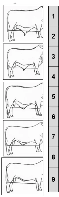
In previous studies sheath area was calculated by photographing each animal in front of a grid of known measurements from a standard distance (Franke and Burnes 1985; Hoogenboezem and Swanepoel 1995). Some authors have used a more subjective measurement (a sheath score) to estimate sheath area (Bertram et al. 1997). This score was assessed using the recording format used in the Australian national BREEDPLAN validation project where 1 = an extreme pendulous sheath to 9 = an excessively trim sheath. Sheath size and shape of sheath are also classified as part of the Brahdex evaluation system in South Africa (Swanepoel and Hoogenboezem 1993) although details of the method were not included in the publication.
Sheath depth has been defined as the vertical distance from the edge of the sheath at the preputial orifice to the abdominal wall (Bertram et al. 1997; McGowan et al. 2002). Other groups have described this as sheath length (Lagos and Fitzhugh 1970; Swanepoel and
Hoogenboezem 1993) or prepuce length (Smith et al. 1981; McGowan et al. 1998). A lot of confusion exists because the term sheath length is described as the distance from the front of the scrotum to the preputial orifice (Smith et al. 1981) which is a completely different
measurement to the others.
As part of the Brahdex evaluation system in South Africa, sheath conformation is classified on a ten point scale (Swanepoel and Hoogenboezem 1993). Seven criteria were used to develop this system and these included the vertical distance from ground to the orifice and the shape of the sheath including navel fold.
slaughtered bulls and the breed difference was significant. Of the Bos taurus breeds examined, polled breeds (Poll Hereford, Poll Shorthorn and Angus) had greater mean prepuce length than horned Hereford and Shorthorn although the differences were not statistically significant in the study. This prepuce measurement was from the tip of the glans penis to the preputial orifice with the penis fully extruded after slaughter. Another method of measuring prepuce length was to determine the length of the epithelium by the use of a probe inserted to the reflection (Long and Hignett 1970; Van Den Berg 1984). Other authors also measured the length of the penile prepuce and total prepuce but the technique was not recorded (Bellenger 1969).
The preputial orifice has been defined to be located about 5cm caudal to the umbilicus and is also only large enough to admit a finger readily (Getty 1975). However, others have quoted an average measurement of 2-4 cm in diameter, although no breed information was given
(Roberts 1971). In separate studies, the internal diameter of the preputial orifice was measured using callipers with a subjectively determined degree of tension against the internal wall (Lagos and Fitzhugh 1970). The external circumference of the sheath at the preputial orifice was measured along the hairline around the preputial orifice. Internal diameter and external
circumference should be related as the diameter and circumference of a circle but base breed differences were found which may have been due to breed differences in hide thickness. No confirmation of this possibility was reported. A similar measurement of the half-circumference of the preputial orifice taken after slaughter with internal callipers was reported (Long and Hignett 1970). Also as part of the Brahdex evaluation system in South Africa orifice diameter was measured (Swanepoel and Hoogenboezem 1993). Circumference of the orifice was also measured with dividers (Van Den Berg 1984).
Preputial eversion at rest was scored by Lagos and Fitzhugh (1970). Bos indicus derived bulls were placed in groups of 35 to 40 and allowed to stand at rest. They were continually observed for 1 hour and classified according to the estimated extent of exposed prepuce. Bulls were not scored when they were urinating as this changed the scoring. The 4 classes were; no
each bull being used in this analysis. They used this score in their analyses and found that the eversion score was related to the sheath depth.
The penis has also been measured in mature bulls and the total length of the flaccid penis was found to vary from about 90 cm, particularly in Bos taurus breeds, to about 110 cm in Bos indicus breeds or crosses (Hofmeyr 1987). Although both Bos taurus and Bos indicus bulls develop preputial prolapse, the difference in length of the penis may still be important as the mechanisms for preputial prolapse may vary between the breeds.
Other measurements included umbilical cord or navel thickness score which was assessed on a scale of 1 = approximately 0.5 cm to 5 = 3.0 cm (Bertram et al. 1997). The 'rosette' or inverted fold of skin in the sheath at the external interface of the umbilical cord was recorded as 0 = absent, 1 = small and 3 = large (an inverted semi circle about 5cm diameter) (Bertram et al.
1997). These were measured as they are closely associated with the sheath and their size may be an indicator of preputial problems.
Sequence of preputial abnormalities
Preputial pathology presents as a variety of clinical conditions due to varying aetiology and pathogenesis (Wolfe 1986). Conditions seen in a study of 172 cases of preputial injuries
included preputial lacerations, abscessation, and stricture with phimosis and preputial prolapse with preputial haematoma (Memon et al. 1988).
Other authors had similar descriptions and Walker and Vaughan (1980) stated that the
pathogenesis of preputial abnormalities is insidious and self aggravating. Small abrasions of the prepuce cause oedema of the prepuce that predisposes to prolapse with subsequent bruising and laceration (Walker and Vaughan 1980; Hofmeyr 1987). This leads to erosions,
abscessation and fibrosis (Walker and Vaughan 1980).
During mating, young bulls may repeatedly mate females in oestrus until the penis and prepuce becomes erythematous and excoriated, creating a traumatic balanoposthitis which may lead to prolapse (Wolfe 1986). In one retrospective study, records on 368 bulls with genital conditions were examined to determine cause and it was determined that the age at which inflammation of the prepuce occurred was during the most active period of breeding activity (Amaya Posada 1979). Lacerations at the reflection of the prepuce onto the penis generally occur during
copulation and some have stated that this injury is seen in hard thrusting young bulls (Rice 1987), which may lead to further problems in high libido bulls that continue to breed and exacerbate the problem.
One sequence observed started with posthitis and ulceration of the prepuce. Infection could then enter the traumatised epithelium leading to inflammation and prolapse. This area is then exposed to further trauma (Donaldson and Aubrey 1960). Other work indicated that laceration with phimosis is the most common type of preputial laceration (Walker and Vaughan 1980).
Other authors stated that phimosis may be congenital or acquired, the latter being more
common. Adhesions within the sheath can also be congenital or acquired and simple adhesions can be severed but multiple adhesions are usually difficult to treat (Gibbons 1956).
Penis and preputial abnormalities may be linked and this is seen when there are successive contractions of the ischiocavernous muscle after tearing the tunica albuginea of the penis forcing blood into the subcutaneous areas forming a haematoma and often causing a
secondary prolapse of the prepuce. Some authors stated that this secondary prolapse is seen more commonly in polled bulls that have poorly developed caudal preputial muscles (Walker and Vaughan 1980). Some highlighted the importance of this secondary problem and stated that haematomas in the region of the penis are exceedingly common (Hofmeyr 1987).
Factors predisposing to preputial prolapse
Breed differences in preputial prolapse.
Studies to date have indicated that breed of bull may influence occurrence of preputial prolapse, indicating that heritability plays a role in the aetiology of abnormalities of the sheath or the associated predisposing mechanisms (Venter and Maree 1978; Memon et al. 1988). The effect of breed is also a contentious issue and findings and ideas vary widely. Lagos and Fitzhugh (1970) highlighted that the apparent variation in the predisposition to prolapse observed among breeds resulted from surveys or experiments that were not designed to eliminate the influences of extraneous sources of variation. More quality research is needed to clarify these breed differences.
The prevalence of preputial problems reported in some breeds only reflects the proportion of that breed in that area (Desrochers et al. 1995). Reference population data are required to determine the significance of breed involvement. Breeds that have been reported to present with preputial problems include: Brahman (Mosaheb et al. 1973; Memon et al. 1988; Bruner and Van Camp 1992), Brangus (Memon et al. 1988; Baxter et al. 1989; Desrochers et al. 1995), Beefmaster (Memon et al. 1988; Baxter et al. 1989), Santa Gertrudis (Lenert 1956; Donaldson and Aubrey 1960; Larson and Bellenger 1971), Angus (Wheat 1951; Milne 1954; Baxter et al.
demonstrates the extent of the issue and confirms the importance of any research in this condition that may clarify the differences in breed incidence.
Bos taurus type bulls were more commonly affected with preputial injury (82.5%) than Bos
indicus bulls (18%) in a study of 51 cases seen in a US veterinary hospital (Desrochers et al. 1995). Individual breeds most affected were; Angus (45%), Simmental (12%) and Brangus (10%). These results are countered by the fact that the predominant breeds seen were Angus (27%), Hereford (22%) and Simmental (17%). It was further stated that the proportion of Bos
taurus breeds (82%) to Bos indicus breeds (18%) affected was in proportion to the breed
population in Kansas. So although the incidence of preputial prolapse varied between individual breeds in this area, Bos indicus and Bos taurus bulls were proportionally affected.
Many researchers presented only population estimates of, rather than specific, total numbers of bulls affected with preputial prolapse from the different breeds. The number of bulls with
preputial injuries from the Brangus, Brahman and Beefmaster breeds was higher than in the nine other breeds included in a survey by Memon et al. (1988). They clarified this by stating that this could be an indication of the breeds' proportion of the total cattle population in the Louisiana and Oklahoma areas as the population data was not available.
Studies showing no difference in incidence of preputial injuries amongst breeds were limited. Most of the 25 head with preputial prolapse obtained from a sample of 550 bulls at an abattoir were Brahman or Brahman derived bulls (Mosaheb et al. 1973). Breed break-up in the total number was not presented and may have given a clearer picture of the breed influence. The cattle in this study came from North Queensland and the Northern Territory where an
increasing proportion of the cattle may be expected to be Brahman infused in 1973 when the paper was written.
Over a four year period in Townsville, cases of posthitis were seen by Donaldson and Aubrey (1960) in 19 Santa Gertrudis bulls, two Santa Gertrudis derived bulls, three Zebu derived bulls, one Hereford bull and one Zebu steer. At the time of the study the population of Santa
Shorthorn and Hereford. Although small numbers were involved and no statistical analysis was presented, the author referred to, but did not directly state, a high incidence in Santa Gertrudis bulls.
Cases of surgical conditions of the prepuce were seen by Hofmeyr (1968) in four Herefords, two Africanders, three Santa Gertrudis, 13 Brahmans, one Jersey and one Friesian. Although not statistically analysed, Africanders were very popular in South Africa in 1963, but Brahmans were present in relatively small numbers and Santa Gertrudis were uncommon (Hofmeyr 1968). After considering the breed proportions and relating this to the proportions presented with surgical conditions of the sheath, Hofmeyr (1968) felt the Brahman breed had a higher incidence of preputial injury that required surgical correction, which indicated a possible weakness of the breed when managed in South Africa, due to harsher conditions. Although bred to thrive under similar environmental conditions as the Africander, the pendulous nature of the preputial skin in Brahman breeds has been maintained. Hofmeyr (1968) acknowledged that the difference between the two breeds may well lie in the fact that the imported representatives of the Brahman breed were obtained from a population that had been selected in a relatively protected physical environment.
Breed differences in preputial eversion.
Preputial eversion is important because it has been anecdotally linked to preputial injuries and most studies show breed differences in preputial eversion tendencies. Preputial eversion has been noted in Brahman (Johnson and Williams 1968; Supple-Kane 1969; Long and Rodriguez Dubra 1972), Brahman derived (Gibbons 1956; Supple-Kane 1969; Long 1969), polled breeds (Monke 1976; Klug et al. 1979; Ott 1986), Hereford (Long 1969) and in horned Friesians (Supple-Kane 1969). Although no population data were given, Supple-Kane (1969) stated that preputial eversion is also very common in the Sahiwal and the Boran in Kenya.