(UDC: 616-001.5-073:537.874.4)
Estimation of wave dispersion and attenuation for the assessment of healing
bones
V. Potsika1, V. Protopappas2, M. Vavva2, K. Raum3, D. Rohrbach3, D. Polyzos2, D.I. Fotiadis1
1
Unit of Medical Technology and Intelligent Information Systems, University of Ioannina, GR 45110 Ioannina, Greece
[email protected], [email protected] 2
Department of Mechanical Engineering and Aeronautics, University of Patras, GR 26500 Patras, Greece
[email protected], [email protected], [email protected] 3
Julius Wolff Institute, Berlin-Brandenburg School for Regenerative Therapies, Charité-Universitätsmedizin Berlin, Augustenburger Platz 1, 13353 Berlin, Germany
[email protected], [email protected]
Abstract
Quantitative ultrasound has recently gained significant interest as a diagnostic tool of the bone healing process. Several multiple scattering theories have been proposed for the investigation of wave scattering in nonhomogeneous media, which however cannot provide realistic dispersion and attenuation estimations for different particle types and volume concentrations. In this study, we use an iterative effective medium approximation (IEMA) (Aggelis et al. 2004) to carry out wave dispersion and attenuation predictions in the callus region at different healing stages. The geometry and the material properties are derived from scanning acoustic microscopy images (SAM) of sheep healing tibia obtained at the third, sixth and ninth postoperative week (Anderson et al. 2008). The callus is assumed to be a composite medium consisted of blood and osseous tissue. The average particle diameters and the volume concentration were 350μm and 44.75% in week 3, 200μm and 38.67% in week 6, 120μm and 22.67% in week 9, respectively. Wave dispersion and attenuation are estimated for frequencies from 24 – 1200 kHz. Group velocity was found to decrease with increasing frequency, while the attenuation coefficient was found to increase in the examined frequency range. The results indicate that the scattering effects are more pronounced in the early healing stages. In conclusion, IEMA can provide reasonable predictions and could be thus exploited for bone healing assessment.
1. Introduction
medium is assumed to be immersed into an infinitely extended effective medium. The frequency dependent wave velocity and attenuation coefficient are calculated by using self-consistent expressions most of which use scattering parameters derived from the solution of the single scattering problem. However, many of these theories are not able to provide reasonable wave dispersion and attenuation predictions for all types of suspensions and a wide range of particle concentrations and wavenumbers.
To this end, an iterative effective medium approximation (IEMA) was presented in(Aggelis
et al. 2004; Laugier et al. 2011), aiming at the accurate evaluation of wave dispersion and attenuation in particulate composites, particle suspensions and emulsions, as well. The effectiveness of the methodology to provide reasonable predictions in composite materials including particles with volume concentrations up to 50% was demonstrated by comparing the numerical results of IEMA with experimental findings.
In this work, IEMA is used to perform wave dispersion and attenuation estimations in the callus region of healing bones at successive healing stages. Callus was assumed to be a porous, composite medium consisting of blood and osseous tissue. The material properties and the geometry of the callus were derived using serial scanning acoustic microscopy (SAM) images representing the third, sixth, ninth postoperative week (Anderson et al. 2008). Numerical calculations of the phase velocity and the attenuation coefficient are conducted in the frequency range from 24 – 1200 kHz. The numerical results show that the scattering effects are more pronounced in the early healing stages indicating that IEMA could provide supplementary information for the assessment of the healing process.
2. Materials and Methods
2.1. Scanning acoustic microscopy images
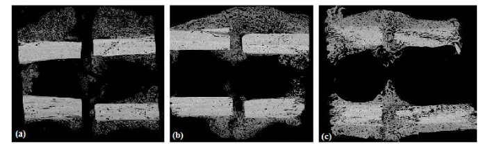
The SAM images illustrated in Fig. 1 were obtained from the right tibia of female Merino sheep and depict embedded longitudinal sections of a 3-mm osteotomy (Anderson et al. 2008). SAM is a non-invasive and non-destructive technique which uses ultrasound waves to detect changes in acoustic impedances occurring at the microstructural level and has been extensively used to investigate the elastic alterations of mineralized callus and cortical tissues (Anderson et al. 2008; Protopappas et al. 2007). Each SAM image represents a healing stage after three, six and nine weeks of consolidation. SAM measurements were derived using a spherically focused 50 MHz
Fig. 1. SAM images representing: a) 3 weeks, b) 6 weeks and c) 9 weeks after the osteotomy.
2.2. The IEMA for particle suspensions
The propagation of a plane wave in nonhomogeneous media can be considered as a sum of a mean wave travelling in the medium with the dynamic effective properties of the composite and fluctuating waves induced by the multiple scattering of the mean wave. Under this consideration a complicated self-consistent multiple scattering condition can be applied to estimate the dynamic effective properties. In order to simplify the calculations, a simple self-consistent condition for composite media was proposed in (Kim et al. 1995):
1 2
(1) ˆ ˆ ˆ (2) ˆ ˆ ˆ
( ; , ) ( ; , )0,
d d
n g d k k n g d k k
(1)
wheren n1
,
2are the volume concentrations of the scatterers and the matrix, respectively,k
ˆ
is thedirection in which a dˆ-polarized plane mean wave propagates andg(1),g(2)are the forward
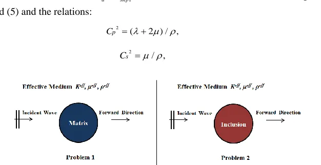
scattering amplitudes derived from the solution of the scattering problems 1 and 2, respectively, as illustrated in Fig. 2. The mean wave is both dispersive and attenuated. The complex
wavenumber kdeff( ) is defined as:
( ), ( ) ( ) d d d eff eff eff i C
k
(2)
where Cdeff( ) , deff( ) are the effective and frequency dependent phase velocity and attenuation
coefficient, respectively, of a longitudinal (d ≡ P) or transverse (d ≡ S) mean wave propagating
with circular frequency ω. In order to calculate Cdeff( ) and deff( ) the nonhomogeneous
medium is replaced by an elastic homogeneous and isotropic material with bulk and shear moduli
eff
K andeff , respectively, calculated using the static mixture model of Christensen (Raum K et
al. 2006):
1 1 2 2 2
2
2 1 2 2 2
4 ( - )( ) 3 , 4 ( - ) ( ) 3
eff n K K K
n K K K
K K (3) 2 2 2
) 2 ) 0
( (
eff eff
A B C
(4)
withA B C, , being functions of 1
,
2,
n1 given in (Raum et al. 2006), while 1, 2 are indices2
/ ,
s
C
(7)
Fig. 2. A plane mean wave propagating in the effective medium and being scattered by: a) a matrix inclusion in problem 1, and b) a particle inclusion in problem 2.
where Cp and Cs are the longitudinal and the shear velocity of the propagated wave, respectively. Next, utilizing the material properties calculated in the first step, we proceed to the second step where the scattering problems 1 and 2, illustrated in Fig. 2, are solved in order to
evaluate the forward scattering amplitudes
(1) (2)
, .
g g
. Subsequently, the scattering amplitudes can be estimated according to the following equation:
1 2
(1) (2)
ˆ ˆ ˆ ˆ ˆ ˆ ˆ ˆ ˆ
( ; , ) ( ; , ) ( ; , ,
d d d
g d k k n g d k k n g d k k)
(8)
and making use of the dispersion relation proposed in (Machado et al. 2010), we can estimate the new effective wavenumber of the mean wave as:
1
1 3
2
1
ˆ ˆ ˆ
3 ( ; , )
,
( )
( ) ( ) d
d step d step
d step
eff eff
eff n g
k
k k
d k k
(9)
where α is the radius of a volume equivalent to the particle sphere. The new complex density
2
(eff)step is calculated based on the (kdeff)step2 and the Eqs. (3), (4), (6), (7). The second step is
repeated with the material properties (3), (4) and the new density(eff)step2 until the
self-consistent condition (Eq. (1)) to be satisfied. Thus, using the Eqs. (3), (9) the frequency dependent, effective phase velocity and attenuation coefficient of the mean wave can be calculated.
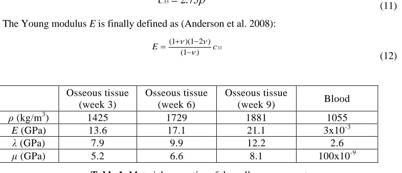
2.3. Material Properties
The material properties of each pixel composing the osseous tissues in Fig. 1 were derived using
empirical equations. The density ρ was calculated using the equation(Tsinopoulos et al. 2000):
2.83
1.02
Z (10)
Then, the elastic constant in the axial direction c33 is calculated via the equation (Tsinopoulos et al. 2000):
3.99 332.75
c
(11)
The Young modulus E is finally defined as (Anderson et al. 2008):
33
(1 )(1 2 ) (1 )
E c
(12)
Osseous tissue (week 3)
Osseous tissue (week 6)
Osseous tissue
(week 9) Blood
ρ (kg/m3) 1425 1729 1881 1055
E (GPa) 13.6 17.1 21.1 3x10⁻³
λ (GPa) 7.9 9.9 12.2 2.6
μ (GPa) 5.2 6.6 8.1 100x10-9
Table 1: Material properties of the callus components.
where ν is the Poisson‟s ratio. The callus tissue is considered isotropic with a Poisson‟s ratio ν = 0.3. The calculated average values of the callus material properties, as well as the material properties of blood are shown in Table 1 (Christensen 1990).
3. Results
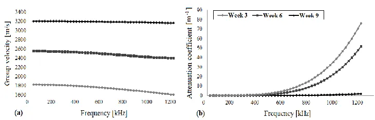
Fig. 3 shows the group velocity and the attenuation coefficient predictions in the frequency range 24 – 1200 kHz for each healing stage. In Fig. 3a, the group velocity was found to decrease: a) from 1826 – 1609 m/s in week 3, b) from 2555 – 2403 m/s in week 6, and c) from 3202 – 3167 m/s in week 9. Fig. 3b shows an exponential increase of the attenuation coefficient with increasing frequency: a) from 0.06 – 76.36 m-1 in week 3, b) from 0 – 51.86 m-1 in week 6, and c) from 0 – 1.91 m-1 in week 9.
4. Discussion
In the present study, we used an iterative methodology to carry out wave dispersion and attenuation predictions in the callus region of healing long bones based on SAM images representing successive healing stages.
A negative dispersion is exhibited in all the examined healing stages. A similar phase velocity behavior has been previously observed by several research groups (Vavva et al. 2008; Waterman and Truell 1961) studying the porous structure of cancellous bone. Haїt et al. (Vavva et al. 2008) suggested that this phenomenon is attributed to the coupling of multiple scattering and absorption effects due to the heterogeneity of the medium. Another possible explanation according to (Waterman and Truell 1961) is that negative dispersion can arise when signals consisted of overlapped fast and slow waves are analyzed as a single longitudinal wave.
Fig. 3.Estimation of: a) the group velocity and b) attenuation coefficient for each healing stage.
a gradual decrease of the particles‟ volume concentration and diameter. Thus, the role of scattering and absorption effects is more pronounced during the early healing stages. Our findings indicate that IEMA provides reasonable results and could be used for the evaluation of bone healing.
However, the presented results concern exclusively the callus tissue, thus neglecting the cortical bone. In our future work the effective material properties and the attenuation coefficient calculated from IEMA will be incorporated to numerical models of healing long bones using the boundary element method in order to simulate wave propagation.
5. Conclusions
In this work, we presented group velocity and attenuation predictions in healing long bones based on an iterative effective medium approximation. For the first time, wave dispersion and attenuation were quantitatively determined in the composite geometry of the callus at different healing stages. This study constitutes a starting point for the systematic investigation of the scattering effects induced by the porous nature of callus at different healing stages. However, the results should be interpreted with caution as further theoretical and numerical research is needed.
Извод
Одређивање дисперзије и слабљења таласа за процену процеса
зарастања костију
V. Potsika1, V. Protopappas2, M. Vavva2, K. Raum3, D. Rohrbach3, D. Polyzos2, D.I. Fotiadis1
1
Unit of Medical Technology and Intelligent Information Systems,University of Ioannina, GR 45110 Ioannina, Greece
[email protected], [email protected] 2
Department of Mechanical Engineering and Aeronautics, University of Patras, GR 26500 Patras, Greece
[email protected], [email protected], [email protected] 3
Julius Wolff Institute, Berlin-Brandenburg School for Regenerative Therapies, Charité-Universitätsmedizin Berlin, Augustenburger Platz 1, 13353 Berlin, Germany
Резиме
Квантитативни ултразвук завређује све више пажње као дијагностичко средство у процесу зарастања костију. Неколико теорија вишеструког расејања предложене су за испитивање расипања таласа у нехомогеним медијима, које, међутим, не могу да пруже реалистичне процене дисперзије и слабљења за различите врсте честица и запреминске концентрације. У овом истраживању, користимо итеративну ефикасну апроксимацију медија (IEMA) (Aggelis et al. 2004) како бисмо извршили предвиђања расипања и слабљења у регији калуса у различитим фазама излечења. Геометрија и материјална својства изведене су из слика скенинг акустичне микроскопије (SAM) потколенице код оваца, у фази излечења, добијених у трећој, шестој и деветој седмици након операције (Anderson C C et al. 2008). Претпоставља се да је калус композитни медиј који се састоји од крви и коштаног ткива. Просечни пречници честица и запреминска концентрација били су 350μm и 44.75% у трећој седмици, 200μm и 38.67% у шестој седмици, 120μm и 22.67% у деветој седмици, респективно. Расипање и слабљење таласа се процењују за фреквенције од 24 – 1200 kHz. Увидели смо да се групна брзина смањује са повећањем фреквенције, док се коефицијент слабљења повећава у испитиваном опсегу фреквенције. Резултати указују на то да су ефекти расипања израженији у ранијим фазама лечења. Закључак је да IEMA може пружити оправдана предвиђања и стога може бити коришћена за процену излечења костију.
References
Aggelis D. G., Tsinopoulos, S. V., Polyzos D., (2004). An iterative effective medium approximation for wave dispersion and attenuation predictions in particulate composites,
suspensions and emulsions, J. Accoust. Soc. Am., 9, pp. 3443–3452.
Anderson C. C., Marutyan K. R., Holland M. R., Wear K. A., Millera J. G., (2008). Interference between wave modes may contribute to the apparent negative dispersion observed in
cancellous bone, J. Acoust. Soc. Am., 124, 1781–1789.
Christensen R. M., (1990). A critical evaluation for a class of micromechanics models, J. Mech.
Phys. Solids, 38, pp. 379-404.
Dodd S. P., Cunningham J. L., Miles A. W., Gheduzzi S., Humphrey V. H., (2006). An in vitro study of ultrasound signal loss across simple fractures in cortical bone mimics and bovine
cortical bone samples, Bone, 40, pp. 656–661.
Foldy L. L., (1945). The multiple scattering of waves, Phys. Rev., 67, pp. 107–119.
Haїat G., Lhemery A., Renaud F., Padilla F., Laugier P., Naili S., (2008). Velocity dispersion in
trabecular bone: influence of multiple scattering and of absorption, J. Acoust. Soc. Am., 124,
pp. 4047–4058.
Kim J. Y., Ih J. G., Lee B. H., (1995). Dispersion of elastic waves in random particulate
composites, J. Acoust. Soc. Am., 97, pp. 1380–1388.
Laugier P. and Haiat G., (2011). Bone quantitative ultrasound, Springer Dordrecht Heidelberg London New York: Science+Business Media B.V., pp. 409–440.
Machado C. B., Albuquerque Pereira W. C., Talmant M., Padilla F., Laugier P., (2010). Computational evaluation of the compositional factors in fracture healing affecting
ultrasound axial transmission measurements, Ultrasound in Med. & Biol., 36, pp. 1314–
1326.
Preininger B., Checa S., Molnar F. L., Fratzl P., Duda G. N., Raum K., (2011). Spatial-temporal mapping of bone structural and elastic properties in a sheep model following osteotomy,
Lett., 9, pp. 193-200.
Vavva M. G., Protopappas V. C., Gergidis L. N., Charalambopoulos A., Fotiadis D. I., Polyzos D., (2008). The effect of boundary conditions on guided wave propagation in
two-dimensional models of healing bone, Ultrasonics, 48, pp. 598–606.
Waterman P. C. and Truell R., (1961). Multiple scattering of waves, J. Math. Phys., 2, pp.