Open Access
Research
CapZ-lipid membrane interactions: a computer analysis
James Smith*, Gerold Diez, Anna H Klemm, Vitali Schewkunow and
Wolfgang H Goldmann
Address: Friedrich-Alexander-University of Erlangen-Nuremberg Center for Medical Physics and Technology, Biophysics Group Henkestrasse 91, 91052 Erlangen, Germany
Email: James Smith* - [email protected]; Gerold Diez - [email protected]; Anna H Klemm - [email protected]; Vitali Schewkunow - [email protected]@biomed.uni-erlangen.de; Wolfgang H Goldmann - [email protected] * Corresponding author
Abstract
Background: CapZ is a calcium-insensitive and lipid-dependent actin filament capping protein, the main function of which is to regulate the assembly of the actin cytoskeleton. CapZ is associated with membranes in cells and it is generally assumed that this interaction is mediated by polyphosphoinositides (PPI) particularly PIP2, which has been characterized in vitro.
Results: We propose that non-PPI lipids also bind CapZ. Data from computer-aided sequence and structure analyses further suggest that CapZ could become partially buried in the lipid bilayer probably under mildly acidic conditions, in a manner that is not only dependent on the presence of PPIs. We show that lipid binding could involve a number of sites that are spread throughout the CapZ molecule i.e., alpha- and beta-subunits. However, a beta-subunit segment between residues 134–151 is most likely to be involved in interacting with and inserting into lipid membrane due to a slighly higher ratio of positively to negatively charged residues and also due to the presence of a small hydrophobic helix.
Conclusion: CapZ may therefore play an essential role in providing a stable membrane anchor for actin filaments.
Background
The actin cytoskeleton is a major component in determin-ing and maintaindetermin-ing the shape of animal cells and is responsible for various motile phenomena. It is regulated by actin-binding proteins that are controlled by a variety of signalling molecules including the well-characterized polyphosphoinositides (PPIs). One of the capping pro-teins is the calcium-insensitive CapZ, which is regulated by phosphatidylinositol 4,5 bisphosphate (PIP2) [1-4]. This protein regulates the spatial and temporal growth of the actin filament by capping its barbed (and fast grow-ing) end.
CapZ proteins have been isolated from various species, and sequence studies demonstrate extensive homology among Drosophila, Saccharomyces, Dictyostelium, Acan-thamoeba, Caenorhabditis and vertebrates. The protein is composed of two subunits, labelled alpha and beta. The alpha-subunits range between 32 kDa and 36 kDa; the beta-subunits are generally smaller, ranging between 28 kDa and 32 kDa. To date, actin binding has only been ascribed to the beta-subunit [5], although both subunits are required for capping activity [6]. Although they show low sequence identity, alignments of the subunits reveal regions of functionally conserved residues, suggesting the Published: 16 August 2006
Theoretical Biology and Medical Modelling 2006, 3:30 doi:10.1186/1742-4682-3-30
Received: 09 April 2006 Accepted: 16 August 2006
This article is available from: http://www.tbiomed.com/content/3/1/30
© 2006 Smith et al; licensee BioMed Central Ltd.
presence of common motifs or putative epitopes for inter-molecular binding. A structural analogy between the alpha- and beta-subunits was confirmed in a recent crys-tallographic study of CapZ from chicken muscle that revealed a striking resemblance in the fold of the two sub-units [7].
Spatial and temporal localization studies in non-muscle cells have not always produced a consistent picture: in one case the distribution is nuclear, while chicken CapZ is concentrated in epithelial cell-cell junction complexes. Yeast capping proteins are found at the membrane in regions generally rich in actin [8]. In muscle cells, CapZ is present at the Z-line independently of actin and probably binds to other protein partners in this region [9].
Here we report that CapZ has the potential to bind to lip-ids (other than PIP2) and could therefore interact with, or embed into, lipid regions consisting of phospholipids, glycolipids, cholesterol and/or long-chain fatty acids. Our computational analysis indicates that the C-terminal half of CapZ beta-subunit could contribute to lipid interac-tion/insertion. CapZ may therefore play an essential role in providing a stable membrane anchor for actin fila-ments.
Methods
The search for highly hydrophobic or amphipathic seg-ments within the CapZ sequence includes the construc-tion of plots of the average hydrophobicity and of the average hydrophobic moment [10]. The normalized 'con-sensus' scale of Eisenberg et al. [11] was taken as the hydrophobicity scale for amino acids. The number of amino acids examined together (also known as the win-dow size) determined the type of segment under investi-gation.
To detect lipid membrane binding and hydrophobic motifs, and potentially antigenic regions, a window size of 11 residues was employed. The algorithm for detecting putative lipid-binding hydrophobic polypeptide sequence segments discriminates between surface-seeking and transmembrane regions. Computationally, this is per-formed by constructing and interpreting plots for the aver-age hydrophobicity <H> and the averaver-age hydrophobic moment <μH> of selected polypeptide segments using a normalized 'consensus' scale [11-13]. According to Eisen-berg et al. [11], various regions in a polypeptide can be divided by boundary lines, conditional on the values of <H> and <μH>, giving three alpha-helical properties: transmembrane, lipid surface-seeking and globular. In general, transmembrane helical regions have a low <μH> and high <H> whereas surface-seeking helical regions have a high <μH> and average <H> [10]. In this work, we used two ratios to assay for surface-seeking propensity, r
sur-face and rtm, relating respectively to the transition from a
globular to a surface-seeking property and from a globular to a transmembrane property. These two ratios depend on <μH> and <H>, where rsurface = <μH>/(0.603 - 0.392<H>) and rtm =<H>/0.51. Three conditions exist, depending on the Eisenberg plot [11]: (1) if rsurface and rtm are both less than or equal to 1.0, then the polypeptide region is glob-ular; (2) if either rsurface or rtm is greater than 1.0 and the other less than or equal to 1.0, then the larger ratio deter-mines the characteristic property; (3) if both values are greater than 1.0, then the region is said to be surface-seek-ing.
An amphipathic helical region was defined by the simple requirement for an effective interaction between an alpha-helix and acidic lipids. The interaction motif is suitable for amino acid segments with a length of 18 residues, which would represent five complete turns of an ideal alpha-helix. When projected on to a plane, the consecutive resi-dues of an ideal helix are spaced with a periodicity of 3.6 at 100 degree intervals. For the amphiphatic helical anal-ysis, a matrix incorporating information about the distri-bution of physico-chemically different residues was employed. This matrix also included information regard-ing amphiphatic structure. This approach is based on a previous treatment by Hazelrig et al. [14]. With an amino acid window size of 18, the results were plotted above the middle residue of the window.
Hydrophobic moments of alpha-helices and beta-strands were calculated, assuming periodicities in the hydropho-bicity of 3.6 and 2.0 residues, respectively. The entire proc-ess yields several candidate sites that relate to sequence and conformational motifs for each candidate protein sequence. The two protein sequences used were obtained from the NCBI database: residues 1 to 286 from the alpha-subunit from NP006126, and residues 1 to 272 from the beta-subunit from NP004921, both from Homo sapiens. The lipid-binding properties of each candidate site can subsequently be evaluated using a variety of in vitro tech-niques.
Here, the experimentally-supported lipid-binding sites for
Homo sapiens CapZ correlated with regions in the high-res-olution crystal coordinates obtained from Gallus gallus
and deposited in the Protein Data Bank (PDB code 1IZN). Over the range of sequences used there was almost 100% identity between the CapZ subunits from Homo sapiens
Results
The secondary structure analysis of the CapZ sequence was started with the search for segments with maximum hydrophobic and amphipathic character. The most hydro-phobic segments and the most amphipathic helical seg-ments were found in the amino-terminal region of the protein between residues 113–130 and 225–242 both in the alpha-subunit and between residues 134–151 and 215–232 both in the beta-subunit.
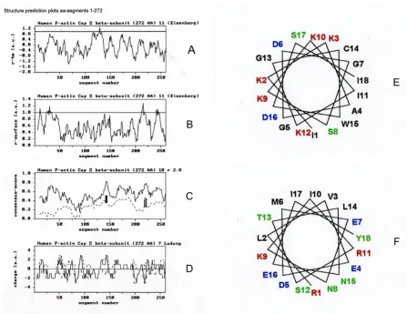
Figures 1 and 2 represent the structure prediction plots calculated for the CapZ primary sequence residues 1–286 (for the alpha-subunit) and 1–272 (for the beta-subunit). The plots (a+b) of the rtm and rsurface ratio profiles evaluate the hydrophobic or amphipathic alpha-helical stretches. For these calculations an amino acid window size of 11 was used. The plot in (c) represents the matrix calcula-tions for an amphipathic alpha-helix motif. At a window size of 18 residues, the consensus score of the existing
Structure prediction plots of CapZ alpha-subunit (residues 1–286) using matrix analyses according to Tempel et al. [10]
Figure 1
Structure prediction plots of CapZ alpha-subunit (residues 1–286) using matrix analyses according to Tempel et al. [10]. (A) Hydrophobicity, (B) Hydrophobic moment and (C) Probability of residues for CapZ alpha-subunit. Secondary structures (D) were calculated according to Eisenberg et al. [11] using a window of 11 residues. The secondary structure analyses of 113–130 (ADGGLKSWRESCDSALRA) and 225–242 (KEFIKIIENAENEYQTAI) are shown in (E) and (F), respectively. The two methods were carried out as follows: The 1st method relies only on the average amino acid composition of secondary structural seg-ments (helix, sheet, coil) in a learning set of proteins, which showed an alpha-content of 55.2%, beta-content of zero, and a coil-content of 44.8% for (E); and an alpha-content of 100% and beta- and coil-contents of zero for (F). The 2nd method relies on composition fluctuations in the secondary structural segments (helix, sheet, coil) of a learning set of proteins, which showed an alpha-content of 38.1%, a content of zero, and a coil-content of 61.9% for (E); and an alpha-content of 93.8%, a beta-content of 6.2%, and coil-beta-content of zero for (F) [27-28].
A
B
C
E
F
sequence (continuous line) and the average consensus score of 400 sequence randomizations (dotted line) are plotted for every segment. For any segment, the standard deviation (SD) of the randomizations is denoted by a ver-tical bar in the SD, where factor Γ was greater than 3.0. The quantitative distribution of charged amino acids within 7-residue segments in (d) are marked by the continuous and discontinuous lines of positively and negatively charged residues.
Results from the plots in Figures 1 and 2(a–d) from resi-dues 1–286 for the alpha-subunit and resiresi-dues 1–272 for the beta-subunit indicate two possible lipid binding regions in each: residues 113–130 and 225–242, and res-idues 134–151 and 215–232, respectively. Secondary structure analysis points to alpha-helical structures. No transmembrane binding domain is discernible in the alpha-subunit; therefore, the polypeptide sequence repre-sents a helical motif with more amphipathic character. If
Structure prediction plots for CapZ beta-subunit (residues 1–272) using matrix analyses according to Tempel et al. [10]
Figure 2
Structure prediction plots for CapZ beta-subunit (residues 1–272) using matrix analyses according to Tempel et al. [10]. (A) Hydrophobicity, (B) Hydrophobic moment and (C) Probability of residues for CapZ beta-subunit. Secondary structures (D) were calculated according to Eisenberg et al. [11] using a window of 11 residues. The secondary structure analyses of 134–151 (IKKAGDGSKKIKGCWDSI) and 215–232 (RLVEDMENKIRSTLNEIY) are shown in (E) and (F), respectively. The two methods were carried out as follows: The 1st method relies only on the average amino acid composition of secondary structural seg-ments (helix, sheet, coil) in a learning set of proteins, which showed an alpha-content of zero, beta-content of zero, and a coil-content of 100% for (E); and an alpha-coil-content of 67.2% and beta-coil-content of 32.8%, and coil-coil-content of zero for (F). The 2nd method relies on composition fluctuations in the secondary structural segments (helix, sheet, coil) of a learning set of proteins, which showed an alpha-content of 16.4%, a beta-content of zero, and a coil-content of 83.6% for (E); and an alpha-content of 100%, beta- and coil-contents of zero, for (F) [27-28].
A
B
C
E
F
there were lipid binding, the expectation would be near-parallel orientations of the alpha-helical axes with the plane of the membrane, so that the hydrophobic/ uncharged amino acids of the alpha- subunit would inter-act hydrophobically with lipid chains.
Specifically, the segment 113–130 in the alpha-subunit shows a high ratio of positively and negatively charged amino acids that form the hydrophilic side of the amphip-athic helix. The hydrophobic helix shows seven non-polar and three polar amino acids and would be poorly-equipped for lipid binding/insertion. The segment 225– 242 in the alpha subunit, however, shows high contents of positively and negatively charged and polar amino acids, and could interact strongly with the hydrophilic (and hydrogen-bonding) side of the opposite amphip-athic helix. The hydrophobic side of the helix contains six non-polar and one polar amino acid, including a strongly hydrophobic amino acid (phenylalanine, F). This gives this helix its predominantly amphipathic character. The glutamic acids (deprotonated at pH 7.0) at positions 11 and 13 would seem to make the helix unsuitable for sur-face binding to a negatively-charged lipid layer.
The segment 134–151 in the beta-subunit shows a slightly higher ratio of positively to negatively charged amino acids on the hydrophilic side of the short amphipathic helical region within the beta-strands, whereas the hydro-phobic helical side contains seven non-polar and one polar amino acid. This distribution of positively charged amino acids would be more favourable for surface bind-ing to negatively charged lipid layers. The segment 215– 232 in the beta-subunit shows a similar amphipathic charge distribution to segment 225–242 in the alpha-sub-unit; however, the (negatively charged) glutamic acid at position 7 probably makes any surface binding to lipid unfavourable.
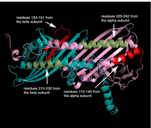
The recent crystal of CapZ shows two subunits that are structurally analogous creating a pseudo-two-fold symme-try perpendicular to the long axis of the molecule (Figure 3). Each subunit contains three domains and an addi-tional carboxyl-terminal extension. Three anti-parallel helices (helices 1–3) that form the amino-terminal domain are in an up-down-up arrangement. The middle domain is composed of four beta-strands (strands 1–4) for the alpha-subunit and three (strands 1–3) for the beta-subunit, containing two reverse turns. The carboxyl-termi-nal domain comprises an anti-parallel beta-sheet formed by five consecutive beta-strands (strands 5–9), flanked on one side by a shorter amino-terminal helix (helix 4) and a long carboxyl-terminal helix (helix 5). The beta-strands of each subunit form a single 10-stranded anti-parallel beta-sheet in the centre of the molecule. The sequence impli-cated in lipid binding, amino acid residues 134–151 in
the beta-subunit, forms largely beta-sheet that is probably flexible and solvent-accessible despite contributing resi-dues to the strong dimer interface (for example, via lysine 136).
Discussion
Recently, it has been reported that when gelsolins (cal-cium-dependent actin-binding proteins) are presented with high lipid concentrations they can bind as many as ten PtdIns(4,5)P2 molecules [17]. The value of the molar ratio between gelsolin and PtdIns(4,5)P2 has been conten-tious, complicated by differences between studies in the state or presentation of the lipid. However, when pre-sented as a minor component with other lipids (i.e. cho-lesterol), one PtdIns(4,5)P2 binds one gelsolin, close to
the physiological situation of 0.3–1.5%, which then allows it to associate with the plasma membrane [18].
Furthermore, it has been reported that polyphosphoi-nositides (PPI) form aggregates within the bilayer under the influence of certain proteins [19] and there may be many possible modes of binding to PPI and other lipids. The finding that several sites within gelsolin can be cross-linked to PPI analogues would seem to support this view [20]. Together with our present data, indicating that CapZ could bind non-PPI lipids with high affinity, it seems likely that CapZ may bind up to four PtdIns(4,5)P2, if they are available, through direct hydrogen-bonding interac-tions with the binding sites; however at lower PtdIns(4,5)P2 concentrations these sites may be occupied by other lipids. This is in agreement with observations by differential scanning calorimetry, film balance and spec-troscopy, which have shown that proteins require a net negative charge created by lipids other than PPIs, a hydro-phobic interface or indeed PPI for membrane interaction/ insertion [17].
CapZ has been found to be associated with both mem-branes and actin filaments in activated macrophages and platelets [21,22]. This is a surprise since PtdIns(4,5)P2 has been assumed to be the binding partner of CapZ and yet this lipid dissociates the CapZ-actin complex [23,24]. It is possible that the binding sites for the CapZ-actin complex in macrophages and platelet membranes are lipids other than PPIs and that these do not dissociate the complex. It has been reported that binding of gelsolin or indeed fil-amin (a dimeric actin cross-linking protein) to phosphati-dylglycerol/phosphatidylcholine small unilaminar vesicles does not inhibit the nucleation of actin polymer-ization or cross-linking.
peptides derived from PPI-binding regions of, for example gelsolin, Arp2/3, talin etc. have this capacity in isolation [25]. The authors have also found that such peptides can incorporate into phosphatidylglycerol/phosphatidylcho-line small unilaminar vesicles in the absence of PPIs [25]. The importance of hydrophobic interactions between these proteins and PPIs has been suggested by molecular dynamics studies in which the PPIs are to some extent pulled out from the bilayer [26].
In conclusion, a number of sites in CapZ have been pro-posed to bind lipids and these tend to be located in linker regions between the discrete domains of the protein. The main sites appear to be in the linker regions, 134–151 and 215–232 in the beta-subunit and secondary sites have been identified within the alpha-subunit. We suggest fur-ther that the first region 134–151 in the beta-subunit becomes inserted between lipid heads and perhaps into the core of a lipid bilayer.
The four predicted lipid-binding sites of CapZ alpha- and beta-subunits
Figure 3
Publish with BioMed Central and every scientist can read your work free of charge "BioMed Central will be the most significant development for disseminating the results of biomedical researc h in our lifetime."
Sir Paul Nurse, Cancer Research UK
Your research papers will be:
available free of charge to the entire biomedical community
peer reviewed and published immediately upon acceptance
cited in PubMed and archived on PubMed Central
yours — you keep the copyright
Submit your manuscript here:
http://www.biomedcentral.com/info/publishing_adv.asp
BioMedcentral
Acknowledgements
This work was funded by the Deutsche Forschungsgemeinschaft (DFG; Is25/8-1 to WHG) and North Atlantic Treaty Organization (NATO; CLG 978417 to WHG). We thank Drs. G. Isenberg and M. Tempel for valuable discussions.
References
1. Isenberg G, Aebi U, Pollard TD: An actin-binding protein from Acanthamoeba regulates actin filament polymerization and interactions. Nature 1980, 288:455-459.
2. Kilimann MW, Isenberg G: Actin filament capping protein from bovine brain. EMBO J 1982, 1:889-894.
3. Hartmann H, Noegel AA, Eckerskorn C, Rapp S, Schleicher M: Cal-cium-dependent F-actin capping proteins. Cap32/34, a Cap-ping Protein from Dictyostelium discoideum, does not share sequence homologies with known Actin-Binding Proteins. J Biol Chem 1989, 264:12639-12647.
4. Nachmias VT, Golla R, Casella JF, Barron-Casella EA: Cap Z, a cal-cium insensitive capping protein in resting and activated platelets. FEBS Lett 1996, 378:258-262.
5. Hug C, Miller TM, Torres MA, Casella JF, Cooper JA: Identification and Characterization of an Actin-Binding Site of CapZ. J Cell Biol 1992, 116:923-931.
6. Kim K, Yamashita A, Wear MA, Maeda Y, Cooper JA: Capping pro-tein binding to actin in yeast: biochemical mechanism and physiological relevance. J Cell Biol 2004, 164:567-580.
7. Yamashita A, Maeda K, Maeda Y: Crystal structure of CapZ: structural basis for actin filament barbed end capping. EMBO J 2003, 22:1529-1538.
8. Amatruda JF, Cooper JA: Purification, Characterization and Immunofluorescence Localization of Saccharomyces cerevi-siae Capping Protein. J Cell Biol 1992, 117:1067-1076.
9. Schafer DA, Korshunova YO, Schroer TA, Cooper JA: Differential localization and sequence analysis of capping protein beta-subunit isoforms of vertebrates. J Cell Biol 1994, 127:453-465. 10. Tempel M, Goldmann WH, Isenberg G, Sackmann E: Interaction of
the 47-kDa talin fragment and the 32-kDa vinculin fragment with acidic phospholipids: a computer analysis. Biophys J 1995,
69:228-241.
11. Eisenberg D, Schwarz E, Komaromy M, Wall R: Analysis of mem-brane and surface protein sequences with the hydrophobic moment plot. J Mol Biol 1984, 179:125-142.
12. Deber DM: The Hydrophobicity Threshold for Peptide Inser-tion into Membranes. Current Topics in Membranes 2002,
52:465-479.
13. Kyte J, Doolittle RF: A simple method for displaying the hydro-pathic character of a protein. J Mol Biol 1982, 157:105-132. 14. Hazelrig JB, Jones MK, Segrest JP: A mathematically defined
motif for the radial distribution of charged residues on apol-ipoprotein amphipathic α-helices. Biophys J 1993, 64:1827-1832.
15. Guex N, Peitsch MC: SWISS-MODEL and the
Swiss-Pdb-Viewer: an environment for comparative protein modeling.
Electrophoresis 1997, 18:2714-2723.
16. DeLano WL: The PyMOL Molecular Graphics System San Carlos, CA: DeLano Scientific; 2002.
17. Tuominen EKJ, Holopainen JM, Chen J, Prestwich GD, Bachiller PR, Kinnunen PKJ, Janmey PA: Fluorescent phosphoinositide deriva-tives reveal specific binding of gelsolin and other actin regu-lator proteins to mixed lipid bilayers. Eur J Biochem 1999,
263:85-92.
18. Mere J, Chahinian A, Maciver S, Fattoum A, Bettache N, Benyamin Y, Roustan C: Gelsolin binds to polyphosphoinositide-free lipid vesicles and simultaneously to actin microfilaments. Biochem J 2005, 386:47-56.
19. Gambhir A, Hangyas-Mihalyne G, Zaitseva I, Cafiso DS, Wang J, Mur-ray D, Pentyala SN, Smith SO, McLaughlin S: Electrostatic seques-tration of PIP2 on phospholipid membranes by basic/
aromatic regions of proteins. Biophys J 2004, 86:2188-2207. 20. Feng L, Mejillano M, Yin HL, Chen J, Prestwich GD: Full-contact
domain labelling: identification of a novel phosphoinositide binding site on gelsolin that requires the complete protein.
Biochemistry 2001, 40:904-913.
21. Hartwig JH, Bokoch GM, Carpenter CL, Janmey PA, Taylor LA, Toker A, Stossel TP: Thrombin receptor ligation and activated Rac uncap actin filament barbed ends through phosphoinositide
synthesis in permeabilized human platelets. Cell 1995,
82:643-653.
22. Hartwig JH, Chambers KA, Stossel TP: Association of gelsolin with actin and cell membranes of macrophages and plate-lets. J Cell Biol 1989, 108:467-479.
23. Janmey PA, Stossel TP: Modulation of gelsolin function by phos-phatidylinositol 4,5-bisphosphate. Nature 1987, 325:362-364. 24. Janmey PA, Iida K, Yin HL, Stossel TP: Polyphosphoinositide
micelles and polyphosphoinositide-containing vesicles disso-ciate endogenous gelsolin-actin complexes and promote actin assembly from the fast-growing end of actin filaments blocked by gelsolin. J Biol Chem 1987, 262:12228-12236. 25. Scott DL, Diez G, Goldmann WH: Protein-Lipid Interactions:
Correlation of a predictive algorithm for lipid-binding sites with three-dimensional structural data. Theoretical Biology and Medical Modelling 2006, 3:17.
26. Liepina I, Czaplewski C, Janmey PA, Liwo A: Molecular dynamics study of a gelsolin-derived peptide binding to a lipid bilayer containing phosphatidylinositol 4,5-bisphosphate. Biopolymers
2003, 71:49-70.
27. Eisenhaber E, Imperiale F, Argos P, Frömmel C: Prediction of sec-ondary structural content of proteins from their amino acid composition alone. I. New analytic vector decomposition methods. Proteins: Structure, function, design 1996, 25(N2):157-168.