Original Research Article
Assessment of the salivary glucose as a non-invasive
test for diabetic patients
Kajol Thapa
1*, Saroj Kunwar
1, Sonu Thapa
1, Asmita Phuyal
1, Sahil Rupakheti
2INTRODUCTION
Diabetes mellitus is a metabolic disorder that interferes with metabolism of carbohydrate leading to the condition where glucose is not fully used.1 Glucose is the component of the blood that is exchangeable across the salivary gland epithelial in the proportion to its concentration in blood.2
Type 2 diabetes mellitus is described as a global epidemic fuelled by population growth, ageing, obesity, changing lifestyles and urbanization. The impacts of the diabetes has been felt in both the developed and developing countries, due to mortality and morbidity associated with its complications in the renal, retinal and vascular system.3 Though non-invasive in vivo
investigation for many years, no any such technology has been approved.
The present study was conducted to estimate glucose level in saliva and serum sample and to find correlation ship between salivary and serum glucose in diabetic (case) and healthy individuals (control).
METHODS
The present study is a comparative cross-sectional study. Ethical clearance was taken from IRC (International Research Committee) of NHRC (Nepal Health Research Council) before conducting the study. The total 211 participant were included in the study among which, 105 were diabetic (case group) and 106 were healthy
ABSTRACT
Background: Over the recent year there has been a startling rise in the number of people suffering from diabetes mellitus. Estimation of blood glucose levels has been an essential laboratory investigation for screening and monitoring of diabetes. Saliva is one of the secretions in human body whose collection is easy and non-invasive. Salivary glucose hence could serve as an easy and non-invasive tool.
Methods: Institutional review committee of NHRC gave us permission to carry out this study. 105 subjects with Type 2 diabetes mellitus who attended the OPD at Star hospital, Sanepa, Lalitpur, Nepal and 106 healthy individuals were consented to participate in this study. Glucose was measured by the GOD-POD (Glucose oxidase peroxidase) methods using the semi-autoanalyser and salivary glucose was compared with corresponding blood glucose levels.
Results: A significant positive correlation of fasting salivary glucose level and fasting blood glucose level was observed in healthy (r=0.241, p=0.001) and in diabetic patients (r=0.202, p=0.001).
Conclusions: The study implies a potential for saliva in monitoring and screening of diabetes mellitus.
Keywords: Fasting blood glucose, Fasting salivary glucose, Diabetes mellitus, Noninvasive
1Department of Biochemistry, Modern Technical College, Sanepa, Lalitpur, Nepal 2Department of Medicine, Star Hospital, Lalitpur, Nepal
Received: 10 February 2019
Accepted: 12 March 2019
*Correspondence:
Kajol Thapa,
E-mail: [email protected]
Copyright: © the author(s), publisher and licensee Medip Academy. This is an open-access article distributed under the terms of the Creative Commons Attribution Non-Commercial License, which permits unrestricted non-commercial use, distribution, and reproduction in any medium, provided the original work is properly cited.
1 year from August 2017 to August 2018. The written consent was taken from each group of the participant before enrolling them in the study. For case group, sample was accepted from the patients who were diagnosed by type-II diabetes mellitus from the medicine department of Star hospital, Sanepa, Lalitpur. For control group, sample from the healthy individual from the community of Lalitpur who were above 35 years and had a healthy medical background were accepted. Sample from the subject with a systemic disease, severe complication of diabetes, recent infection like tuberculosis, Hepatitis, oral mucosal lesions, dental wear and also the physiological conditions like pregnancy were excluded from the study. Convenient sampling technique was used and sample size was calculated by using formula for quantitative variable.4
Sample size (n) = n = (1.96)2 x (5.01)2 / (0.95)2 = 3.84 x 26.01 / 0.9 = 110
Where,
n = required sample L = allowable errors
Z= 1.96 for 0.05 significance level (95% confidence interval)
Estimated prevalence5 is 19%
Allowable error of 5% (L= 0.95)
Collection of the samples
Saliva collection
Firstly, a written consent was taken from the participant and all the procedure were explained briefly. A structured questionnaire was conducted and important information of the entire participant was recorded. Detailed history of participant revealed that most of the participants were diagnose after 35 years. Many of them were taking oral medication, some were taking insulin and few of them were not taking any medication for the controlling the blood glucose.
Fasting un-stimulated mixed saliva was collected. Initially, the entire participant were requested and instructed to rinse the mouth with water and split for two times; in order to prevent the contamination by oral flora and dilution of saliva. After that the subjects, were allowed to sit in upright position and labeled wide mouth sterile container was given. The participant were instructed to keep their mouth open to pool the saliva on the floor of mouth then the patient were asked to split in the container gradually at the end of every 60 second for five minutes. After the collection of saliva the container was immediately transferred in the Ice-bag and then to
refrigerator at 2-8⁰C; to minimize the utilization of the glucose by bacteria.
Blood collection
Blood was collected by the venipuncture method from the median cubital vein; fasting blood sample (2.5 ml) was collected in fluoride tube.
Steps of blood collection
First the written consent was taken from each participant
Then the subjects were allowed to seat comfortably
Tourniquet was applied to palpate the vein
The site was sterilized using the rectified spirit
At the angle of 30-40⁰ the vein was penetrated with skin tight by another hand
The piston of the syringe was drawn slowly to prevent the hemolysis of blood
Tourniquet was removed slowly from another hand
The needle was removed and the blood was transfer to the vials
The specimen was labeled properly with the patient identification number
And finally the needle was incinerated.
Analysis of serum and salivary glucose
The collected saliva was transferred into the sterile glass tube and was centrifuged at 3000rpm for 10 minutes to obtain the clear supernatant. While, the fluoride tube containing blood was also centrifuge at 3000rpm for 5min; to obtain a serum. Then 1000µl of glucose reagent was pipette in 3 tube followed by 10µl of standard in first tube, 10µl of supernatant of saliva in second tube and finally 10µl of serum in third tube. Glucose was then estimated by the glucose oxidase - peroxidase method using Beacon-liquizyme. The concentration of the glucose was then measured spectrophotometically. The glucose oxidation method was calibrated from the solution of glucose which was prepared and diluted to make series of standards. For serum analysis five standards of 100 mg/dl, 200 mg/dl, 300 mg/dl, 400 mg/dl and 500 mg/dl was taken. On the flip side, for salivary glucose five standards of 5 mg/dl, 10 mg/dl, 15 mg/dl, 20 mg/dl and 25 mg/dl was taken to ensure the quality of test procedure.
Statistical analysis
show the correlation between salivary glucose and serum glucose between two groups.
RESULTS
Total 211 participants were enrolled in the study, among which 105 were the diabetic (case) and 106 were the healthy individual (control). The total number of the female participant in the study was 133 and that of male participant was 78. The mean age of the diabetic and control group was (58.20±12.27) and (57.75±10.65) respectively (p=0.77).
The mean fasting serum glucose in the case and control was (142.07±43.37 mg/dl) and (87.81±16.16 mg/dl) respectively. The difference between mean serum glucose in case and control was statistically significant with p value (p<0.05). The mean fasting salivary glucose in the case was (4.88±1.699 mg/dl) and is significantly higher than the mean fasting salivary glucose of control (3.369±0.765 mg/dl) (p<0.05).
Table 1: Comparison of mean of fasting salivary and serum glucose in case and control.
Biochemical parameters Diabetic (n=105) Control (n=106) P value
Mean salivary glucose 4.88±1.69 3.36±0.756 0.005
Mean serum glucose 142.07±43.37 87.81±16.16 0.001
Independent-t test was applied; P value less than 0.05 is considered to be significant.
Table 2: Correlation between fasting salivary glucose and serum glucose in case and control.
Correlations Salivary glucose Serum glucose
Case Salivary glucose R 0.450**
P 0.001
Serum glucose R 0.450**
P 0.001
Control Salivary glucose R 0.491**
P 0.001
Serum glucose R 0.491**
P 0.001
Pearson Correlation test was applied. ** Correlation is significant at P value less than 0.05.
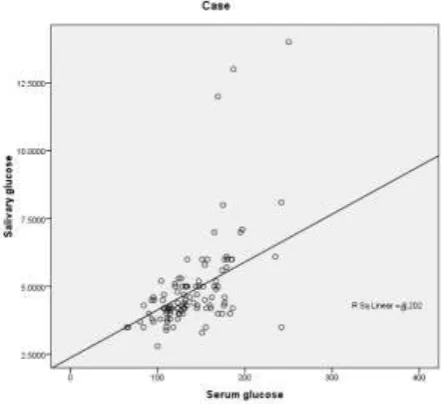
Figure 1: Correlation between the fasting salivary glucose and fasting serum glucose in case.
The correlation between the salivary glucose and serum glucose was observed using the Pearson correlation coefficient test. The correlation coefficient (r) between
was statistically significant at (p=0.001, r=0.202). Similarly, the correlation coefficient between fasting serum glucose and fasting salivary glucose in control group was also statistically significant at (p=0.001, r=0.241) (Table 2).
The linear graph shows the positive correlation between salivary and serum glucose in diabetic and non-diabetic group (Figure 1) (Figure 2).
DISCUSSION
Diabetes mellitus is a complex multi-system disorder which is characterized by relative or absolute insufficiency of insulin secretion or concomitant resistance to the metabolic action of insulin on target tissues. It is the most common endocrine disorder with potentially devastating complications that affects all age groups worldwide.3
The management of the diabetes mellitus has been the major challenging issue in this 21st century where, incidence of diabetes is increasing rapidly day by day at a higher frequency. The key aspect for the management of diabetes mellitus can be; maintaining the glucose level in the normal range, frequently monitoring the glucose level in the blood and finding the cost effective way for production of insulin by recombinant technology among which judiciously monitoring glucose is the important aspect for management of diabetes mellitus.
The quantitative estimation of fasting salivary glucose and fasting serum glucose collected at the same time were proceed, the obtained value were scrutinized to find the possibility of using saliva in reflecting glucose concentration in blood. The result of the study confirmed that salivary glucose is higher in diabetic group when compared with healthy individuals, which is in concordance to the study done by Agrawal et al,Akasapu et al, Amer et al, Darwazeh et al.3,6-8
It is possibly due to the increase permeability of the basement membrane and leakage of the microvasculature in the diabetes mellitus, which allows the easy access for the glucose in the saliva. The complications of the diabetes results due to the micro-vascular changes in blood vessels and change in the basement membrane in diabetic patients.9 Many theories have been put forth to explain the micro-vascular alterations. To summarize, hyperglycemia leads to increased advance glycosylation end products, commonly known as ―AGEs‖. These AGEs crosslink proteins such as collagen and extracellular matrix proteins, leading to alteration of basement membrane which inturn results to endothelial dysfunction. This alters the microvasculature structure and makes it more permeable. Other products such as sorbitol, diacylglycerol, and fructose 6-phosphate, which are formed because of the chronic hyperglycemia, also lead to the basement membrane alterations by altering the extracellular matrix proteins. The end result is the leaky basement membrane, which explains the increase passage of glucose from the blood into the saliva in diabetes mellitus.10
Also, the study by Darwazeh et al, concluded that individuals with diabetes who have denture wears had
higher salivary glucose level than the individuals who do not have, which suggested that there could be the additional contributory factors that may rise glucose in the saliva apart from the basement membrane permeability.8 Also, Vasconcelos et al reported non-significant difference in the mean salivary glucose level in diabetic patients (14.03±16.76 mg/dl) and the control group (6.35±6.02 mg/dl) with (p=0.036).11
Although, Yamaguchi et al, suggested to use sub-mandibular or sub-lingual salivary sample instead of parotid saliva for estimating blood glucose level as daily variations in the correlation between BGL and parotid saliva glucose were too large.12
Similar to our study, positive correlation between the salivary glucose and serum glucose were found in the study done by Agrawal et al,Akasapu et al, Abhikshyeet et al.3,6,10
Conversely lack of the correlation between salivary glucose and plasma glucose was observed by Marchetti et al, in fasting state in both diabetic and non diabetic and that indicates the degree of the metabolic control does not effects the way of the salivary gland handle the glucose.13 Also the study, Forba et al and Carda et al shows no significant correlation between the salivary and plasma glucose.14,15 In those studies old method of the glucose estimation were used, which was not appropriate. Also the researcher England et al expressed doubt regarding replacement of the serum as the sample with parotid secretion in the diagnosis of diabetic mellitus because of the lower level of the glucose concentration in the saliva as compared to the serum.16
The finding of our present study, proves the presence of the glucose in the saliva of the diabetic and non diabetic participant which is in concordance to Naik et al,Aydin et al, Bernardi et al, Ben-Aryen et al.17-20 on the flip side to our study Amer et al, did not find glucose even in a small concentration in the saliva of the healthy subjects.7 Study by the Smriti et al who used the un-stimulated saliva similar to our study, categorized participant into three different group, group A includes 60 subject known to have diabetic mellitus who were on medication, group B includes 60 subjects known to have diabetic mellitus who were not in medication and the group C includes 60 subjects who were healthy controls (non diabetic).21 The result demonstrated that there was a significant difference in the mean salivary glucose level among three study groups, the mean fasting salivary glucose was highest in diabetic not under medication (11.68±1.97 mg/dl) followed by diabetic on medication (9.68±2.48 mg/dl) with least being in control (6.50±0.47 mg/dl)
glucose estimation by the use of saliva can be the most valuable substitute for previously establish different invasive test.
CONCLUSION
We wind up our study with a conclusion that, the salivary glucose rises simultaneously with the rise of the serum glucose in the patient with Diabetes mellitus. Our result emphatically shows the positive statistical co-relation between the fasting salivary glucose and the fasting serum glucose thus, assessment of the salivary glucose can be established as a non-invasive tool for diagnosis and monitoring the diabetes mellitus.
ACKNOWLEDGEMENTS
The study is the outcome of dedicated efforts of many individuals and we are proud privilege to show our gratitude to all of them. We are immensely thankful to Star Hospital for providing us an opportunity to conduct our study. We are also thankful to Professor Dr. Keshab Parajuli for sharing ideas and motivating us during our study. Lastly, we offer heartily regards to all the participants for their kind involvement and cooperation.
Funding: No funding sources Conflict of interest: None declared
Ethical approval: The study was approved by the Institutional Ethics Committee
REFERENCES
1. Burtis, Bruns, Teitz. Kidney function test. Teitz Textbook of clinical chemistry & Molecular Diagnostic. Philadephia: Elsevier publishers; 2013: 1415-1435.
2. Singh. Comparison of the salivary and plasma glucose level in the type –II Diabetics Patient’s. Res Adv Dent. 2014;3(1):263-8.
3. Akasapu A, Hegde U, Niti P, Correlation of the blood glucose levels with salivary glucose levels in Type 2 Diabetes Mellitus. Current trends Biomedical Engineering Biosci. 2017;6(1):555679. 4. Charan J, Biswas T. How to calculate sample size
for different study design in medical research? Indian J Psychological Med. 2013;35(2)121-6. 5. Gyawali B, Sharma R, Neupane D, Mishra S,
Teijlingen E, Kallestrup P. Prevalence of type 2 diabetes in Nepal: a systematic review and meta analysis from 2000 to 2014. Glob Health Action. 2015;8:29088.
6. Agrawal RP, Sharma N, Rathore MS, Gupta VB, Jain S, Agarwal V, et al, Noninvasive method for the Glucose level Estimation by Saliva. J Diabetes Metab. 2013;4:5.
7. Amer S, Yousuf M, Siddiqui PQ, Alam J. Salivary glucose concentrations in patients with diabetes mellitus—a minimally invasive technique for
monitoring blood glucose levels. Pak J Pharm Sci. 2001;14(1):33-37.
8. Darwazeh AM, MacFarlane TW, McCuish A, Lamey PJ. Mixed salivary glucose levels and candidal carriage in patients with diabetes mellitus. J Oral Pathol Med. 1991;20:280-3.
9. Sreedevi, Shashikanth MC, Shambulingappa P. Comparison of plasma glucose and salivary glucose in diabetic patients. J Indian Acad Oral Med Radiol. 2008;20(1):9-13.
10. Abhikshyeet P, Ramesh V, Oza N. Glucose estimation in the salivary secretion of diabetes mellitus patients. Diabetes Metab Syndr Obes. 2012;5:149-154.
11. Vasconcelos AC, Soares MS, Almeida PC, Soares TC. Comparative study of the concentration of salivary and blood glucose in type 2 diabetic patients. J Oral Sci 2010;52(2):293-8.
12. Yamaguchi M, Mitsumori M, Kano Y. Development of noninvasive procedure for monitoring the blood glucose levels using saliva. Eng Med Biol Soc. 1998;(4):1763-6.
13. Marchetti P, Tognarelli M, Giannarelli R, Grossi C, Picaro L, di Carlo A, et al. Decreased salivary glucose secretory rate: usefulness for detectionOf diabeteic patients with autonomic neuropathy. Diabetes Res Clin Pract. 1989;7(3):181-6.
14. Forbat LN, Collins RE, Maskell GK, Sönksen PH. Glucose concentrations in parotid fluid and venous blood of patients attending a diabetic clinic. J R Soc Med. 1981;74(10):725-8.
15. Carda C Mosquera-Lloreda N Salom L, Gomez de Ferraris ME, Peydró A. Structural and functional salivary disorders in type 2 diabetic patients. Med Oral Patol Oral Cir Bucal. 2006;11(4):E309-14. 16. Englander HR, Jeffay AI, Fuller JB, Chauncey HH.
Glucose concentrations in blood plasma and parotid saliva of individuals with and without diabetes mellitus. J Dent Res. 1963;42:1246.
17. Naik VV, Satpathy Y, Pilli GS, Mishra MN. Comparison and cor-relation of glucose levels in serum and saliva of patients with dia-betes mellitus. Indian J Pub Health Res Dev. 2011;2(1):103-105. 18. Aydin S. A comparison of ghrelin, glucose,
alpha-amylase and protein levels in saliva from diabetics. J Biochem Mol Biol. 2007;40(1):29-35.
19. Bernardi MJ, Reis A, Loguercio AD, Kehrig R, Leite MF, Nicolau J. Study of the buffering capacity, pH and salivary flow rate in type 2 well-controlled and poorly well-controlled diabetic patients. Oral Health Prev Dent. 2007;5(1):73-78.
20. Ben-Aryeh H, Cohen M, Kanter Y, Szargel R, Laufer D. Salivary composition in diabetic patients. J Diabet Complications. 1988;2(2):96-99.
22. Kaufman E, Lamster IB. The diagnostic applications of saliva-a review. Crit Rev Oral Biol Med. 2002;13(2):197-212.