Copyright © 2001, American Society for Microbiology. All Rights Reserved.
Selection of a Teicoplanin-Resistant
Enterococcus faecium
Mutant
during an Outbreak Caused by Vancomycin-Resistant
Enterococci with the VanB Phenotype
MAGDALENA KAWALEC,1MAREK GNIADKOWSKI,1JOLANTA KE˛DZIERSKA,2
ALEKSANDER SKOTNICKI,3JANUSZ FIETT,1ANDWALERIA HRYNIEWICZ1*
Sera & Vaccines Central Research Laboratory, 00-725 Warsaw,1and Bacteriology Laboratory, University Hospital,2
and Hematology Clinics of Collegium Medicum, Jagiellonian University,331-501 Cracow, Poland
Received 7 June 2001/Returned for modification 11 August 2001/Accepted 15 September 2001
Vancomycin-resistant enterococci (VRE) have recently become an increasing problem in hospitals in Poland, being responsible for a growing number of nosocomial outbreaks. In this work, we have analyzed the second outbreak of VRE with the VanB phenotype to be identified in the country. It was caused by clonal dissemination of a single strain of vancomycin-resistantEnterococcus faecalis(VRES) and horizontal transmission of vanco-mycin resistance genes among several vancovanco-mycin-resistantEnterococcus faecium(VREM) strains. Two similar restriction fragment length polymorphism types of thevanB gene cluster characterized VRES and VREM isolates, and they both contained the samevanB2 variant of thevanB gene. Two vancomycin-susceptibleE.
faecium(VSEM) isolates, recovered from the same wards during the outbreak, proved to be related to certain
VREM isolates and could represent endemic strains that had acquired vancomycin resistance. One VSEM and four VREM isolates, all identified in the same patient, belonged to a single clone, although they revealed remarkable diversity in terms of susceptibility, PFGE patterns, plasmid content, and number ofvanB gene cluster copies. Most probably they reflected the dynamic evolution of anE. faeciumstrain in the course of infection of a single patient. One of the VREM isolates turned out to be resistant to teicoplanin, which coincided with the use of this antibiotic in the patient’s therapy. ItsvanBgene variant differed by a single mutation from that found in other isolates; however, it also lacked a large part of the vanB gene cluster, including the regulatory genesvanRBand -SB, and the vancomycin-inducible promoterPYB. Expression of the resistance genesvanHB, -B, and -XBwas constitutive in the mutant, and this phenomenon was responsible for its unusual phenotype.
Vancomycresistant enterococci (VRE) usually cause in-fections in severely debilitated, immunocompromised patients who undergo prolonged antimicrobial therapy (9, 12, 34, 36). Of the six different VRE phenotypes known to date (23, 31, 32, 35, 40, 44), phenotypes VanA and VanB are of the highest clinical importance as they are most frequently observed in two predominant enterococcal species, Enterococcus faecalis and
Enterococcus faecium (12, 36, 37). Although the majority of
nosocomial VRE outbreaks have been attributed to VanA organisms, outbreaks caused by VanB VRE have also been reported several times. They were found to result from either the clonal spread of resistant strains (13, 18, 47, 49) or the horizontal transfer of resistance determinants (10, 11, 42, 51) or both (28).
The VanB phenotype originally described in 1989 (45) is characterized by its high-level resistance to vancomycin and susceptibility to teicoplanin (44). It is determined by a cluster of genes—vanRB, -SB, -YB, -W, -HB, -B, and -XB—which reside
within composite transposons such as Tn1547or similar ele-ments (21, 43). These transposons are found in chromosomal or plasmid DNA (42), and they may be horizontally transmit-ted either by themselves, together with other mobile elements
(42, 43), or by plasmid conjugation (10). Resistance to vanco-mycin depends upon the products ofvanHB, -B, and -XBgenes,
and their expression is regulated by a two-component system, which consists of proteins encoded by vanRBand -SBgenes
(VanRB-VanSB) (1, 4, 20). GenesvanYB, -W, -HB, -B, and -XB
are transcribed together from promoterPYB, which is located
upstream ofvanYB, and this process is activated by VanRBin
its phosphorylated form. VanRBphosphorylation is catalyzed
by the VanSBsensor histidine kinase in response to its
stimu-lation by vancomycin; switching off the resistance genes is, on the other hand, due to phosphatase activity of VanSB, which
dephosphorylates VanRBin the absence of vancomycin. Since
teicoplanin fails to interact with VanSB, VRE strains with the
phenotype VanB demonstrate susceptibility to this glycopep-tide (1, 4, 6, 7, 20).
This work presents an analysis of a VRE outbreak which occurred in 2000 in a Polish hospital, during which a VanB teicoplanin-resistantE. faeciumstrain was selected.
MATERIALS AND METHODS
Clinical isolates.Thirteen vancomycin-resistant enterococcal isolates (sixE. faecalisisolates [VRES] and sevenE. faeciumisolates [VREM]) were recovered from infected or colonized patients in two hematological wards of the University Hospital in Cracow between March and September 2000 (Table 1). All of the VRES isolates were identified in blood, urine, or sputum samples collected from different patients. Four out of the seven VREM isolates (3123, 3124A, 3124B, and 3128) were cultured from the clinical specimens (blood and urine) of a single patient, and three of them (3123, 3124A, and 3124B) were discerned in the same blood sample based on differences in their colony morphology or susceptibility.
* Corresponding author. Mailing address: Sera & Vaccines Central Research Laboratory, ul. Chełmska 30/34, 00-725 Warsaw, Poland. Phone: (48) 22-841 33 67. Fax: (48) 22-841 29 49. E-mail: waleria @urania.il.waw.pl.
4274
on May 15, 2020 by guest
http://jcm.asm.org/
The remaining three VREM isolates were identified in the urine of another infected patient and in stool samples from two other patients. Carriage testing was performed as described previously (28).
Two vancomycin-susceptibleE. faecium(VSEM) isolates from different pa-tients were involved in the study, and these were all vancomycin-susceptible enterococci identified as infection agents in the wards at that time. One of the patients was that from whom multiple VREM isolates were recovered; more-over, the VSEM isolate was cultured from the same blood sample as the three VREM isolates mentioned above (isolate 3122).
Genus identification of the isolates was performed according to the method of Facklam and Collins (22), and species were identified using the API ID32 STREP test (bioMe´rieux, Charbonnieres-les-Bains, France), supplemented by potassium tellurite reduction, motility, and pigment production tests (22).
Antimicrobial susceptibility testing.MICs of different antimicrobial agents were evaluated by the agar dilution method according to NCCLS guidelines (38). The following agents were tested: penicillin, ampicillin, streptomycin, gentami-cin, tetracycline, and chloramphenicol (Polfa Tarchomin, Warsaw, Poland); van-comycin (Eli Lilly, Indianapolis, Ind.); teicoplanin (Marion Merrell, Denham, United Kingdom); ciprofloxacin (Bayer, Wuppertal, Germany); linezolid (Phar-macia & Upjohn, Kalamazoo, Mich.); and quinupristin-dalfopristin (Rhone Pou-lenc Rorer, Paris, France).E. faecalisATCC 29212,Staphylococcus aureusATCC 29213, and theE. faecalisV583 VanB standard strain (21, 45) were used as reference strains.
Resistance transfer (mating).The vancomycin resistance transfer experiment was performed using the filter-mating procedure (30), withE. faecalisFA2-2 (15) orE. faecium64/3 (50) strains as recipients. Transconjugants were selected on BHI agar (Oxoid, Basingstoke, United Kingdom) plates supplemented with rifampin (64g/ml; Polfa Tarchomin), fusidic acid (64g/ml; Leo Pharmaceu-tical Products, Ballerup, Denmark), and vancomycin (32g/ml).
PFGE typing.For the pulsed-field gel electrophoresis (PFGE) typing, total DNA of the isolates was purified according to the procedure of Clark et al. (14) and digested with theSmaI restriction enzyme (MBI Fermentas, Vilnius, Lithua-nia). PFGE was performed in a CHEF DRII system (Bio-Rad, Hercules, Calif.), under conditions described by de Lencastre et al. (17). The results were inter-preted in accordance with the criteria set by Tenover et al. (48).
Detection of thevanBgene and thevanSB-vanYBregion.Total DNA of the
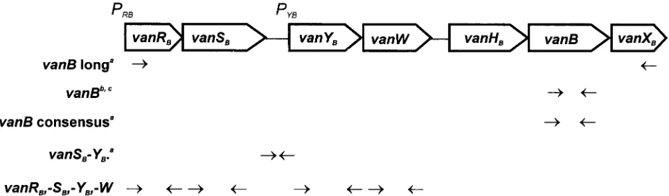
isolates was purified with the use of a Genomic DNA Prep Plus kit (A&A Biotechnology, Gdan´sk, Poland). ThevanBgene was detected by specific PCR with two different pairs of primers, primersvanB(14) and primersvanB consen-sus (16), and thevanSB-vanYBregion was amplified using primers proposed by Dahl et al. (16) (Fig. 1). Genomic DNA of theE. faecalisV583 VanB standard strain (21, 45) was used as a positive control.
[image:2.587.39.551.84.257.2]Sequencing ofvanBgene-specific PCR products.vanBgene-containing PCR products of approximately 1.1 kb, obtained for selected VRE isolates, were sequenced as previously described (29), with the addition of two internal primers: 5⬘-CGATCCGCACTACATCGG-3⬘and 5⬘-AACGGCGATGCCCGCAT-3⬘.
[image:2.587.52.535.555.697.2]FIG. 1. Dislocation of PCR primers used in the study with respect to the scheme of the Tn1547 vanBgene cluster present in theE. faecalisV583 VanB reference strain (20, 21, 43, 45). Superscript letters:a, reference 16;b, reference 14;c, in study isolates the upstreamvanBprimer annealed within thevanHBgene and not in thevanBgene.
TABLE 1. Selected clinical data for isolates in this study
Isolate
no. Phenotype Sourceb
Date of isolation
(mo/yr) Mating
c PFGE L-PCRd MIC (g/ml) of
e:
PEN AMP GEN STR VAN TEC CHL TET CIP LNZ Q-D
2496 VRES i (blood) 03/00 ⫺ A RFLP-4 32 2 ⬎1,024 ⬎2,048 256 0.25 32 32 128 2 ND 2492 VRES i (urine) 06/00 ⫺ A RFLP-4 16 4 ⬎1,024 ⬎2,048 256 0.25 32 32 256 2 ND 2493 VRES i (blood) 07/00 ⫺ A RFLP-4 2 0.5 ⬎1,024 ⬎2,048 256 0.25 32 32 256 2 ND 3127 VRES i (blood) 08/00 ⫺ A RFLP-4 32 2 ⬎1,024 ⬎2,048 256 0.25 32 64 128 2 ND 3130 VRES i (sputum) 09/00 ⫺ A RFLP-4 32 2 ⬎1,024 ⬎2,048 256 0.25 32 64 128 2 ND 3135 VRES i (sputum) 09/00 ⫺ A RFLP-4 32 8 ⬎1,024 ⬎2,048 256 0.25 64 64 128 2 ND 2499 VREM c 05/00 ⫺ a1 RFLP-2 ⬎128 64 ⬎1,024 ⬎2,048 512 0.25 16 0.5 128 2 0.5 2498 VREM i (urine) 07/00 ⫺ b RFLP-2 ⬎128 64 ⬎1,024 ⬎2,048 256 0.5 8 1 256 2 1
2497 VREM c 07/00 ⫺ a2 — ⬎128 64 ⬎1,024 ⬎2,048 512 32 32 0.5 128 2 1
3123a VREM i (blood) 08/00 ⫺ c1 RFLP-2 128 128 ⬎1,024 ⬎2,048 256 0.5 64 64 64 2 0.5
3124Aa VREM i (blood) 08/00 ⫹ c2 RFLP-2 ⬎128 128 128 ⬎2,048 256 0.5 8 64 64 2 1
3124Ba VREM i (blood) 08/00 ⫹ c3 RFLP-2 128 128 128 ⬎2,048 128 0.25 8 64 64 2 1
3128a VREM i (urine) 08/00 ⫹ c4 RFLP-2 ⬎128 64 64 ⬎2,048 512 0.25 8 64 64 2 1
2494 VSEM i (blood) 05/00 ⫺ a3 ND ⬎128 128 128 2,048 2 0.25 8 1 256 2 1
3122a VSEM i (blood) 08/00 ⫺ c5 ND ⬎128 128 64 ⬎2,048 2 0.25 8 64 64 2 0.5
aIsolates recovered from a single patient. bi, infection; c, carriage.
c⫹and⫺indicate isolates that produced and did not produce transconjugants, respectively.
dRFLP types ofvanBgene clusters amplified by L-PCR; —, lack of L-PCR product; ND, not determined.
eAbbreviations: PEN, penicillin; AMP, ampicillin; GEN, gentamicin; STR, streptomycin; VAN, vancomycin; TEC, teicoplanin; CHL, chloramphenicol; TET,
tetracycline; CIP, ciprofloxacin; LNZ, linezolid; Q-D, quinupristin-dalfopristin.
on May 15, 2020 by guest
http://jcm.asm.org/
RFLP analysis of thevanBgene cluster.Restriction fragment length polymor-phism (RFLP) of long PCR (L-PCR) products containing clusters ofvanRB, -SB, -YB, -W, -HB, -B, and -XBgenes was studied as reported previously (29) with the use of primersvanBlong for L-PCR (16) (Fig. 1).DraI andPagI (isoschizomer ofBspHI) restriction enzymes (MBI Fermentas) were used in the analysis and genomic DNA of theE. faecalisV583 VanB standard strain (21, 45) was included as a positive control.
Analysis of thevanBgene cluster location.Undigested total DNAs of the isolates were separated by PFGE and blotted onto a Hybond-N⫹membrane (Amersham Pharmacia Biotech, Little Chalfont, United Kingdom) for hybrid-ization with thevanBgene cluster probe. The L-PCR amplicon of thevanB
cluster of theE. faecalisV583 VanB reference strain (16, 21, 45) was used as the probe. Probe labeling, hybridization, and signal detection were performed with the ECL Random-Prime labeling and detection system (Amersham Pharmacia Biotech). DNA ofE. faecalisV583 (21, 45) was used as a positive control.
Detection ofvanAandvanRB, -SB, -YB, and -Wgenes.Total DNAs of selected
isolates were tested for the presence ofvanAandvanRB, -SB, -YB, and -Wgenes. ThevanAgene was detected by PCR with two different pairs of primers (14, 19), whereas the remaining genes were amplified with primers designed according to the Tn1547sequence (reference 20; GenBank accession no. U35369) (Fig. 1). Sequences of these primers were as follows:vanRB1, 5⬘-CTTGTCGAGGATG ATG-3⬘; vanRB2, 5⬘-CCTCCAATCGGTAACC-3⬘; vanSB1, 5⬘-GTCGGTGT AACGGCAAC-3⬘;vanSB2, 5⬘-GCTGGTTGTTTGCCTC-3⬘;vanYB1, 5⬘-GAA TCATCACAAACGGC-3⬘; vanYB2, 5⬘-CTCTGTCTTGTCTGGC-3⬘; vanW1, 5⬘-GATTGACACAGCGCTTC-3⬘; vanW2, 5⬘-CTCCTGAATATCCACAC-3⬘. Amplicons ofvanRB, -SB, -YBand -Wgenes, obtained for theE. faecalisV583 VanB reference strain (21, 45), were subsequently used as probes in the dot blot hybridization (46) with DNAs of the analyzed isolates. Probe labeling, hybrid-ization and signal detection were performed as described above. DNAs isolated from theE. faeciumBM4147 VanA standard strain (3) and theE. faecalisV583 VanB standard strain (21, 45) were used as controls.
Analysis ofvanBgene expression by RT-PCR.Expression of thevanBgene was studied in selected VREM isolates. Total RNA was purified from 10-ml cultures of the isolates grown overnight in Todd-Hewitt broth (THB) (Oxoid), THB with vancomycin (8g/ml), and THB with teicoplanin (8g/ml). RNA was obtained by using a Total RNA Prep Plus kit (A&A Biotechnology), treated with DNase I (amplification grade; Sigma Chemical Company, St. Louis, Mo.), and subjected to reverse transcription (RT) with thevanBlong downstream primer (16) (Fig. 1). The SuperScript II reverse transcriptase (Gibco BRL Life Technologies Inc., Karlsruhe, Germany) was used in the reaction according to the manufacturer’s protocol, and this was subsequently followed by specific PCR with thevanB
consensus pair of primers (16). Total RNAs extracted from theE. faecalisV583 VanB standard strain (21, 45) and the VSEM 2494 clinical isolate were used as controls. RNA preparations were tested for the lack of DNA contamination by PCR that had not been preceded by RT. RT-PCR products were analyzed by agarose gel electrophoresis.
Nucleotide sequence accession numbers.Nucleotide sequences ofvanHB-vanB
regions of VREM 2497 and 3123 isolates will appear in the EMBL database under accession numbers AJ306726 and AJ306727, respectively.
RESULTS
Antimicrobial susceptibility testing of VRE isolates.MICs of various antimicrobials evaluated for VRE isolates are pre-sented in Table 1. All the isolates demonstrated high-level resistance to vancomycin (MICs, 128 to 512g/ml) and sus-ceptibility to teicoplanin (MICs, 0.25 to 0.5g/ml), except for a single VREM isolate (isolate 2497) which showed resistance to both glycopeptides (vancomycin MIC, 512g/ml; teicopla-nin MIC, 32g/ml). Isolates were found to be uniformly re-sistant to ciprofloxacin and to high concentrations of at least streptomycin out of the two aminoglycosides tested (strepto-mycin and gentamicin). Resistance to other antimicrobials was also widely spread in the studied VRE population. All VREM isolates were resistant to penicillin and ampicillin, and the majority of VRES isolates demonstrated resistance to penicil-lin, chloramphenicol, and tetracycline. All VRE isolates re-vealed susceptibility to linezolid, and all VREM isolates were susceptible to quinupristin-dalfopristin. The four VREM
iso-lates recovered from a single patient (3123, 3124A, 3124B, and 3128) demonstrated similar MIC patterns; however, significant differences were observed in the case of isolate 3123, which was characterized by clearly increased MICs of gentamicin and chloramphenicol compared to the other isolates in this group. PFGE typing of VRE isolates. Results of the analysis are shown in Table 1. All the VRES isolates produced identical PFGE banding patterns (PFGE type A) in contrast to the VREM isolates, which could be classified into three distinct types with subtypes (48) (PFGE types a1 and a2, b, and c1 to c4). Two isolates of PFGE subtypes a1 and a2 (2499 and 2497, respectively) were those recovered from different carriers, whereas all four VREM isolates of PFGE subtypes c1 to c4 were those collected from a single patient (3123, 3124A, 3124B, and 3128).
Susceptibility and PFGE typing of VSEM isolates. Suscep-tibility testing and PFGE typing of the two VSEM isolates (2494 and 3122) were performed along with those of the VRE isolates, and the results are shown in Table 1. The VSEM isolates also revealed a multidrug resistance phenotype with resistance to penicillin, ampicillin, and ciprofloxacin and to high concentrations of streptomycin. Isolate 3122 was addition-ally resistant to tetracycline, similarly to the only tetracycline-resistant VREM isolates, all collected from the same patient (3123, 3124A, 3124B, and 3128). Moreover, when compared to isolates 3124A, 3124B, and 3128, isolate 3122 differed only with respect to the vancomycin MIC.
The VSEM isolates produced PFGE patterns that were sim-ilar to those of the VREM isolates. Isolate 2494 could be classified into PFGE type a (subtype a3), together with VREM isolates 2499 and 2497. Isolate 3122 represented another sub-type of PFGE sub-type c (subsub-type c5), which also included all the VREM isolates recovered from the same patient (3123, 3124A, 3124B, and 3128).
Vancomycin resistance transfer and susceptibility of transcon-jugants.Results of mating are presented in Table 1. Only three VREM isolates (3124A, 3124B, and 3128) produced vancomy-cin-resistant transconjugants, and the efficiency of conjugation was low, ranging from 10⫺7to 10⫺9 recombinants per donor
cell. Susceptibility testing revealed that no other resistance determinants were cotransferred with those of vancomycin re-sistance.
Detection of the vanB gene and the vanSB-vanYB region.
Presence of thevanBgene was checked in the VRE isolates by PCR with two different pairs of primers,vanB(14) andvanB
consensus (Fig. 1) (16). In all VRE isolates, PCR with primers
vanB, which are specific for thevanB1gene variant (16), pro-duced amplicons of about 1.1 kb instead of the 433 bp expected
for vanB1 and observed in the case ofvanB1-containing E.
faecalisV583 (14, 21, 45). On the other hand, primersvanB
consensus, which amplify a 484-bp fragment of allvanBgene variants known to date (16), yielded a product of about 500 bp (results not shown). The same pattern of vanB gene PCR products obtained with the two pairs of primers had previously been observed with VREM isolates from a hospital in Warsaw (29).
For all but one of the VRE isolates, PCR of the vanSB
-vanYBregion (which includes thePYBpromoter [Fig. 1])
pro-duced amplicons of approximately 300 bp that corresponded well to the size of 309 bp expected for the Tn1547transposon
on May 15, 2020 by guest
http://jcm.asm.org/
present in theE. faecalisV583 strain (16, 21, 45). The only exception was the VREM 2497 isolate, for which novanSB
-vanYB-specific PCR product was observed (results not shown).
Sequencing of vanB gene-containing PCR products. The 1.1-kbvanBgene-containing amplicons, obtained in PCR with
vanBprimers (14) for isolates VRES 2496, VREM 2497, and VREM 3123, were subjected to direct DNA sequencing. As had been found previously in a VREM isolate from a hospital in Warsaw (29), PCR products encompassed the 896-bp frag-ment of thevanBcoding region starting from its 5⬘end and the 201-bp fragment of the vanHB gene that is located directly
upstream ofvanB. This indicated that the forward primer of thevanBpair (14) annealed to a sequence present within the
vanHBgene, instead ofvanB. In the VRES 2496 and VREM
3123 isolates the analyzed sequence of 1,045 bp was identical to the corresponding region of the VREM strain from Warsaw (29) and contained thevanB2(24) variant of thevanBgene originally identified in the United States (GenBank accession no. U94526) (39). The sequence found in the VREM 2497 isolate revealed a single base pair difference when compared to the others. An A-to-G mutation in position 388 of thevanB
gene causes a Met-to-Val substitution at position 130 of the protein sequence with respect toE. faecalisV583 (21).
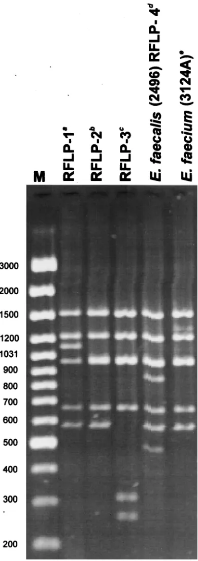
RFLP analysis of vanB gene clusters.DNA molecules en-compassing thevanBgene cluster region (Fig. 1) were ampli-fied by L-PCR, and products of the expected size of about 6 kb were obtained for all but one of the VRE isolates. Results of the DraI/PagI (BspHI) RFLP study of these amplicons are shown in Table 1; Fig. 2 presents RFLP patterns characteristic for representative VRES and VREM isolates together with those specific forE. faecalisV583, RFLP-1 (16, 21, 45), and all VREM VanB strains identified in Poland previously, RFLP-2 (16, 29) and a novel one, here designated RFLP-3 (28). All the VRES isolates containedvanBgene clusters of the same RFLP type, which turned out to be unique when compared to the types described previously (16, 28) and was designated RFLP-4. On the contrary, the VREM isolates possessed a cluster variant of the RFLP-2 type, observed before in VRE isolates from several countries (16), including the VREM strain from Warsaw (29). RFLP-2 and RFLP-4 differed from each other by the presence of two additional DNA bands in RFLP-4. The only isolate that failed to produce the 6-kb am-plicon in L-PCR was VREM 2497, for which an approximately 1.3-kb product was observed. This product, however, was also generated in the presence of the upstreamvanBlong primer alone and did not contain thevanB gene as revealed by hy-bridization (results not shown).
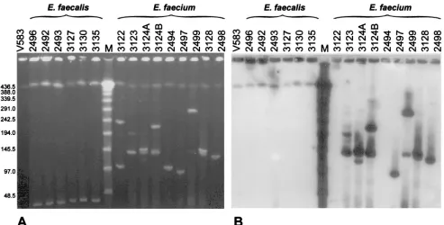
Location of vanB gene clusters. The isolates’ undigested total DNA was separated by PFGE and hybridized with the
vanBgene cluster probe. Results of the analysis are shown in Fig. 3. In all isolates PFGE visualized a DNA band of the same migration and the biggest size observed, which most probably represented their chromosomal DNA. It is also highly likely that plasmid molecules formed other bands seen in the gel, which in the case ofE. faecium isolates demonstrated a re-markable diversity in size. In all the VRES isolates thevanB
cluster probe hybridized with the putative chromosomal band, whereas in all the VREM isolates hybridization occurred with plasmid DNA. The hybridizing plasmids were of various sizes; however, all PFGE type c isolates from a single patient (3123,
3124A, 3124B, and 3128) possessed one of thevanB cluster-containing plasmids of the same migration rate (similar to the
ladder 150-kb band). Several of the VREM isolates were characterized by two (or more) hybridizing plasmid DNA bands. Detection ofvanA, andvanRB, -SB, -YB, and -Wgenes in the
teicoplanin-resistant VREM isolate.The only teicoplanin-re-sistant VREM isolate, 2497, which also turned out to be neg-ative in thevanB gene cluster L-PCR and the vanSB-vanYB
region PCR was subjected to a more detailed analysis. It was checked for the presence of thevanAgene; however, specific PCR did not yield any product (result not shown). Subse-quently the isolate was tested for a set of genes of thevanB
cluster (Fig. 1), including the regulatory genes vanRB and
vanSB. PCR failed to amplify any products specific forvanRB
(expected size, 634 bp),vanSB(expected size, 898 bp),vanYB
(expected size, 664 bp), and vanW (expected size, 803 bp) genes that were obtained with DNA from a related VREM isolate with a clear VanB phenotype, isolate 2499, and theE.
faecalis V583 VanB reference strain (21, 45) (results not
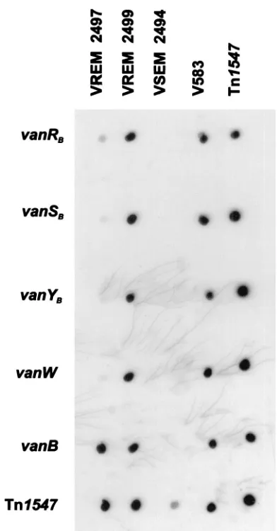
shown). In order to confirm these results, dot blot hybridiza-tion was performed, in which amplicons ofvanRB, -SB, -YB, -W,
and -Bgenes ofE. faecalisV583 were used as probes. DNA of isolate 2497 hybridized only with thevanBprobe, whereas all the probes interacted with DNA of isolate 2499 (Fig. 4).
Expression analysis of the vanB gene in the teicoplanin-resistant VREM isolate.The absence of the regulatory genes
vanRBand vanSBindicated the possibility of constitutive
ex-pression of the resistance genesvanHB, vanB, and vanXB in
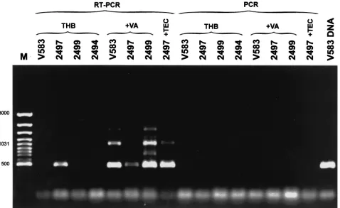
VREM isolate 2497. In order to check this hypothesis, VREM isolates 2497 (teicoplanin resistant) and 2499 (with the typical VanB phenotype) and the E. faecalis V583 VanB standard strain (21, 45) were grown in the absence and presence of vancomycin. Isolate 2497 was also grown with teicoplanin, and the VSEM 2494 isolate was grown in a nonsupplemented me-dium. Total RNA preparations of the cultures were subjected to RT-PCR, in which the downstreamvanBlong primer com-plementary to thevanXBgene (16) (Fig. 1) was used for RT,
andvanBconsensus primers, annealing to thevanBgene (16), were utilized in the subsequent PCR step. Results are shown in Fig. 5. The only non-glycopeptide-supplemented culture, in which the RT-PCR product of the expected size of approxi-mately 500 bp was observed, was that of isolate 2497. It was also obtained in cultures of isolates 2497, 2499, andE. faecalis
V583 grown with vancomycin and of isolate 2497 which was grown in the presence of teicoplanin. The length of the other major RT-PCR band of about 1 kb corresponded to the ex-pected product size (1,069 bp), which could be amplified from the upstreamvanBconsensus and the downstreamvanBlong primer. Lack of any PCR product in reactions that were not preceded by RT excluded the possibility of amplification as a result of DNA contamination of RNA preparations, and spec-ificity of the reaction was confirmed by PCR failure in the culture of VSEM isolate 2494.
DISCUSSION
In mid-1999, the first VRE isolates with the VanB pheno-type were identified in Poland, which resulted from indepen-dent selection events in two separate hospitals in Warsaw (28, 29). The data presented in this work document the third
on May 15, 2020 by guest
http://jcm.asm.org/
dence of these microorganisms being reported in the country and describe the second Polish VanB VRE outbreak that oc-curred in 2000 in two hematology units of the University Hos-pital in Cracow (located about 300 km south of Warsaw). In contrast to the previously analyzed VRE epidemics, caused by VanA VREM in Gdan´sk (27) and by VanB VREM strains in Warsaw (28), this outbreak was due to bothE. faeciumandE.
faecalis. The isolates collected during the investigation
dem-onstrated resistance to multiple antimicrobials, which is a com-mon characteristic of VRE (8, 10, 12, 36), including all VRE isolates collected before in Poland (27, 28, 29). Except for a single VREM isolate, isolate 2497 (discussed below in detail), they all revealed the typical VanB phenotype with resistance to vancomycin and susceptibility to teicoplanin (1, 44). They were also frequently resistant to penicillins, aminoglycosides at high concentrations, ciprofloxacin, tetracycline, and chlorampheni-col, and the only drugs with in vitro activity against all the isolates were quinupristin-dalfopristin (E. faecium) and lin-ezolid.
Identification of the VanB phenotype was confirmed by PCR detection of thevanBgene with primersvanBconsensus (16) and amplification of the unusually sized product (approx-imately 1.1 kb) with primersvanB(14) suggested that the gene variant present in the outbreak isolates was notvanB1 (16). Such a vanB gene amplicon has already been observed in a VREM strain from a Warsaw hospital (29) and was revealed to originate from the annealing of the upstream vanB primer within the adjacentvanHBgene. Sequence analysis of the
am-plified fragment demonstrated that selected VRES (isolate 2496) and VREM (isolate 3123) isolates contained avanB2
gene variant identical to that identified in the Warsaw strain (29) and in an isolate from the United States, in which it was originally described (39). The corresponding region in VREM isolate 2947 differed from the remaining ones only by a single mutation, and it represented a novel, though closely related,
vanB2 variant. These data, together with the results of the
RFLP analysis ofvanBgene clusters (discussed below), sug-gested that there may have been an epidemiological link be-tween the VRE isolates analyzed here and the VREM strain identified in Warsaw about 9 months earlier. The direct trans-mission of the Warsaw strain to the hospital in Cracow seems rather unlikely, as indicated by the different PFGE patterns produced by VREM isolates from the two institutions (results not shown). However, it is possible that transmission of the particular variant of VanB phenotype determinants was medi-ated by another strain or, more probably, that their reservoir is widely spread in Poland.
The RFLP analysis of vanB gene clusters present in out-break isolates demonstrated that all the VREM isolates con-tained the same cluster RFLP type, which had been identified before as RFLP-2 in VRE from Norway, Sweden, the United Kingdom, Germany, and the United States (16) and was also observed in the VREM strain from the Warsaw hospital (29). On the other hand, the VRES isolates were found to contain another polymorph of the region, which seems to represent a novel type (RFLP-4); however, it differed from RFLP-2 only by the presence of two additional restriction fragments. This observation, together with thevanBPCR and sequencing data, suggested that one of thevanBgene cluster variants evolved from the other due to DNA recombination (insertion or
dele-FIG. 2. RFLP analysis of thevanBgene cluster with the use ofDraI andPagI (BspHI) restriction enzymes. Lane M, GeneRuler 100-bp DNA Ladder Plus (MBI Fermentas). Superscript letters:a, RFLP-1 is a polymorph type of the original Tn1547 vanBcluster present inE. faecalisV583 (16, 20, 45);b, RFLP-2 was originally described by Dahl et al. (16) and here is represented by the cluster present in the VREM 8533 isolate from a hospital in Warsaw (29);c, RFLP-3 was identified by Kawalec et al. in another Warsaw hospital (28) and is represented here by the cluster of the VREM isolate 8284; d,E. faecalis 2496 represents all study VRES isolates;e,E. faecium3124A is a represen-tative of all study VREM isolates.
on May 15, 2020 by guest
http://jcm.asm.org/
[image:5.587.67.262.76.628.2]tion of a DNA fragment). It is, however, impossible to reveal whether the two gene cluster variants were introduced inde-pendently into E. faecalisand E. faeciumpopulations in the hospital or whether any of these was transmitted from one species to the other, which was then followed by its modifica-tion.
All the VRES isolates were found to be indistinguishable by PFGE, which indicated that the infections of six patients with this microorganism were due to clonal dissemination of a sin-gle strain. Contrarily, the VREM isolates collected from two infected patients represented two distinct PFGE types (types b and c); moreover, other isolates from two colonized patients, though related to each other, were classified as another type (type a). These data revealed that the spread of VREM in the hospital was mostly nonclonal and could, therefore, be medi-ated by horizontal transfer of VanB resistance genes among nonrelated strains. The hypothesis was supported by the fact that some VREM isolates (PFGE type c isolates 3124A, 3124B, and 3128) produced vancomycin-resistant transconju-gants in mating experiments. The hybridization study of the
vanB gene cluster probe with the undigested total DNA of isolates separated by PFGE demonstrated that in VRES,van
genes were located in chromosomal DNA, whereas in VREM isolates they resided within plasmid molecules. These plasmids were of different sizes in isolates belonging to different PFGE types; moreover, some VREM isolates contained more than onevanBgene cluster-carrying plasmid. (In some isolates the multiple hybridizing bands could also represent different forms of the same plasmid.) These data suggested that thevanBgene cluster was located within an active transposon, which could be inserted into various DNA replicons. It was probably spread
amongE. faecium strains by horizontal transfer mediated by the element itself or, less likely, by conjugation of plasmids, followed by their multiple rearrangements.
Two VSEM isolates identified as etiologic agents of infec-tions during the time of the VRE outbreak in the same wards were found by PFGE to be related to VREM isolates, and they also demonstrated susceptibility patterns similar to those of their VREM counterparts. They belonged to two different PFGE types discerned among E. faecium isolates: type a (VSEM isolate 2494), which included two VREM isolates from two other colonized patients (isolates 2497 and 2499), and type c (VSEM isolate 3122), which also grouped four VREM iso-lates from the same patient (3123, 3124A, 3124B, and 3128). It may be suggested that, as with previous reports (28, 41, 47), the VSEM isolates analyzed here represented endemic hospital strains which had acquired vancomycin resistance genes. How-ever, a small number of VSEM isolates identified in the wards at the time did not allow us to study in detail the endemic vancomycin-susceptible enterococcus background of the out-break, and it cannot be ruled out that the two VSEM isolates appeared due to a loss of VanB resistance determinants by VREM strains.
[image:6.587.46.539.83.333.2]An interesting group of isolates was formed by five closely related PFGE type cE. faeciumisolates collected from a single patient, four of which (VSEM 3122 and VREM 3123, 3124A, and 3124B) were identified in the same blood sample, with the remaining one (VREM 3128) being recovered from urine. Apart from the difference in vancomycin susceptibility (VSEM versus VREM) and variations in their PFGE patterns (five subtypes), these isolates also revealed some heterogeneity in susceptibility to other antimicrobials, plasmid profile, and
FIG. 3. Analysis of location ofvanBgene clusters by hybridization of thevanBcluster probe with undigested and PFGE-separated DNA of study isolates. (A) PFGE gel; (B) hybridization. Lanes contain DNA of the indicated isolates, DNA of theE. faecalisV583 VanB reference strain (lanes V583) (45), or aladder PFGE marker (lanes M) (New England BioLabs, Beverly, Mass.).
on May 15, 2020 by guest
http://jcm.asm.org/
number ofvanB gene cluster copies. It is very likely that all these observations document evolutionary changes which oc-curred in the originally homogeneousE. faeciumstrain in the course of infection of a single patient. Similar data were pre-viously obtained in the study of VREM incidence in the War-saw hospital (29). Interestingly, all the isolates were recovered 23 days after the end of the weeklong vancomycin therapy of the patient, who was also treated with piperacillin-tazobactam and ceftazidime along with vancomycin. At the time of theE.
faeciumisolation, the patient was on prolonged therapy with
cefepime and clindamycin. Such a profile of antibiotic treat-ment could create favorable conditions for the selection and evolution of enterococcal strains.
The most striking part of the outbreak analysis was the
identification of a teicoplanin-resistant VREM isolate, isolate 2497, of PFGE type a that was related to two other isolates, VREM 2499 of the typical VanB phenotype and VSEM 2494. Isolate 2497 was recovered from a patient carrier who had been hospitalized for 6 months before the strain’s isolation and had undergone prolonged antimicrobial therapy. The patient, among others, was treated twice with vancomycin and 4 days prior to identification of the isolate teicoplanin was introduced into therapy (together with amikacin and colistin). PCR anal-ysis excluded the VanA phenotype in the isolate, whereas the
vanBgene was amplified with primersvanBconsensus (16) and
vanB(14) as in all other isolates. The unusual PCR product of about 1.1 kb containing parts ofvanHBand vanBgenes
(am-plified with primersvanB) was found to differ only by a single nucleotide substitution compared to those obtained for isolates VREM 3123 and VRES 2496. However, the isolate failed to amplify the entire vanB gene cluster and the spacer region located between thevanSBandvanYBgenes (which includes
the vancomycin-induciblePYBpromoter), which prompted us
to investigate in greater detail the organization and expression of its glycopeptide resistance determinant.
PCR and hybridization revealed that isolate 2497 did not contain the four genes that are located upstream of thevanHB
gene within the wild-typevanBgene cluster, namely,vanRB,
-SB, -YB, and -W(16, 20, 43). The most important finding was
the lack of genes vanRB and -SB, which code for the
two-component VanRB-VanSBsystem that activates transcription
ofvanYB, -W, -HB, -B, and -XBgenes from thePYBpromoter in
the presence of vancomycin but not teicoplanin (20). There-fore, it was hypothesized that the deletion of a large part of the
vanBgene cluster, includingvanRBand -SBgenes and thePYB
promoter, resulted in constitutive expression of resistance genes vanHB, -B, and -XB, and, subsequently, in teicoplanin
resistance. In order to check this hypothesis the RT-PCR ex-periment was carried out, and it indeed demonstrated that the genes were transcribed in this isolate in the absence of glyco-peptides. The promoter sequence responsible for the effect remains to be determined. In the work of Evers and Courvalin, S1 nuclease mapping suggested that a weak promoter might be present immediately upstream of the vanHB gene, but this
result was poorly reproducible and was not confirmed by other approaches (20).
Teicoplanin-resistant VanB mutants have been rarely ob-served among clinical isolates of enterococci. Hayden et al. described a VanB VREM isolate for which the MIC of teico-planin was 64g/ml and which constitutively expressed a 41-kDa membrane protein that was vancomycin inducible in the
E. faecalisV583 reference strain (26). However, the protein
was not studied in detail, and it is not known what modifica-tions of thevanBgene cluster were responsible for the pheno-type. On the other hand such mutants have been often ob-tained in the laboratory, including selection in an animal model, and all those studied down to the molecular level were found to result from point mutations in thevanSBgene (2, 4,
5, 6, 7, 25, 33). Some of these changes caused the VanSBability
to recognize teicoplanin as the expression inducer, whereas others abolished its phosphatase activity, which led to the per-manent phosphorylation of VanRBand constitutive
transcrip-tion of the resistance genes. Finally, VanSBnull mutants were
[image:7.587.64.260.70.445.2]revealed to express the heterogeneous inducible phenotype
FIG. 4. Analysis of presence ofvanRB, -SB, -YB, and -Wgenes in the
teicoplanin-resistant VREM 2497 isolate by dot blot hybridization with a set of specific probes. DNAs of particular isolates are dislocated in columns: VREM 2497, the teicoplanin-resistant isolate; VREM 2499, the isolate related to 2497 with the typical VanB phenotype; VSEM 2494, the VSEM isolate related to VREM 2497 and 2499; V583, theE. faecalis V583 VanB standard strain (45); Tn1547, L-PCR product encompassing thevanBgene cluster from theE. faecalisV583 strain. Rows represent hybridization with probes specific for corresponding genes; the Tn1547probe was the labeled L-PCR product described above.
on May 15, 2020 by guest
http://jcm.asm.org/
that probably resulted from taking over the VanSBfunctions by
a host kinase (2, 4, 5, 6, 7). Deletion mutants lacking the whole VanRB-VanSBregulatory system and thePYBpromoter have
not been, however, observed to date.
The data presented in this work document a VanB VRE outbreak, which even though it did not involve many patients (10 patients in all) turned out to be very complex and so reflected well the dynamics of VRE epidemiology. It was caused by VRES as well as VREM which could exchange vancomycin resistance determinants or acquire them indepen-dently. The VRE spread was both clonal (VRES) and non-clonal (VREM), and horizontal transmission of vanB gene clusters probably occurred due to the conjugative functions of the transposable elements containing the clusters. The VREM strains were undergoing diversification on the level of chromo-somal and plasmid DNA, and evolution of one of them could be observed in the course of infection of a single patient. Treatment of one of the patients with teicoplanin was probably responsible for the selection of a VREM strain variant, which due to the deletion of a large part of thevanBgene cluster was also resistant to teicoplanin. This finding, together with data from other laboratories, indicates that various genetic changes may determine resistance to teicoplanin in VanB enterococci and thus limit its use against these microorganisms (2, 6, 33, 36).
ACKNOWLEDGMENTS
We thank Ewa Sadowy and Andrew Hazlewood for critical reading of the manuscript and Andrzej Pałucha for very helpful discussions and the SuperScript II enzyme. Also we are very thankful to Patrice Cour-valin who kindly providedE. faeciumBM4147 andE. faecalisV583, Wolfgang Witte for theE. faecium64/3 strain, and Wolfgang Haas for
E. faecalisFA2-2.
This work was partially financed by the U.S.-Poland Maria Skło-dowska-Curie Joint Fund II (MZ/NIH 98-324) and the program SPUB-M-INCO-COPERNICUS (4 PR UE/P-05/DZ 112/2000) of the Polish Committee for Scientific Research (KBN).
REFERENCES
1.Arthur, M., and P. Courvalin.1993. Genetics and mechanisms of glycopep-tide resistance in enterococci. Antimicrob. Agents Chemother.37:1563– 1571.
2.Arthur, M., F. Depardieu, and P. Courvalin.1999. Regulated interactions between partner and non-partner sensors and response regulators that con-trol glycopeptide resistance gene expression in enterococci. Microbiology 145:1849–1858.
3.Arthur, M., C. Molinas, F. Depardieu, and P. Courvalin.1993. Character-ization of Tn1546, a Tn3-related transposon conferring glycopeptide resis-tance by synthesis of depsipeptide peptidoglycan precursors inEnterococcus faeciumBM4147. J. Bacteriol.175:117–127.
4.Arthur, M., and R. Quintiliani, Jr.2001. Regulation of VanA- and VanB-type glycopeptide resistance in enterococci. Antimicrob. Agents Chemother. 45:375–381.
[image:8.587.55.533.76.369.2]5.Aslangul, E., M. Baptista, B. Fantin, F. Depardieu, M. Arthur, P. Courvalin, et al.1997. Selection of glycopeptide-resistant mutants of vanB-type Entero-coccus faecalisBM4281in vitroand in experimental endocarditis. J. Infect. Dis.175:598–605.
FIG. 5. RT-PCR analysis ofvanBgene expression in the teicoplanin-resistant VREM 2497 isolate. M, GeneRuler 100-bp DNA Ladder Plus (MBI Fermentas); V583 DNA, product of PCR withvanBconsensus primers (16) performed with DNA of theE. faecalisV583 VanB reference strain (45). RT-PCR lanes refer to RNA preparations that were subjected to both RT and PCR; PCR lanes refer to control reactions in which the RT step was omitted. Abbreviations: THB, refers to reactions performed on RNAs extracted from cultures grown in the absence of glycopeptides; ⫹VA and⫹TEC, refer to cultures supplemented with vancomycin and teicoplanin, respectively. Strain designations: V583, theE. faecalisV583 VanB standard strain (45); 2497, the teicoplanin-resistant VREM isolate; 2499, isolate related to 2497 VREM with the typical VanB phenotype; 2494, isolate related to 2497 and 2499 VSEM isolate.
on May 15, 2020 by guest
http://jcm.asm.org/
6.Baptista, M., F. Depardieu, P. Courvalin, and M. Arthur.1996. Specificity of induction of glycopeptide resistance genes inEnterococcus faecalis. Antimi-crob. Agents Chemother.40:2291–2295.
7.Baptista, M., P. Rodrigues, F. Depardieu, P. Courvalin, and M. Arthur. 1999. Single-cell analysis of glycopeptide resistance gene expression in teico-planin-resistant mutants of VanB-typeEnterococcus faecalis. Mol. Microbiol. 32:17–28.
8.Bell, J. M., J. C. Paton, and J. Turnidge.1998. Emergence of vancomycin-resistant enterococci in Australia: phenotypic and genotypic characteristics of isolates. J. Clin. Microbiol.36:2187–2190.
9.Boyce, J. M., M. S. Favero, R. P. Gaynes, D. A. Goldmann, W. R. Jarvis, G. Pugliese, and R. A. Weinstein. 1998. Populations at risk and routes of transmission, p. 15–16.InG. Pugliese and R. A. Weinstein (ed.), Issues and controversies in prevention and control of VRE. Etna Communications, Chicago, Ill.
10.Boyce, J. M., S. M. Opal, J. W. Chow, M. J. Zervos, G. Potter-Bynoe, C. B. Sherman, R. L. C. Romulo, S. Fortna, and A. A. Medeiros.1994. Outbreak of multidrug-resistant Enterococcus faeciumwith transferable vanBclass vancomycin resistance. J. Clin. Microbiol.32:1148–1153.
11.Carias, L. L., S. D. Rudin, C. J. Donskey, and L. B. Rice.1998. Genetic linkage and cotransfer of a novelvanB-containing transposon (Tn5382) and a low-affinity penicillin-binding protein 5 gene in a clinical vancomycin-resistantEnterococcus faeciumisolate. J. Bacteriol.180:4426–4434. 12.Cetnikaya, Y., P. Falk, and C. G. Mayhall.2000. Vancomycin-resistant
en-terococci. Clin. Microbiol. Rev.13:686–707.
13.Chow, J. W., A. Kutitza, D. M. Shlaes, M. Green, D. F. Sahm, and M. J. Zervos.1993. Clonal spread of vancomycin-resistantEnterococcus faecium
between patients in three hospitals in two states. J. Clin. Microbiol.31:1609– 1611.
14.Clark, N., R. Cooksey, B. Hill, J. Swenson, and F. C. Tenover.1993. Char-acterization of glycopeptide-resistant enterococci from U.S. hospitals. Anti-microb. Agents Chemother.37:2311–2317.
15.Clewell, D. B., P. K. Tomich, M. C. Gawron-Burke, A. E. Franke, Y. Yagi, and F. Y. An.1982. Mapping ofStreptococcus faecalisplasmids pAD1 and pAD2 and studies relating to transposition of Tn917. J. Bacteriol.152:1220– 1230.
16.Dahl, K. H., G. Skov Simonsen, Ø. Olsvik, and A. Sundsfjord.1999. Heter-ogeneity invanBgene cluster of genetically diverse clinical strains of van-comycin-resistant enterococci. Antimicrob. Agents Chemother. 43:1105– 1110.
17.De Lencastre, H., E. P. Severina, R. B. Roberts, B. N. Kreiswirth, A. Tomasz, and the BARG Initiative Pilot Study Group.1996. Testing the efficacy of a molecular surveillance network: methicillin-resistantStaphylococcus aureus
(MRSA) and vancomycin-resistantEnterococcus faecium(VREF) genotypes in six hospitals in the metropolitan New York City area. Microb. Drug Resist.2:343–351.
18.Donskey, C. J., J. R. Schreiber, M. R. Jacobs, R. Shekar, R. A. Salata, S. Gordon, C. C. Whalen, F. Smith, L. B. Rice, and the Northeast Ohio Van-comycin-Resistant Enterococcus Surveillance Program.1999. A polyclonal outbreak of predominantly VanB vancomycin-resistant enterococci in north-east Ohio. Clin. Infect. Dis.29:573–579.
19.Dutka-Malen, S., S. Evers, and P. Courvalin.1995. Detection of glycopep-tide resistance genotypes and identification to the species level of clinically relevant enterococci by PCR. J. Clin. Microbiol.33:24–27.
20.Evers, S., and P. Courvalin.1996. Regulation of VanB-type resistance gene expression by the VanSB-VanRBtwo-component regulatory system in
En-terococcus faecalisV583. J. Bacteriol.178:1302–1309.
21.Evers, S., P. E. Reynolds, and P. Courvalin.1994. Sequence of thevanBand
ddlgenes encoding D-alanine:D-lactate and D-alanine:D-alanine ligases in vancomycin-resistantEnterococcus faecalisV583. Gene140:97–102. 22.Facklam, R., and M. D. Collins.1989. Identification ofEnterococcusspecies
isolated from human infections by a conventional test scheme. J. Clin. Mi-crobiol.27:731–734.
23.Fines, M., B. Perichon, P. Reynolds, D. F. Sahm, and P. Courvalin.1999. VanE, a new type of acquired glycopeptide resistance inEnterococcus fae-calisBM4405. Antimicrob. Agents Chemother.43:2161–2164.
24.Gold, H. S., S. U¨ nal, E. Cercenado, C. Thauvin-Eliopulos, G. M. Eliopulos, C. B. Wennerstein, and R. C. Moellering, Jr.1993. A gene conferring resis-tance to vancomycin but not teicoplanin in isolates ofEnterococcus faecalis
andEnterococcus faeciumdemonstrates homology withvanB,vanA, and
vanCgenes of enterococci. Antimicrob. Agents Chemother.37:1604–1609. 25.Gutmann, L., D. Billot-Klein, S. Al-Obeid, I. Klare, S. Francoual, E. Collatz,
and J. Van Heijenoort.1992. Inducible carboxypeptidase activity in vanco-mycin-resistant enterococci. Antimicrob. Agents Chemother.36:77–80. 26.Hayden, M. K., G. M. Trenholme, J. E. Schultz, and D. F. Sahm.1993.In
vivodevelopment of teicoplanin resistance in a VanBEnterococcus faecium
isolate. J. Infect. Dis.167:1224–1227.
27.Kawalec, M., M. Gniadkowski, and W. Hryniewicz.2000. Outbreak of van-comycin-resistant enterococci in a hospital in Gdan´sk, Poland, due to hori-zontal transfer of different Tn1546-like transposon variants and clonal spread of several strains. J. Clin. Microbiol.38:3317–3322.
28.Kawalec, M., M. Gniadkowski, M. Zaleska, T. Ozorowski, L. Konopka, and
W. Hryniewicz.2001. Outbreak of vancomycin-resistantEnterococcus fae-ciumof the phenotype VanB in a hospital in Warsaw, Poland: probable transmission of the resistance determinants into an endemic vancomycin-susceptible strain. J. Clin. Microbiol.39:1781–1787.
29.Kawalec, M., M. Gniadkowski, U. Zielin´ska, W. Kłos, and W. Hryniewicz. 2001. A vancomycin-resistantEnterococcus faeciumstrain carrying thevanB2
gene variant in a Polish hospital. J. Clin. Microbiol.39:811–815. 30.Klare, I., E. Collatz, S. Al.-Obeid, J. Wagner, A. C. Rodloff, and W. Witte.
1992. Glykopeptidresistenz beiEnterococcus faeciumaus Besiedlungen und Infectionen von Patienten aus Intensivstationen Berliner Kliniken und einem Transplantationszentrum. Z. Antimikrob. Antineoplast. Chemother. 10:45–53.
31.Leclercq, R., E. Derlot, J. Duval, and P. Courvalin.1988. Plasmid-mediated resistance to vancomycin and teicoplanin inEnterococcus faecium. N. Engl. J. Med.319:157–161.
32.Leclercq, R., S. Dutka-Malen, J. Duval, and P. Courvalin.1992. Vancomycin resistance genevanC is specific toEnterococcus gallinarum. Antimicrob. Agents Chemother.36:2005–2008.
33.Lefort, A., M. Baptista, B. Fantin, F. Depardieu, M. Arthur, C. Carbon, and P. Courvalin.1999. Two-step acquisition of resistance to the teicoplanin-gentamicin combination by VanB-typeEnterococcus faecalisin vitro and in experimental endocarditis. Antimicrob. Agents Chemother.43:476–482. 34.Maki, D. G., and W. A. Agger.1988. Enterococcal bacteremia: clinical
fea-tures, the risk of endocarditis and management. Medicine67:248–269. 35.McKessar, S. J., A. M. Berry, J. M. Bell, J. D. Turnidge, and J. C. Paton.
2000. Genetic characterization ofvanG, a novel vancomycin resistance locus ofEnterococcus faecalis. Antimicrob. Agents Chemother.44:3224–3228. 36.Murray, B. E.1997. Vancomycin-resistant enterococci. Am. J. Med.102:
284–293.
37.Murray, B. E. 1998. Diversity among multidrug-resistant enterococci. Emerg. Infect. Dis.4:37–47.
38.National Committee for Clinical Laboratory Standards.2000. Methods for dilution antimicrobial susceptibility tests for bacteria that grow aerobically; approved standard, M7–A5. NCCLS, Wayne, Pa.
39.Patel, R., J. R. Uhl, P. Kohner, M. K. Hopkins, J. M. Steckelberg, B. Kline, and F. R. I. Cockerill.1988. DNA sequence variation withinvanA,vanB,
vanC-1, andvanC-2/3genes of clinicalEnterococcusisolates. Antimicrob. Agents Chemother.42:202–205.
40.Perichon, B., P. E. Reynolds, and P. Courvalin.1997. VanD-type glycopep-tide-resistant Enterococcus faecium BM4339. Antimicrob. Agents Che-mother.41:2016–2018.
41.Perlada, D. E., A. G. Smulian, and M. T. Cushion.1997. Molecular epide-miology and antibiotic susceptibility of enterococci in Cincinnati, Ohio: a prospective citywide survey. J. Clin. Microbiol.35:2342–2347.
42.Quintiliani, R., Jr., and P. Courvalin.1994. Conjugal transfer of the vanco-mycin-resistance determinantvanBbetween enterococci involves the move-ment of large genetic elemove-ments from chromosome to chromosome. FEMS Microbiol. Lett.119:359–364.
43.Quintiliani, R., Jr., and P. Courvalin.1996. Characterization of Tn1547, composite transposon flanked by the IS16and IS256-like elements, that confers vancomycin resistance inEnterococcus faecalisBM4281. Gene172: 1–8.
44.Quintiliani, R., Jr., S. Evers, and P. Courvalin.1993. ThevanBgene confers various levels of self-transferable resistance to vancomycin in enterococci. J. Infect. Dis.167:1220–1223.
45.Sahm, D. F., J. Kissinger, M. S. Gilmore, B. E. Murray, R. Mulder, J. Solliday, and B. Clarke.1989. In vitro susceptibility studies of vancomycin-resistantEnterococcus faecalis. Antimicrob. Agents Chemother.33:1588– 1591.
46.Sambrook, J., E. F. Fritsch, T. Maniatis.1989. Molecular cloning: a labo-ratory manual, 2nd ed. Cold Spring Harbor Labolabo-ratory Press, Cold Spring Harbor, N.Y.
47.Suppola, J. P., E. Kolho, S. Salmenlinna, E. Tarkka, J. Vuopio-Varkila, and M. Vaara.1999.VanA andvanB incorporate into an endemic ampicillin-resistant vancomycin-sensitiveEnterococcus faeciumstrain: effect on inter-pretation of clonality. J. Clin. Microbiol.37:3934–3939.
48.Tenover, F. C., R. Arbeit, V. Goering, P. Mickelsen, B. M. Murray, D. Pershing, and B. Swaminathan.1995. Interpreting chromosomal DNA re-striction patterns produced by pulsed-field gel electrophoresis: criteria for bacterial strain typing. J. Clin. Microbiol.33:2233–2239.
49.Thal, L., S. Donabedian, B. Robinson-Dunn, J. W. Chow, L. Dembry, D. B. Clewell, D. Alshab, and M. J. Zervos.1998. Molecular analysis of glycopep-tide-resistantEnterococcus faeciumisolates collected from Michigan Hospi-tals over a 6-year period. J. Clin. Microbiol.36:3303–3308.
50.Werner, G., I. Klare, and W. Witte.1997. Arrangement of the vanA gene cluster in enterococci of different ecological origin. FEMS Microbiol. Lett. 155:55–61.
51.Woodford, N., B. L. Jones, Z. Baccus, H. A. Ludlam, and D. F. J. Brown. 1995. Linkage of vancomycin and high-level gentamicin resistance genes on the same plasmid in a clinical isolate ofEnterococcus faecalis. J. Antimicrob. Chemother.35:179–184.