5
X
October 2017
A Survey on Image Enhancement Based Histogram
Equalization Techniques
Amit Gupta1, Vivek Jain2
1
Dept. of Computer Science, SRCEM, Banmore, India
2
Dept. of Computer Science, SRCEM, Banmore, India
Abstract: Image enhancement (IE) is the process of enhancing visual appearance of image in order to make it more effective for computer to process. The look and visibility of image rely on human eyes, which vary from one person to another. Several enhancement methods are used to improve the clarity of image, but different application require different types of techniques for enhancing image quality. In this review paper, we present different enhancement techniques categorized into spatial and frequency domain to classify image quality with its advantage and disadvantage.
Keywords: Image Enhancement; Spatial Domain; Frequency Domain; Histogram Equalization
I. INTRODUCTION
Image processing is a very active area of research in computing environment. In day to day life cycle, several applications include medicine to transportation and industry as well. A necessary requirement of digital image that creates enhanced contrast with hiding information in areas like biomedical image analysis, fault detection and remote sensing. So, the method that process a given image into visually better resultant from original one are called Image Enhancement (IE) [1].
[image:2.612.182.433.369.506.2](a) Original Image (b) Enhanced Image
Fig 1: Showing effects of IE [2]
IE method is the process of improving the visual quality of digital image, without the prior knowledge of source degradation. It is the method of improving the images interpretability or perception of information in human views [3]. The principle work of IE is to build differentiate in a low complexity picture or to discover the concealed points of interest in a picture. At whatever point changing over a picture for digitizing reason some type of debasement happens at the yield.
II. RELATEDWORK
Sundaram et al. [6] proposed a method using Histogram Modified Contrast Limited Adaptive Histogram Equalization (HM CLAHE)
to regulate the level of contrast enhancement, that result is a strong contrast image that brings the location details more relevant to interpretation. In this modified histogram is used as both optimization method and CLAHE. The proposed method is evaluate using Mias mammogram images and performance parameter using Enhancement Measure (EME). The result of proposed is better contrast enhancement with preserving the neighbourhood information on the mammogram images.
Xiaoying Fang et al. [7] proposed an enhancement method to enhance the image fusion result with assessment on sharpness. As we
know that Image enhancement (IE) improves the perception of information. In this algorithm at first an image is taken from a real scene and then it is separated into many regions according to the use for enhancement.
Agarwal et al. [8] proposed a method using “Modified Histogram Based Contrast Enhancement using Homomorphic Filtering” (MH-FIL) on medical images. In all types of medical images, histogram based techniques enhances the low contrast for proper visual. This method work in two parts, in first part enhanced the image global contrast and second part image is sharpen using homomorphic filtering. And this filtering is followed by image normalization. The proposed algorithm result proved as efficient and flexible for medical image enhancement and can be closed a pre-processing step for proper analysis and understanding of medical image.
S.S.Chong [9] proposed an improved form of hyperbolic algorithm contrast enhancement technique suitable for magnetic resonance imaging (MRI). In this method contrast enhancement of image is done by controlled fusion of the gray level stretching on structure. The experimental result of proposed method is better on fatty and granular tissues with contrast and also avoids the over enhancement of image by maintaining the overall image brightness.
Ravinder Kaur et al. [10] in this study, enhancement of image algorithm is based on the weighted filter, histogram equalization (HE) and wavelet transformation to solve this problem. The experimental results shows that proposed approach can enhance the high-contrast images effectively; it not only improves the global brightness and high-contrast of images but also preserves details and remove noise. The other advantage of the proposed method is that it is fully automatic and requires no parameter settings. Therefore, it is useful and suitable for most digital camera users.
Zhijun Yao et al. [11] presented a new image enhancement method namely DSINMHE. In this method, enhancement is controlled by use of histogram modification method and maximizes entropy in the HE process. Subjective evaluation result of DSINMHE is also better over other methods according to natural appearance.
III.TECHNIQUESOFIMAGEENHANCEMENT
Relevant Image enhancement (IE) is used to enhance the visual appearance of image, so that human eyes can easily view or understand image, give ‘better’ input for other image processing techniques. These techniques are defined into two domains:
A. Spatial Domain
This technique directly deals with the image pixels, and the values of pixels are changed to get desired enhancement. The main use of this technique is that they are easy to understand and complexity is low that favor’s real time implementations. Spatial Domain includes techniques like:
1) Log Transformation Technique: In this transformation technique the basic image enhancement of spatial domain can be efficiently used for contrast enhancements of dark images. The log transform is basically a gray level transform and the pixels are changed to gray levels, This transform technique determine the values of low gray level of input image to a wider range of output levels. The general equation of transform is given as S=c log (1+r) where S is output grey level, c is a constant and r is the input
grey level. It is assumed that r ≥ 0 [12].
B. Powers-Law Transformations
In this transformation, image transform is guided by equation known as Gamma correction [13] and the value of gamma Y decides the level of enhancement. Hence this technique is also known as Gamma correction. This transformation techniques is used many display devices such as CRT (TV).
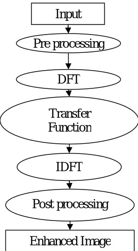
In this domain, an image is firstly transformed into frequency domain. All the enhancement operations of image are executed on Fourier transform then the reverse process of inverse Fourier transforms are executed to get the resultant image. The result of output image pixel values are changed on the basis of applied transformation function. The techniques includes in Frequency domain are DFT, DCT, low-pass filter, high-pass filter, notch filter etc.
1) Low pass filter:In Low pass filtering includes the removal of high frequency components from an image that result is sharp
transitions reduction that are associated with noise. In case of idle low pass filter keeps all the low frequency components and removes all high frequency components that results two issues in low pass filters i.e. blurring and ringing. Basically such problems are affected by the shapes related with the spatial domain filters [13].
[image:4.612.239.375.218.465.2]2) High pass filter: High pass filters are mainly used in image to sharper the image view. It works in same way as low pass filters except uses different convolution kernel and focuses on the fine details of image. This filter improves the sharpening of image, while the excessive use of filter results is degraded image quality [12].
Fig 2: Frequency Transform
D. ADVANTAGE AND DISADVANTAGE OF ENHANCEMENT
Method Advantage Disadvantage
Spatial Domain
The main advantage of this method is easy to understand and are less complex which favor’s real time implementations.[14]
The disadvantage of this method is lack in providing adequate and robustness requirements.
Frequency Domain
The advantages of frequency
domain image enhancement
method includes ease of
computation and view, the
manipulation and frequency
composition of image of the image and the easy applicability of special transformed domain properties.
The main limitation of this method is cannot enhance
every part of image
simultaneously and also difficult to automate the
image enhancement
E. APPLICATION OF IMAGE ENHANCEMENT
Several application areas in which image enhancement is used as:
1) E-learning: The use of image enhancement in the area of e-learning, where enhances the text written on chalkboard are viewed or
present as streamed video to increase the level of text readability.
2) Medical: The application of image enhancement (IE) in medical is to improve the noise and sharpen the image details for visual representation. This makes important tool for reviewing anatomic areas in MRI, ultrasound and x-rays to name a few.
3) Forensic: The use of image enhancement (IE) in forensic is to collect the evidence, identifying and surveillance. The collect images can be used for security videos analysis, fingerprint detection and investigation of crime scene are enhanced so that identification of the culprits and protection of victims [4].
4) Other areas: Several areas of image enhancement (IE) are Medical imaging, Satellite imaging, Aerial imaging, remote sensing and Digital camera application, Astrophotography, Fingerprint matching, etc.
IV.HISTOGRAMEQUALIZATIONTECHNIQUES
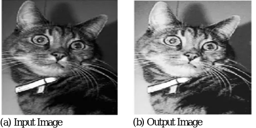
A. Histogram Equalization
Histogram is generally a graphic representation of the distribution of data. The histogram defines how certain times a specific gray level (intensity) appears in an image. Histogram equalization technique is used for enhancing the contrast and adjustment in image processing. The technique of histogram can be used in many application areas such as object tracking, speech recognition, and medical image processing such as providing better view of bone structure in x-ray images, improving the foreground and background of photographs in terms of both brightness and darkness [15].
[image:5.612.118.459.338.498.2](a) Input Image (b) Output Image
Fig 3: Histogram Equalization
B. Adaptive Histogram Equalization (AHE)
AHE technique is used to improve or enhance the contrast in images. Histogram equalization (HE) only focuses on local contrast place of overall contrast. AHE technique is applicable for all methods. In AHE techniques, those images exhibit with regions that are lighter and darker and contrast in such regions will not enhanced are properly enhanced image regions by use [15].
(a) Input Image (b) Output Image
[image:5.612.178.437.577.707.2]C. Brightness Preserving BI- Histogram Equalization
This technique separates image histogram into two parts. The intensity of partition is defined by the mean brightness value of input that is average intensity of each pixel that forms an input image. BBHE levels the sub-pictures autonomously on the premise of suitable histograms with in the imperative, such samples in the correct set are mapped into the range from the base gray-level to the info mean and the examples in the last set are mapped into the range from the mean of the most extreme gray-level. Subsequently, the resultant balanced sub-pictures are encompassed by each other around the info mean, which has a result of saving mean brightness [16].
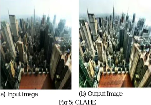
V. CLAHE
CLAHE represents the Contrast limited adaptive histogram equalization. This method does not need any predicted weather information for the processing of fogged image. In this method firstly, the image is taken by camera in foggy environment and converted from RGB (red, green and blue) color space is converted to HSV (hue, saturation and value) color space.
[image:6.612.182.425.234.404.2](a) Input Image (b) Output Image
Fig 5: CLAHE
The image transform is done because the color sense of humans is similar to HSV colors; CLAHE processed the secondary value component of image without effecting hue and saturation. The original histogram is cropped and the cropped pixels are redistributed to each gray-level. In CLAHE every pixel value is decreased to maxima of client selectable. Finally, the handled picture in HSV shading space is changed over back to RGB shading space [16].
VI.DYNAMICHISTOGRAMEQUALIZATION(DHE)
The DHE method performs well over traditional HE, so that it can enhance an image without making any changing property for details in the given image. DHE separates the input image histogram into much number of sub-histograms until it confirms that no dominating portion is present in any of the newly created sub-histograms. After that, all sub histogram must go through histogram equalization and is permissible to occupy a specified gray level range in the enhanced obtained image. The obtained IE is general better by DHE with controlled dynamic scope of gray-levels and taking out the likelihood of the low histogram segments being packed that may make some portion of the picture have washed out appearance [19].
VII. PERFORMANCE MEASUREMENT
A. Peak Signal to Noise Ratio (PSNR):
PSNR is the evaluation standard of the reassembled image quality and is the most wanted feature. It can be calculate in decibels (dB) and it is given by
Where the value 255 is the maximum possible value that can be attained by the image signal. The higher value of PSNR shows the better reassembled image.
B. Entropy (En):
C. Mean Square Error (MSE)
It is defined as the average square difference between reference signals to distorted signal. It can be evaluate by adding up the squared difference pixel-by-pixel and dividing by the total pixel count. Suppose m x n is a noise free monochrome image X, and Y is defined as the noisy approximation. Then the mean square error between these two signals is defined as:
D. Signal-To-Noise Ratio (SNR)
It is defined as the ratio between signal power to noise power and evaluate in terms of decibels. . Higher the SNR value betters the reconstructed image.
Consider r(x,y) be the original image and t(x,y) is enhanced image. The noise estimation in enhanced fundus image is analyzed
by-VIII. CONCLUSION
Image enhancement plays a significant role in image processing. Image enhancement improves the image to provide better representation and information. The main focus of enhancement method is to produce images without severe side effects at the same time maintain input mean brightness. In this paper presents a study on Image Enhancement with its techniques and advantage in spatial and frequency domain In future work, we can use the multilevel hierarchal clustering on medical x-ray images.
REFERENCES
[1] Ravindra Pal Singh, Manish Dixit and Sanjay Silakari “Image Contrast Enhancement using GA and PSO: A Survey” Sixth International Conference on Computational Intelligence and Communication Networks IEEE, 2014.
[2] Shikha Mahajan, Richa Dogra “A Review on Image Enhancement Techniques” International Journal of Engineering and Innovative Technology (IJEIT) Volume 4, Issue 11, May 2015.
[3] Rajesh Garg, Bhawna Mittal, Sheetal Garg, ”Histogram Equalization Techniques for Image Enhancement”, IJECT Vol. 2,Issue 1,March2011.
[4] Komal Viji , Yaduvir Singh, “Comparison between Different Techniques of Image Enhancement” International Journal of VLSI and signal Processing Applications, Vol.1,Issue 2,May 2011 ,(112-117).
[5] Drakshaveni.G1, Dr. Prasad Naik Hamsavath2 “Comparative Analysis of Image Enhancement Techniques to be used in Medical Images -A Survey” International Journal of Latest Research in Engineering and Technology (IJLRET) PP. 42-51.
[6] Sundaram, M., K. Ramar, N. Arumugam, and G. Prabin. "Histogram based contrast enhancement for mammogram images." In Signal Processing, Communication, Computing and Networking Technologies (ICSCCN), 2011 International Conference on, pp. 842-846. IEEE, 2011.
[7] X. Fang, J. Liu, W. Gu and Y. Tang, "A Method to Improve the Image Enhancement Result based on Image Fusion," 2012.
[8] Agarwal, T.K. et al. “ Modified Histogram based contrast enhancement using Homomorphic Filtering for medical images”, Advance Computing Conference (IACC), 2014 IEEE International on 1-22 Feb. 2014
[9] S.S.Chong et al. Faculty of Engineering & Technolgy, Multimedia University, Melaka, Malaysia “Modified HL Contrast Enhancement Technique for Breast Mr Image”, 2013 IEEE International Conference on Signal and Image Processing Applications (ICSIPA).
[10] Ravinder Kaur, Taqdir “Image Enhancement Techniques- A Review” International Research Journal of Engineering and Technology (IRJET) Volume: 03 Issue: 03 | March-2016
[11] Zhijun Yao; Zhongyuan Lai; Chun Wang “Image Enhancement Based on Equal Area Dualistic Sub-image and Non-parametric Modified Histogram Equalization Method” International Symposium on Computational Intelligence and Design (ISCID) IEEE 2016.
[12] Rashmi Choudhary, Sushopti Gawade “Survey on Image Contrast Enhancement Techniques” International Journal of Innovative Studies in Sciences and Engineering Technology (IJISSET)| Volume: 2 Issue: 3 | March 2016
[13] Shikha Mahajan, Richa Dogra “A Review on Image Enhancement Techniques” International Journal of Engineering and Innovative Technology (IJEIT)|Volume 4, Issue 11, May 2015
[14] Sargun and Shashi B. Rana “A Review of Medical Image Enhancement Techniques for Image Processing” International Journal of Current Engineering and Technology Vol.5, No.2 (April 2015).
![Fig 1: Showing effects of IE [2]](https://thumb-us.123doks.com/thumbv2/123dok_us/8304040.855419/2.612.182.433.369.506/fig-showing-effects-of-ie.webp)