comment
reviews
reports
deposited research
interactions
information
refereed research
Protein family review
Fibroblast growth factors
David M Ornitz* and Nobuyuki Itoh
Addresses: *Department of Molecular Biology and Pharmacology, Washington University Medical School, 660 S. Euclid Avenue, St. Louis, MO 63110, USA. Department of Genetic Biochemistry, Kyoto University Graduate School of Pharmaceutical Sciences, Yoshida-Shimoadachi, Sakyo, Kyoto 606-8501, Japan.
Correspondence: David M Ornitz. E-mail: [email protected]
Summary
Fibroblast growth factors (FGFs) make up a large family of polypeptide growth factors that are found
in organisms ranging from nematodes to humans. In vertebrates, the 22 members of the FGF family
range in molecular mass from 17 to 34 kDa and share 13-71% amino acid identity. Between vertebrate
species, FGFs are highly conserved in both gene structure and amino-acid sequence. FGFs have a high
affinity for heparan sulfate proteoglycans and require heparan sulfate to activate one of four
cell-surface FGF receptors. During embryonic development, FGFs have diverse roles in regulating cell
proliferation, migration and differentiation. In the adult organism, FGFs are homeostatic factors and
function in tissue repair and response to injury. When inappropriately expressed, some FGFs can
contribute to the pathogenesis of cancer. A subset of the FGF family, expressed in adult tissue, is
important for neuronal signal transduction in the central and peripheral nervous systems.
Published: 9 March 2001
GenomeBiology2001, 2(3):reviews3005.1–3005.12
The electronic version of this article is the complete one and can be found online at http://genomebiology.com/2001/2/3/reviews/3005
© BioMed Central Ltd (Print ISSN 1465-6906; Online ISSN 1465-6914)
Gene organization and evolutionary history
Gene organization
The prototypical Fgf genes contain three coding exons (Figure 1), with exon 1 containing the initiation methionine, but several Fgf genes (for example, Fgf2 and Fgf3) have additional 5 transcribed sequence that initiates from upstream CUG codons [1,2]. The size of the coding portion of Fgfgenes ranges from under 5 kb (in Fgf3and Fgf4) to over 100 kb (in Fgf12). In several Fgfsubfamilies, exon 1 is subdi-vided into between two and four alternatively spliced sub-exons (denoted 1A-1D in the case of Fgf8). In these Fgf genes, a single initiation codon (ATG) in exon 1A is used. This gene organization is conserved in humans, mouse and zebrafish, but its functional consequences are poorly under-stood. Other subfamilies of Fgfs (such as Fgf11-14) have alternative amino termini, which result from the use of alter-native 5 exons. It is not known whether a common 5
untranslated exon splices to these exons or whether alterna-tive promoter and regulatory sequences are used.
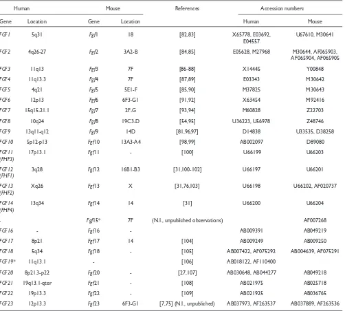
In the mouse, there are at least 22 Fgfgenes [3,9], and the locations of 16 have been identified (Table 1). Many of the mouse Fgfgenes are scattered throughout the genome, but as in the human, Fgf3, Fgf4 and Fgf19 are closely linked (within 80 kb on chromosome 7F) and Fgf6and Fgf23are closely linked on chromosome 6F3-G1.
Evolutionary history
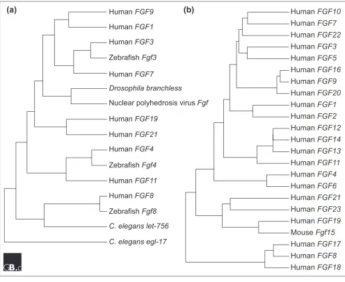
Fgfs have been identified in both invertebrates and verte-brates [3]. Interestingly, an Fgf-like gene is also encoded in the nuclear polyhedrosis virus genome [10]. Fgf-like sequences have not been found in unicellular organisms such as Escherichia coli and Saccharomyces cerevisiae. Although the Drosophila and Caenorhabditis elegans genomes have been sequenced, only one Fgfgene ( branch-less) has been identified in Drosophila[11] and two (egl-17 and let-756) have been identified in C. elegans [12,13], in contrast to the large number of Fgfgenes identified in verte-brates. The evolutionary relationship between invertebrate and vertebrate Fgfsis shown in Figure 2a.
The Fgfgene expansion has been hypothesized to be coinci-dent with a phase of global gene duplications that took place during the period leading to the emergence of vertebrates [14]. Across species, most orthologous FGF proteins are highly conserved and share greater than 90% amino-acid sequence identity (except human FGF15 and mouse Fgf19; see below). To date, four Fgfs(Fgf3, 8, 17and 18) have been identified in zebrafish, seven (Fgf3, Fgf(i), Fgf(ii), Fgf8, 9 and 20) in Xenopus (Fgf(i) and Fgf(ii) are most closely related to Fgf4and Fgf6[15]) and seven (Fgf2, 4, 8, 12, 14, 18and 19) in chicken [3].
The apparent evolutionary relationships of the 22 known human FGFs are shown in Figure 2b. Vertebrate FGFs can be classified into several subgroups or subfamilies. Members of a subgroup of FGFs share increased sequence similarity and biochemical and developmental properties. For example, members of the FGF8 subfamily (FGF8, FGF17, and FGF18) have 70-80% amino acid sequence identity, similar receptor-binding properties and some overlapping sites of expression (for example, the midbrain-hindbrain junction) [16,17]. Members of FGF subgroups are not closely linked in the genome, however, indicating that the subfamilies were gener-ated by gene-translocation or by genome-duplication events, not by local duplication events.
Human FGF15and mouse Fgf19have not been identified. Human FGF19 is evolutionarily most closely related to mouse Fgf15(51% amino acid identity; Figure 2b) [18] and both the human FGF19and mouse Fgf15 genes are closely linked to the human and mouse Fgf3and Fgf4 genes on orthologous regions of human chromosome 11q13 and mouse chromosome 7F (N.I., unpublished observations). These findings indicate that human FGF19 may be the human ortholog of mouse Fgf15. Because all other Fgf orthologs share greater than 90% amino acid identity, it remains possible that the true orthologs of these genes have not been identified, have been lost or have diverged during vertebrate evolution.
Characteristic structural features
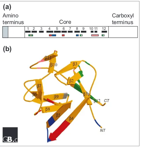
[image:2.609.56.298.84.363.2]FGFs range in molecular weight from 17 to 34 kDa in verte-brates, whereas the DrosophilaFGF is 84 kDa. Most FGFs share an internal core region of similarity, with 28 highly conserved and six identical amino-acid residues [19]. Ten of these highly conserved residues interact with the FGF recep-tor (FGFR) [20]. Structural studies on FGF1 and FGF2 iden-tify 12 antiparallel >strands in the conserved core region of the protein (Figure 3) [21,22]. FGF1 and FGF2 have a >trefoil structure that contains four-stranded >sheets arranged in a triangular array (Figure 3b; reviewed in [23]). Two >strands (strands >10 and >11) contain several basic amino-acid residues that form the primary heparin-binding site on FGF2. Regions thought to be involved in receptor binding are dis-tinct from regions that bind heparin (Figure 3) [21-24].
Figure 1
Gene structure of selected members of the
Fgf
family. Only
the portion of each gene containing coding exons is shown.
Constitutively expressed exons are in black; alternatively
spliced exons are in gray.
Fgfs 1, 2, 4
and
9
contain the
proto-typic three-exon organization. For
Fgf1
, 5’ untranslated exons
are not shown; inclusion of these exons extends the gene by
approximately 69 kb [78].
Fgf8
is an example of a gene with 5’
alternative splicing, and
Fgf13
demonstrates alternatively used
5’ exons separated by over 30 kb. References:
Fgf1
[78];
Fgf2
[79];
Fgf4
[80];
Fgf8
[52];
Fgf9
[81];
Fgf13
[76].
ATG
Fgf8
Fgf9 1 2 3
>24 kb
~10 kb Stop
~57 kb 5.7 kb
~30 kb
2 3 3.1 4 5
1a
1b
Fgf13 (Fhf2)
Stop Stop
2 kb
Fgf4
Stop 2 3 1
4.9 kb 11.4 kb
2 3
1
Stop
Fgf1
Fgf2
>17.5 kb
>16 kb
2 3
1
Stop
1A1B1C 1D 2 3
comment
reviews
reports
deposited research
interactions
information
refereed research
Localization and function
Localization
Subcellular localization and secretion
Most FGFs (FGFs 3-8, 10, 15, 17-19, and 21-23) have amino-terminal signal peptides and are readily secreted from cells. FGFs 9, 16 and 20 lack an obvious amino-terminal signal peptide but are nevertheless secreted [25-27].FGF1 and FGF2 also lack signal sequences, but, unlike FGF9, are not secreted; they can, however, be found on the cell surface and within the extracellular matrix. FGF1 and FGF2 may be released from damaged cells or could be released by an exocytotic mecha-nism that is independent of the endoplasmic-reticulum-Golgi
[image:3.609.54.556.116.568.2]pathway [28]. FGF9 has been shown to contain a non-cleaved amino-terminal hydrophobic sequence that is required for secretion [29,30]. A third subset of FGFs (FGF11-14) lack signal sequences and are thought to remain intracellular [31-34]. It is not known whether these FGFs interact with known FGFRs or function in a receptor-independent manner within the cell. FGF2 and FGF3 have high-molecular-weight forms that arise from initiation from upstream CUG codons [2,14,35]. The additional amino-terminal sequence in these proteins contains nuclear-localization signals, and the pro-teins can be found in the nucleus; the biological function of nuclear-localized FGF is unclear.
Table 1
Chromosomal localizations of FGFs in human and mouse
Human Mouse References Accession numbers
Gene Location Gene Location Human Mouse
FGF1 5q31 Fgf1 18 [82,83] X65778, E03692, U67610, M30641
E04557
FGF2 4q26-27 Fgf2 3A2-B [84,85] E05628, M27968 M30644, AF065903,
AF065904, AF065905
FGF3 11q13 Fgf3 7F [86-88] X14445 Y00848
FGF4 11q13.3 Fgf4 7F [87,89] E03343 M30642
FGF5 4q21 Fgf5 5E1-F [85,90] M37825 M30643
FGF6 12p13 Fgf6 6F3-G1 [91,92] X63454 M92416
FGF7 15q15-21.1 Fgf7 2F-G [93,94] M60828 Z22703
FGF8 10q24 Fgf8 19C3-D [54,95] U36223, U56978 Z48746
FGF9 13q11-q12 Fgf9 14D [81,96,97] D14838 U33535, D38258
FGF10 5p12-p13 Fgf10 13A3-A4 [98,99] AB002097 D89080
FGF11 17p13.1 Fgf11 - [100] U66199 U66203
(FHF3)
FGF12 3q28 Fgf12 16B1-B3 [31,100-102] U66197 U66201
(FHF1)
FGF13 Xq26 Fgf13 X [31,76,103] U66198 U66202, AF020737
(FHF2)
FGF14 13q34 Fgf14 14 [31] U66200 U66204
(FHF4)
- Fgf15* 7F (N.I., unpublished observations) AF007268
FGF16 - Fgf16 - AB009391 AB049219
FGF17 8p21 Fgf17 14 [104] AB009249 AB009250
FGF18 5q34 Fgf18 - [105] AB007422, AF075292 AB004639, AF075291
FGF19* 11q13.1 - [106] AB018122, AF110400
FGF20 8p21.3-p22 Fgf20 - [27,107] AB030648, AB044277 AB049218
FGF21 19q13.1-qter Fgf21 - [108] AB021975 AB025718
FGF22 19p13.3 Fgf22 - [109] AB021925 AB036765
FGF23 12p13.3 Fgf23 6F3-G1 [7,75] (N.I., unpublished) AB037973, AF263537 AB037889, AF263536
Developmental expression patterns and function
The 22 members of the mammalian FGF family are differen-tially expressed in many, if not all, tissues, but the patterns and timing of expression vary. Subfamilies of FGFs tend to have similar patterns of expression, although each FGF also appears to have unique sites of expression. Some FGFs are expressed exclusively during embryonic development (for example, Fgf3, 4, 8, 15, 17 and 19), whereas others are expressed in embryonic and adult tissues (for example, Fgf1, 2, 5-7, 9-14, 16, 18, and 20-23).
Function
The expression patterns of FGFs (see above) suggest that they have important roles in development. FGFs often signal
[image:4.609.57.553.86.492.2]directionally and reciprocally across epithelial-mesenchymal boundaries [36]. The integrity of these signaling pathways requires extremely tight regulation of FGF activity and receptor specificity. For example, in vertebrate limb develop-ment, mesenchymally expressed Fgf10 in the lateral-plate mesoderm induces the formation of the overlying apical ecto-dermal ridge; the ridge subsequently expresses Fgf8, which signals back to the underlying mesoderm [37]. This direc-tional signaling initiates feedback loops and, along with other signaling molecules, regulates the outgrowth and patterning of the limb. Importantly, the differential expression of the alternative splice forms of the receptors in the apical ectoder-mal ridge and underlying mesoderm is such as to limit or prevent autocrine signaling within a given compartment.
Figure 2
Evolutionary relationships within the FGF family.
(a)
Apparent evolutionary relationships between FGFs from vertebrates,
invertebrates and a virus. Amino-acid sequences of nine representative FGFs were chosen from human and compared with
FGFs from
Drosophila
,
C. elegans
, zebrafish and
Autographa californica
nuclear polyhedrosis virus.
(b)
Apparent evolutionary
relationships of the 22 known human and murine FGFs. Sequences were aligned using Genetyx sequence analysis software
and trees were constructed from the alignments using the neighbor-joining method.
Human
FGF10
Human
FGF7
Human
FGF22
Human
FGF3
Human
FGF5
Human
FGF16
Human
FGF9
Human
FGF20
Human
FGF1
Human
FGF2
Human
FGF12
Human
FGF14
Human
FGF13
Human
FGF11
Human
FGF4
Human
FGF6
Human
FGF21
Human
FGF23
Human
FGF19
Mouse
Fgf15
Human
FGF17
Human
FGF8
Human
FGF18
Human
FGF9
Human
FGF1
Human
FGF3
Zebrafish
Fgf3
Human
FGF7
Drosophila branchless
Nuclear polyhedrosis virus
Fgf
Human
FGF19
Human
FGF21
Human
FGF4
Zebrafish
Fgf4
Human
FGF11
Human
FGF8
Zebrafish
Fgf8
C. elegans let-756
C. elegans egl-17
Studies of the biochemical activities of FGFs have focused on the specificity of interactions between FGFs and FGFRs, on factors that affect the stability of FGFs and on the composition and mechanism of the active FGF-FGFR signaling complex.
Specificity of FGFs for FGF receptors
The FGFR tyrosine kinase receptors contain two or three immunoglobulin-like domains and a heparin-binding sequence [38-40]. Alternative mRNA splicing of the FGFR gene specifies the sequence of the carboxy-terminal half of immunoglobulin-domain III, resulting in either the IIIb or the IIIc isoform of the FGFR [41-43]. This alternative-splic-ing event is regulated in a tissue-specific manner and dra-matically affects ligand-receptor binding specificity [44-48]. Exon IIIb is expressed in epithelial lineages and exon IIIc tends to be expressed in mesenchymal lineages [44,46-48]. In vitro patterns of binding specificity have been determined
for each splice form of FGFR1-3 and for FGFR4, which is not alternatively spliced [49-51]. Ligands specific for these receptor splice forms are expressed in adjacent tissues, resulting in directional epithelial-mesenchymal signaling. For example, epithelially expressed FGFR2b (that is, FGFR2 IIIb isoform) can be activated by FGF7 and FGF10, ligands produced in mesenchymal tissue [49-51]. These ligands show no activity towards mesenchymally expressed FGFR2c. Conversely, FGF8 is expressed in epithelial tissue and acti-vates FGFR2c but shows no activity towards FGFR2b ([49,52] and our unpublished observations). Notably, FGF8 expression is often restricted to epithelial tissue such as the apical ectodermal ridge of the developing limb bud [53,54].
Interaction with heparin or heparan sulfate proteoglycans
[image:5.609.54.297.86.341.2]An important feature of FGF biology involves the interaction between FGF and heparin or heparan sulfate (HS) proteogly-can (HSPG) [19]. These interactions stabilize FGFs to thermal denaturation and proteolysis and may severely limit their diffusion and release into interstitial spaces [55,56]. FGFs must saturate nearby HS-binding sites before exerting an effect on tissue further away, or else must be mobilized by heparin/HS-degrading enzymes. The interaction between FGFs and HS results in the formation of dimers and higher-order oligomers [57-59]. Although the biologically active form of FGF is poorly defined, it has been established that heparin is required for FGF to effectively activate the FGFR in cells that are deficient in or unable to synthesize HSPG or in cells pretreated with heparin/HS-degrading enzymes or inhibitors of sulfation [60-62]. Genetic studies have also shown that mutations in enzymes involved in HS biosynthe-sis affect FGF signaling pathways during development
[19,63]. Additional studies have shown that heparin and/or
HS act to increase the affinity and half-life of the FGF-FGFR complex (reviewed in [40,64]).
A minimal complex containing one FGF molecule per FGFR can form in the absence of HS [24]. Structural studies suggest that HS may bridge FGF2 and the FGFR by binding to a groove formed by the heparan-binding sites of both the ligand and the receptor [24,65]. Binding studies with soluble chimeric FGFRs have identified a second potential FGF-binding site that, in some cases, can interact cooperatively with the primary FGF-binding site [66].
Important mutants
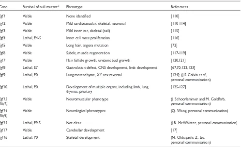
Many members of the Fgf family have been disrupted by homologous recombination in mice. The phenotypes range from very early embryonic lethality to subtle phenotypes in adult mice. The major phenotypes observed in Fgfknockout mice are shown in Table 2. Because FGFs within a subfamily have similar receptor-binding properties and overlapping patterns of expression, functional redundancy is likely to occur. This has been demonstrated for Fgf17and Fgf8, which cooperate to regulate neuroepithelial proliferation in the mid-brain-hindbrain junction [17]. In the case of Fgf knockouts
comment
reviews
reports
deposited research
interactions
information
refereed research
Figure 3
(a)
Structural features of the FGF polypeptide. The amino
terminus of some FGFs contains a signal sequence (shaded).
All FGFs contain a core region that contains conserved
amino-acid residues and conserved structural motifs. The
locations of
>
strands within the core region are numbered
and shown as black boxes. The heparin-binding region (pink)
includes residues in the loop between
>
strands 1 and 2 and
in
>
strands 10 and 11. Residues that contact the FGFR are
shown in green (the region contacting Ig-domain 2 of the
receptor), blue (contacting Ig-domain 3) and red (contacting
the alternatively spliced region of Ig-domain 3). Amino-acid
residues that contact the linker region are shown in gray
[20].
(b)
Three-dimensional structure of FGF2, a
prototypical member of the FGF family. A ribbon diagram of
FGF2 is shown;
>
strands are labeled 1-12 and regions of
contact with the FGFR and heparin are color-coded as in (a)
[22,24]. Image provided by M. Mohammadi.
Core
1 2 3 4 5 6 7 8 9 10 11 12
Amino
terminus
Carboxyl
terminus
=
resulting in early lethality, other functions later in develop-ment will need to be addressed by constructing conditional alleles that can be targeted at specific times and places in development. For example, Fgf8-/- mice die by embryonic
day 9.5 [67]. A conditional allele for Fgf8 targeted to the apical ectodermal ridge has been used to demonstrate an essential role for Fgf8in early limb development [68,69].
Several mutations in Fgf genes have been identified in C.elegans, Drosophila, zebrafish, mouse and human. The C. elegansgene egl-17is required for sex myoblast migration [12], and a null allele of let-756causes developmental arrest of the early larva [13]. The Drosophila branchless gene is required for tracheal branching and cell migration [11]. In zebrafish, acerebellar (ace) embryos lack the cerebellum and the midbrain-hindbrain boundary organizer. The ace gene encodes the zebrafish homolog of Fgf8[70]. Interest-ingly, zebrafish aussichtmutant embryos, which overexpress Fgf8, also have defects in development of the central nervous system [71].
In the mouse, the angora mutation, which affects hair growth, was found to be allelic with Fgf5 [72]. A mouse mutant with a Crouzon-syndrome-like craniofacial dysmor-phology phenotype was found to result from an insertional
mutation in the Fgf3/Fgf4locus [73]. Recently, positional cloning of the autosomal dominant hypophosphataemic rickets gene identified missense mutations in human FGF23 [74]. A recent paper demonstrates that this disease is caused by a gain-of-function mutation [75]. The chromo-somal location (Xq26) and tissue-specific expression pattern of Fgf13(also called Fhf2) suggests that it may be a candidate gene for Borjeson-Forssman-Lehmann syn-drome, an X-linked mental retardation syndrome [76].
Frontiers
Issues most studied
[image:6.609.59.557.428.731.2]FGFs have been intensely studied for nearly 30 years. Most of the early work focused on the mechanisms that regulate stability, secretion, export and interactions with heparin and on the mechanisms and consequences of signal transduction in various types of cells. More recent work has focused on the mechanisms regulating receptor specificity and receptor activation, the structure of the FGF-FGFR-HS complex, and the identification of new members of the FGF family. Func-tional studies have begun to address the role of FGFs in cell biology, development and physiology. Initial studies focused on the regulation of cell proliferation, migration and differ-entiation; more recent work has addressed the negative
Table 2
FGF knockout mice
Gene Survival of null mutant* Phenotype References
Fgf1 Viable None identified [110]
Fgf2 Viable Mild cardiovascular, skeletal, neuronal [110-114]
Fgf3 Viable Mild inner ear, skeletal (tail) [115]
Fgf4 Lethal, E4-5 Inner cell mass proliferation [116]
Fgf5 Viable Long hair, angora mutation [72]
Fgf6 Viable Subtle, muscle regeneration [117-119]
Fgf7 Viable Hair follicle growth, ureteric bud growth [120,121]
Fgf8 Lethal, E7 Gastrulation defect, CNS development, limb development [67,70,122,123]
Fgf9 Lethal, P0 Lung mesenchyme, XY sex reversal [124]; (J.S. Colvin et al.,
personal communication)
Fgf10 Lethal, P0 Development of multiple organs, including limb, lung, [125-127] thymus, pituitary
Fgf12 Viable Neuromuscular phenotype (J. Schoorlemmer and M. Goldfarb,
(Fhf1) personal communication)
Fgf14 Viable Neurological phenotypes (Q. Wang, personal communication)
(Fhf4)
Fgf15 Lethal, E9.5 Not clear (J.R. McWhirter, personal communication)
Fgf17 Viable Cerebellar development [17]
Fgf18 Lethal, P0 Skeletal development (N. Ohbayashi, Z. Liu,
personal communication)
effect of FGFs and FGFRs on proliferation of some cell types, which was surprising as FGFs were thought to promote pro-liferation. In vitrostudies have now been complemented by gene targeting in mice. The knockout approach has been fairly successful in identifying primary phenotypes but will be challenged by the need to address redundancy amongst the 22 FGFs and to study their developmental and physio-logical functions after the point of lethality of the null allele.
Unresolved questions
A major unresolved question concerns the mechanism(s) reg-ulating FGF activity in vivoin the presence of cell-surface and extracellular-matrix HSPG. Current hypotheses predict that tissue-specific heparan fragments of defined sequence (and particularly of defined sulfation pattern) will differentially regulate FGFs by controlling their diffusion in the extracellu-lar matrix and their ability to activate specific receptors [77]. These issues will be resolved by determining the sequence of tissue-specific HS and by demonstrating whether specific HS sequences can modulate the binding specificity of FGFs beyond that determined by the specific FGFR and its alterna-tive splice form in the presence of heparin.
A second area of research will aim to elucidate the develop-mental roles of all the FGFs, first alone and then in various combinations. This will include determining whether a single FGF with a defined developmental function interacts with one or multiple FGFRs. A third major frontier will be to elucidate the physiological roles of FGFs that are expressed in adult tissues. This will again involve testing combinations of FGFs in cases in which knockouts are viable and designing condi-tional alleles in cases of embryonic lethality. Major areas being considered include neuronal and cardiovascular physiology, neuronal regeneration and homeostasis and tissue repair.
The last major frontier will be to elucidate the primary roles of FGFs in genetic diseases and cancer. Several FGFs were initially cloned from human and animal tumors. Future work will be required to determine whether FGF activation is itself an etiological agent in primary human tumors or whether it is a progression factor in the pathogenesis of cancer. As functional roles for FGFs are elucidated in embry-onic development, it is expected that various human birth defects and genetic diseases will be attributed to mutations in Fgf genes. These studies will probably lead to the develop-ment of pharmacogenetic agents to treat these diseases. Because a large number of skeletal diseases are caused by mutations in Fgfr genes, it is anticipated that mutations in some Fgf genes will also be involved in skeletal pathology.
Acknowledgements
This work was supported by NIH grants CA60673 and HD35692 and by a grant from the American Heart Association (to D.M.O.) and by the Grant-in-Aid for Scientific Research from the Ministry of Education, Science and Culture, Japan and a grant from the Human Frontier Science Program, France (to N.I.).
References
1. Kiefer P, Acland P, Pappin D, Peters G, Dickson C: Competition between nuclear localization and secretory signals deter-mines the subcellular fate of a single CUG-initiated form of FGF3.EMBO J1994, 13:4126-4136.
This is one of first papers to show that an FGF can initiate from an upstream CUG codon and can be localized in the nucleus.
2. Arnaud E, Touriol C, Boutonnet C, Gensac MC, Vagner S, Prats H, Prats AC: A new 34-kilodalton isoform of human fibroblast growth factor 2 is cap dependently synthesized by using a non-AUG start codon and behaves as a survival factor.Mol Cell Biol1999,19:505-514.
Identification of a high-molecular-weight form of FGF2 that initiates from an upsteam CUG codon and is localized in the nucleus.
3. GenBank [http://www.ncbi.nlm.nih.gov/Genbank/index.html]
DNA sequences of FGFs listed in Table 1 can be accessed through this website.
4. HUGO Gene Nomenclature Database
[http://www.gene.ucl.ac.uk/nomenclature/]
This database contains nomenclature and mapping information for human genes.
5. Cytokine Family cDNA Database
[http://cytokine.medic.kumamoto-u.ac.jp/CFC/FGF/FGF.html]
This database contains nomenclature and mapping information for the FGF family.
6. LocusLink [http://ncbi.nlm.nih.gov/LocusLink/]
This database contains updated mapping information for the FGF family with links to a variety of other databases.
7. Yamashita T, Yoshioka M, Itoh N: Identification of a novel fibroblast growth factor, FGF-23, preferentially expressed in the ventrolateral thalamic nucleus of the brain. Biochem Biophys Res Commun2000, 277:494-498.
FGF23 is the most recent reported member of the FGF family. This paper summarizes the chromosomal localization and apparent evolu-tionary relationships of human FGFs.
8. Kelley MJ, Pech M, Seuanez HN, Rubin JS, O’Brien SJ, Aaronson SA:
Emergence of the keratinocyte growth factor multigene family during the great ape radiation.Proc Natl Acad Sci USA 1992, 89:9287-9291.
This paper identifies a human multigene family composed of amplified and dispersed FGF7gene copies and suggests models for the evolution of the great apes and humans.
9. Mouse Genome Informatics[http://www.informatics.jax.org]
This website provides information on mouse gene sequences, map position and function.
10. Ayres MD, Howard SC, Kuzio J, Lopez-Ferber M, Possee RD: The complete DNA sequence of Autographa californica nuclear polyhedrosis virus.Virology1994, 202:586-605.
The complete nucleotide sequence of the genome of the baculovirus
Autographa californicanuclear polyhedrosis virus (AcNPV) revealed that this genome encodes an FGF-like protein.
11. Sutherland D, Samakovlis C, Krasnow MA: branchless encodes a
DrosophilaFGF homolog that controls tracheal cell migra-tion and the pattern of branching.Cell1996, 87:1091-1101.
This paper identifies a Drosophilahomolog (branchless, bnl) of mam-malian FGFs, which activates the breathless FGF receptor tyrosine kinase. This FGF pathway regulates tracheal branching patterns by guiding tracheal cell migration during primary branch formation and by activating cytoplasmic branching at the ends of primary branches.
12. Burdine RD, Chen EB, Kwok SF, Stern MJ: egl-17 encodes an invertebrate fibroblast growth factor family member required specifically for sex myoblast migration in
Caenorhabditis elegans. Proc Natl Acad Sci USA 1997, 94: 2433-2437.
The C. elegansgene egl-17is a member of the Fgffamily and one of the first known functional invertebrate FGFs. EGL-17 acts as a ligand for EGL-15 (FGFR) to regulate sex myoblast migration.
13. Roubin R, Naert K, Popovici C, Vatcher G, Coulier F, Thierry-Mieg J, Pontarotti P, Birnbaum D, Baillie D, Thierry-Mieg D: let-756, a C. elegansFgf essential for worm development.Oncogene1999,
18:6741-6747.
Null mutations in C. elegans let-756lead to developmental arrest in early larval stages; let-756is thus an essential gene for C. elegans devel-opment.
14. Coulier F, Pontarotti P, Roubin R, Hartung H, Goldfarb M, Birnbaum D: Of worms and men: an evolutionary perspective on the
comment
reviews
reports
deposited research
interactions
information
fibroblast growth factor (FGF) and FGF receptor families.J Mol Evol1997, 44:43-56.
This review compares FGF and FGF receptor sequences in vertebrates and nonvertebrates and concludes that the FGF and FGF receptor fam-ilies have evolved through phases of gene duplications, one of which may have coincided with the emergence of vertebrates. The review includes all four Fgfrsand 11 Fgfs.
15. Isaacs HV, Tannahill D, Slack JM:Expression of a novel FGF in the Xenopusembryo. A new candidate inducing factor for mesoderm formation and anteroposterior specification.
Development 1992, 114:711-720.
Identification of novel XenopusFGFs.
16. Maruoka Y, Ohbayashi N, Hoshikawa M, Itoh N, Hogan BLM, Furuta Y: Comparison of the expression of three highly related genes, Fgf8, Fgf17and Fgf18, in the mouse embryo.Mech Dev 1998, 74:175-177.
This paper compares the expression pattern of the Fgf8 subfamily which includes Fgf8, 17and 18. Expression is examined during early development and in the developing brain and limbs.
17. Xu JS, Liu ZH, Ornitz DM: Temporal and spatial gradients of Fgf8 and Fgf17 regulate proliferation and differentiation of midline cerebellar structures. Development 2000, 127: 1833-1843.
This is the first paper to demonstrate cooperation between two Fgfs
during vertebrate development.
18. Nishimura T, Utsunomiya Y, Hoshikawa M, Ohuchi H, Itoh N:
Structure and expression of a novel human FGF, FGF-19, expressed in the fetal brain. Biochim Biophys Acta 1999,
1444:148-151.
Identification of human FGF19as a possible homolog of mouse Fgf15. Both of these genes are most closely related to FGFs21 and 23.
19. Ornitz DM: FGFs, heparan sulfate and FGFRs: complex inter-actions essential for development.BioEssays2000, 22:108-112.
This review article addresses the developmental implications of the interactions between FGFs, FGFRs and heparan sulfate proteoglycans. The paper by Lin et al.[63], which demonstrates that heparan sulfate is essential for FGF function during Drosophiladevelopment, is highlighted.
20. Plotnikov AN, Hubbard SR, Schlessinger J, Mohammadi M: Crystal structures of two FGF-FGFR complexes reveal the determi-nants of ligand-receptor specificity.Cell2000, 101:413-424.
The analysis of two different ligand-receptor crystal structures is used to determine elements that regulate ligand-binding specificity.
21. Zhu X, Komiya H, Chirino A, Faham S, Fox GM, Arakawa T, Hsu BT, Rees DC: Three-dimensional structures of acidic and basic fibroblast growth factors.Science1991, 251:90-93.
This paper presents one of the first structures of FGF1 and FGF2.
22. Eriksson AE, Cousens LS, Weaver LH, Matthews BW: Three-dimensional structure of human basic fibroblast growth factor.Proc Natl Acad Sci USA1991, 88:3441-3445.
This paper presents one of the first structures of FGF2.
23. Faham S, Linhardt RJ, Rees DC: Diversity does make a differ-ence: fibroblast growth factor-heparin interactions.Curr Opin Struct Biol1998,8:578-586.
A review that models the formation of an FGF-FGFR-proteoglycan complex, based on the structure of FGF in complex with fragments of heparin.
24. Plotnikov AN, Schlessinger J, Hubbard SR, Mohammadi M: Struc-tural basis for FGF receptor dimerization and activation.Cell 1999, 98:641-650.
Crystal structure of FGF2 complexed with FGFR1 showing extensive interaction between immunoglobulin domains 2 and 3 and the interdo-main linker of the FGFR.
25. Miyake A, Konishi M, Martin FH, Hernday NA, Ozaki K, Yamamoto S, Mikami T, Arakawa T, Itoh N: Structure and expression of a novel member, FGF-16, of the fibroblast growth factor family.Biochem Biophys Res Commun1998, 243:148-152.
This paper identifies Fgf16as a homolog of Fgf9.
26. Miyamoto M, Naruo K, Seko C, Matsumoto S, Kondo T, Kurokawa T: Molecular cloning of a novel cytokine cDNA encoding the ninth member of the fibroblast growth factor family, which has a unique secretion property.Mol Cell Biol 1993, 13: 4251-4259.
This paper identifies FGF9 as a new member of the FGF family without a classical signal peptide.
27. Ohmachi S, Watanabe Y, Mikami T, Kusu N, Ibi T, Akaike A, Itoh N:
FGF-20, a novel neurotrophic factor, preferentially
expressed in the substantia nigra pars compacta of rat brain.Biochem Biophys Res Commun2000,277:355-360.
This paper identifies Fgf20 as a homolog of Fgf9 andFgf16.
28. Mignatti P, Morimoto T, Rifkin DB: Basic fibroblast growth factor, a protein devoid of secretory signal sequence, is released by cells via a pathway independent of the endoplas-mic reticulum-Golgi complex.J Cell Physiol1992, 151:81-93.
FGF2 lacks a signal peptide but is known to act extracellularly. This study shows that FGF2 can be released via an exocytotic mechanism independent of the endoplasmic reticulum-Golgi pathway.
29. Miyakawa K, Hatsuzawa K, Kurokawa T, Asada M, Kuroiwa T, Imamura T: A hydrophobic region locating at the center of fibroblast growth factor-9 is crucial for its secretion. J Biol Chem1999, 274:29352-29357.
FGF9 lacks an amino-terminal signal sequence but is secreted from expressing cells. This study shows that nascent FGF9 polypeptides translocate into endoplasmic reticulum without peptide cleavage via a co-translational pathway in which both an amino terminus and a central hydrophobic domain is important.
30. Revest JM, DeMoerlooze L, Dickson C: Fibroblast growth factor 9 secretion is mediated by a non-cleaved amino-terminal signal sequence.J Biol Chem2000, 275:8083-8090.
FGF9 lacks a recognizable amino-terminal signal sequence, although it is efficiently secreted. This study shows that FGF9 enters the endoplasmic reticulum and traverses the Golgi complex in a similar manner to other constitutively secreted proteins. They demonstrate that the first 28 amino acids of FGF9 can function as an efficient non-cleaved signal peptide.
31. Smallwood PM, Munoz-sanjuan I, Tong P, Macke JP, Hendry SH, Gilbert DJ, Copeland NG, Jenkins NA, Nathans J: Fibroblast growth factor (FGF) homologous factors: new members of the FGF family implicated in nervous system development. Proc Natl Acad Sci USA1996, 93:9850-9857.
This study identifies a novel FGF-related subfamily, termed fibroblast growth factor homologous factors (FHF). FHFs lack a classical signal sequence and contain nuclear localization signals. FHFs are expressed in the developing and adult nervous systems, suggesting a role in nervous system development and function.
32. Yamamoto S, Mikami T, Ohbayashi N, Ohta M, Itoh N: Structure and expression of a novel isoform of mouse FGF homolo-gous factor (FHF)-4.Biochim Biophys Acta1998, 1398:38-41.
Identification of alternative amino terminal coding exon for FHF4/FGF14.
33. Munoz-Sanjuan I, Smallwood PM, Nathans J: Isoform diversity among fibroblast growth factor homologous factors is gen-erated by alternative promoter usage and differential splic-ing.J Biol Chem2000, 275:2589-2597.
Characterization of multiple isoforms of Fhfs1-4/Fgfs 11-14, generated through the use of alternative 5’ exons. Isoforms show different subcel-lular distributions and distinct expression patterns in developing and adult mouse tissues.
34. Wang Q, McEwen DG, Ornitz DM: Subcellular and develop-mental expression of alternatively spliced forms of fibro-blast growth factor 14.Mech Dev2000, 90:283-287.
Identification of isoforms of Fhf4/Fgf14that result from the alternative usage of two different first exons. Isoforms show different subcellular localization and distinct expression patterns in developing and adult mouse tissues. Expression was observed in migrating and post migra-tory neurons.
35. Antoine M, Reimers K, Dickson C, Kiefer P: Fibroblast growth factor 3, a protein with dual subcellular localization, is tar-geted to the nucleus and nucleolus by the concerted action of two nuclear localization signals and a nucleolar retention signal.J Biol Chem1997, 272:29475-29481.
Identification of an FGF3 CUG initiation site. This amino-terminal extended product is localized in both the nucleus/nucleolus and secre-tory pathway. The amino terminus contains both a signal sequence and a nuclear localization signal.
36. Hogan BL: Morphogenesis.Cell1999, 96:225-233.
Review article highlighting the roles of multigene families, such as Fgfs, Bmps, Hedgehogs, Wntsand Egfsin morphogenesis.
37. Martin GR: The roles of FGFs in the early development of vertebrate limbs.Genes Dev1998, 12:1571-1586.
38. Lee PL, Johnson DE, Cousens LS, Fried VA, Williams LT: Purifica-tion and complementary DNA cloning of a receptor for basic fibroblast growth factor.Science1989, 245:57-60.
This is the first cloning of an FGF receptor.
39. Johnson DE, Lee PL, Lu J, Williams LT: Diverse forms of a recep-tor for acidic and basic fibroblast growth facrecep-tors.Mol Cell Biol 1990, 10:4728-4736.
Identification of FGF receptor 1 variants which contain either two or three immunoglobulin-like domains. Identification of putative secreted forms of FGF receptor 1.
40. McKeehan WL, Wang F, Kan M: The heparan sulfate-fibroblast growth factor family: diversity of structure and function. Prog Nucleic Acid Res Mol Biol1998, 59:135-176.
This review presents models for FGF receptor activation by FGF and heparin. Also discussed are developmental roles for FGFs in liver and prostate development.
41. Miki T, Bottaro DP, Fleming TP, Smith CL, Burgess WH, Chan AM, Aaronson SA: Determination of ligand-binding specificity by alternative splicing: two distinct growth factor receptors encoded by a single gene.Proc Natl Acad Sci USA1992, 89: 246-250.
Demonstration that FGFR alternative splicing can regulate ligand binding specificity.
42. Chellaiah AT, McEwen DG, Werner S, Xu J, Ornitz DM: Fibroblast growth factor receptor (FGFR) 3. Alternative splicing in immunoglobulin-like domain III creates a receptor highly specific for acidic FGF/FGF-1. J Biol Chem 1994, 269: 11620-11627.
Identification of an alternative splice form of FGFR3 with very restricted ligand binding properties.
43. Naski MC, Ornitz DM: FGF signaling in skeletal development. Front Biosci1998, 3:D781-D794.
Review article highlighting the functions of the Fgfrswith a focus on roles in skeletal development.
44. Yan G, Fukabori Y, McBride G, Nikolaropolous S, McKeehan WL:
Exon switching and activation of stromal and embryonic fibroblast growth factor (FGF)-FGF receptor genes in prostate epithelial cells accompany stromal independence and malignancy.Mol Cell Biol1993, 13:4513-4522.
Demonstration that tumor progression can be accompanied by a change in Fgfr alternative splicing with consequent change in ligand binding specificity.
45. Orr-Urtreger A, Bedford MT, Burakova T, Arman E, Zimmer Y, Yayon A, Givol D, Lonai P: Developmental localization of the splicing alternatives of fibroblast growth factor receptor-2 (FGFR2).Dev Biol1993, 158:475-486.
Demonstration that alternative Fgfrsplice forms are expressed prefer-entially in epithelial and mesenchymal lineages.
46. Gilbert E, Del Gatto F, Champion-Arnaud P, Gesnel MC, Breathnach R: Control of BEK and K-SAM splice sites in alternative splicing of the fibroblast growth factor receptor 2 pre-mRNA.Mol Cell Biol1993, 13:5461-5468.
Identification of the mechanism regulating tissue-specific usage of alter-native splice forms of Fgfr2.
47. Avivi A, Yayon A, Givol D: A novel form of FGF receptor-3 using an alternative exon in the immunoglobulin domain III. FEBS Lett1993, 330:249-252.
Identification of alternative splice forms of Fgfr3.
48. Scotet E, Houssaint E: The choice between alternative IIIb and IIIc exons of the FGFR-3 gene is not strictly tissue-specific. Biochim Biophys Acta1995, 1264:238-242.
The less stringent alternative splicing choice between exons IIIb and IIIc of FGFR3 is in contrast with the very tissue-specific alternative splicing observed for FGFR2 in which epithelial cells use only the IIIb exon and fibroblasts use only the IIIc exon.
49. Ornitz DM, Xu J, Colvin JS, McEwen DG, MacArthur CA, Coulier F, Gao G, Goldfarb M: Receptor specificity of the fibroblast growth factor family.J Biol Chem1996, 271:15292-15297.
The first comparison of the relative mitogenic activity of a large number of FGFs on cells expressing a single alternatively spliced FGFR 1-3 or FGFR4.
50. Igarashi M, Finch PW, Aaronson SA: Characterization of recom-binant human fibroblast growth factor (Fgf-10) reveals func-tional similarities with keratinocyte growth factor (Fgf-7). J Biol Chem1998, 273:13230-13235.
Demonstrates that the receptor-binding specificity of FGF10 is similar to that of the closely related FGF7.
51. Miki T, Fleming TP, Bottaro DP, Rubin JS, Ron D, Aaronson SA:
Expression cDNA cloning of the KGF receptor by creation of a transforming autocrine loop.Science1991, 251:72-75.
First cloning of a receptor for FGF7/KGF.
52. MacArthur CA, Lawshé A, Xu J, Santos-Ocampo S, Heikinheimo M, Chellaiah AT, Ornitz DM: FGF-8 isoforms activate receptor splice forms that are expressed in mesenchymal regions of mouse development.Development1995, 121:3603-3613.
Identification of alternatively spliced forms of FGF8 and demonstration that these molecules bind the mesenchymal splice forms of Fgfr2.
53. Heikinheimo M, Lawshé A, Shackleford GM, Wilson DB, MacArthur CA: Fgf-8expression in the post-gastrulation mouse suggests roles in the development of the face, limbs and central nervous system.Mech Dev1994, 48:129-138.
Identification of temporal and spatial patterns of Fgf8expression in the developing mouse.
54. Crossley PH, Martin GR: The mouse Fgf8 gene encodes a family of polypeptides and is expressed in regions that direct outgrowth and patterning in the developing embryo. Development1995, 121:439-451.
Identification of alternative splice forms of Fgf8and Fgf8expression in the apical ectodermal ridge.
55. Moscatelli D: High and low affinity binding sites for basic fibroblast growth factor on cultured cells: absence of a role for low affinity binding in the stimulation of plasminogen activator production by bovine capillary endothelial cells.J Cell Physiol1987, 131:123-130.
This paper challenges the role of cell surface heparan sulfate in FGFR activation but establishes a role for cell surface heparan sulfate in binding FGF.
56. Flaumenhaft R, Moscatelli D, Rifkin DB: Heparin and heparan sulfate increase the radius of diffusion and action of basic fibroblast growth factor.J Cell Biol1990, 111:1651-1659.
This paper demonstrates that the extracellular matrix can store FGF and limit its diffusion in tissues.
57. Mach H, Volkin DB, Burke CJ, Middaugh CR, Linhardt RJ, Fromm JR, Loganathan D, Mattsson L: Nature of the interaction of heparin with acidic fibroblast growth factor.Biochemistry1993, 32: 5480-5489.
This paper establishes that FGF1 binds heparin at high density (one molecule every 4-5 saccharide units) and with high affinity (50-140 nM).
58. Herr AB, Ornitz DM, Sasisekharan R, Venkataraman G, Waksman G:
Heparin-induced self-association of fibroblast growth factor 2. Evidence for two oligomerization processes.J Biol Chem1997,
272:16382-16389.
Presents analytical data that indicates that biologically active heparin octasaccharides can induce a monomer-dimer-tetramer assembly of FGF2.
59. Moy FJ, Safran M, Seddon AP, Kitchen D, Bohlen P, Aviezer D, Yayon A, Powers R: Properly oriented heparin-decasaccharide-induced dimers are the biologically active form of basic fibroblast growth factor.Biochemistry1997, 36:4782-4791.
Evidence that a cis-oriented FGF2 dimer is the minimal biologically active structural unit of FGF2 in the presence of a decasaccharide.
60. Ornitz DM, Yayon A, Flanagan JG, Svahn CM, Levi E, Leder P:
Heparin is required for cell-free binding of basic fibroblast growth factor to a soluble receptor and for mitogenesis in whole cells.Mol Cell Biol1992, 12:240-247.
First study to show that heparin, FGF and an FGF receptor binding domain form a trimolecular complex in vitro. This paper also shows that this complex is essential for receptor activation on cells lacking heparan sulfate proteoglycan.
61. Rapraeger AC, Krufka A, Olwin BB: Requirement of heparan sulfate for bFGF-mediated fibroblast growth and myoblast differentiation.Science1991, 252:1705-1708.
The first study to demonstrate that cell surface heparan sulfate is directly involved in FGF cell signaling.
62. Yayon A, Klagsbrun M, Esko JD, Leder P, Ornitz DM: Cell surface, heparin-like molecules are required for binding of basic fibroblast growth factor to its high affinity receptor. Cell 1991,64:841-848.
The first study to demonstrate that heparin is required for FGF to bind to an FGF receptor.
63. Lin XH, Buff EM, Perrimon N, Michelson AM: Heparan sulfate proteoglycans are essential for FGF receptor signaling during Drosophilaembryonic development.Development1999,
126:3715-3723.
The first genetic evidence that heparan sulfate glycosaminoglycans are essential for fibroblast growth factor receptor signaling in a well defined
comment
reviews
reports
deposited research
interactions
information
developmental context. These data support the model in which heparan sulfate facilitates FGF and/or FGF-FGFR oligomerization.
64. Szebenyi G, Fallon JF: Fibroblast growth factors as multifunc-tional signaling factors.Int Rev Cytol1999, 185:45-106.
This is a comprehensive review that highlights all aspects of FGF biology including cell signaling, receptor binding and activation and roles for FGFs in embryonic development.
65. Schlessinger J, Plotnikov AN, Ibrahimi OA, Eliseenkova AV, Yeh BK, Yayon A, Linhardt RJ, Mohammadi M: Crystal structure of a ternary FGF-FGFR-heparin complex reveals a dual role for heparin in FGFR binding and dimerization. Mol Cell 2000,
6:743-750.
This paper presents a diffusion-based crystal structure which shows heparan fragments bound to both FGF and the FGFR in a head-to-head orientation.
66. Chellaiah A, Yuan W, Chellaiah M, Ornitz DM: Mapping ligand binding domains in chimeric fibroblast growth factor recep-tor molecules. Multiple regions determine ligand binding specificity.J Biol Chem1999, 274:34785-34794.
Identification of a second ligand-binding region on the FGF receptor.
67. Meyers EN, Lewandoski M, Martin GR: An Fgf8 mutant allelic series generated by Cre- and Flp-mediated recombination. Nat Genet1998, 18:136-141.
First null, hypomorphic and conditional allele created for an FGF. This study reveals requirements for Fgf8during gastrulation, cardiac, cranio-facial, forebrain, midbrain and cerebellar development.
68. Moon AM, Capecchi MR: Fgf8 is required for outgrowth and patterning of the limbs.Nat Genet2000,26:455-459.
Demonstrates an essential role for Fgf8in limb bud development.
69. Lewandoski M, Sun X, Martin GR: Fgf8 signalling from the AER is essential for normal limb development. Nat Genet 2000,
26:460-463.
Demonstrates an essential role for Fgf8in limb bud development.
70. Reifers F, Bohli H, Walsh EC, Crossley PH, Stainier DY, Brand M: Fgf8is mutated in zebrafish acerebellar(ace) mutants and is required for maintenance of midbrain-hindbrain boundary development and somitogenesis.Development1998, 125: 2381-2395.
Demonstrates a role for Fgf8in midbrain hindbrain development in zebrafish by demonstrating genetic linkage between Fgf8and the acere-bellar(ace) mutation. Demonstrates that aceis probably a null muta-tion in Fgf8.
71. Heisenberg CP, Brennan C, Wilson SW: Zebrafish aussicht mutant embryos exhibit widespread overexpression of ace (fgf8) and coincident defects in CNS development. Develop-ment1999,126:2129-2140.
Identification of a genetic locus that may regulate Fgf8 (ace)expression in zebrafish.
72. Hebert JM, Rosenquist T, Gotz J, Martin GR: FGF5 as a regulator of the hair growth cycle: evidence from targeted and spon-taneous mutations.Cell1994, 78:1017-1025.
Demonstrates that a null allele in the Fgf5gene is allelic with the mouse
angoramutation. Identifies an essential role for FGF5 in regulating the hair growth cycle.
73. Carlton MBL, Colledge WH, Evans MJ: Crouzon-like craniofacial dysmorphology in the mouse is caused by an insertional mutation at the Fgf3/Fgf4 locus.Dev Dyn1998, 212:242-249.
Identification of a retroviral integration in the intragenic region between
Fgf3and Fgf4in the Bulgy-eye (Bey) mutant mouse. Expression of both
Fgf3and Fgf4is up-regulated in the cranial sutures of Bey mice. Pheno-copies some features of Crouzon syndrome.
74. White KE, Evans WE, O’Riordan JL, Speer MC, Econs MJ, Lorenz-Depiereux B, Grabowski M, Meitinger T, Strom TM: Autosomal dominant hypophosphataemic rickets is associated with mutations in FGF23.Nat Genet2000, 26:345-348.
First identification of a missense mutation in an FGF ligand in a human genetic disease. Autosomal dominant hypophosphataemic rickets (ADHR) is characterized by low serum phosphorus concentrations, rickets, osteomalacia, lower extremity deformities, short stature, bone pain and dental abscesses.
75. White KE, Jonsson KB, Carn G, Hampson G, Spector TD, Mannstadt M, Lorenz-Depiereux B, Miyauchi A, Yang IM, Ljunggren O, et al.:The autosomal dominant hypophosphatemic rickets (ADHR) gene is a secreted polypeptide overexpressed by tumors that cause phosphate wasting.J Clin Endocrinol Metab2001,86:497-500. FGF-23, the gene mutated in ADHR, encodes a secreted protein and its mRNA is abundantly expressed by several different oncogenic hypophosphatemic osteomalacia tumors.
76. Gecz J, Baker E, Donnelly A, Ming JE, McDonald-McGinn DM, Spinner NB, Zackai EH, Sutherland GR, Mulley JC: Fibroblast growth factor homologous factor 2 (FHF2): gene structure, expres-sion and mapping to the Borjeson-Forssman-Lehmann syn-drome region in Xq26 delineated by a duplication breakpoint in a BFLS-like patient.Hum Genet1999, 104:56-63.
Identification of linkage between FGF12/FHF2 and Borjeson-Forssman-Lehmann syndrome.
77. Chang Z, Meyer K, Rapraeger AC, Friedl A: Differential ability of heparan sulfate proteoglycans to assemble the fibroblast growth factor receptor complex in situ.FASEB J2000, 14: 137-144.
First paper to demonstrate that tissue-specific heparan sulfate mole-cules can affect patterns of FGF and FGF receptor binding in situ.
78. Madiai F, Hackshaw KV, Chiu IM: Characterization of the entire transcription unit of the mouse fibroblast growth factor 1 (FGF-1) gene. Tissue-specific expression of the FGF-1.A mRNA.J Biol Chem1999, 274:11937-11944.
This paper defines the entire transcriptional unit of the mouse Fgf1
gene and shows that heart is the most abundant source of Fgf1.A mRNA.
79. Abraham JA, Whang JL, Tumolo A, Mergia A, Friedman J, Gospo-darowicz D, Fiddes JC: Human basic fibroblast growth factor: nucleotide sequence and genomic organization.EMBO J1986,
5:2523-2528.
First cloned member of the FGF family.
80. Brookes S, Smith R, Thurlow J, Dickson C, Peters G: The mouse homologue of hst/k-FGF: sequence, genome organization and location relative to int-2. Nucleic Acids Res1989, 17: 4037-4045.
This paper idenitifies the mouse Fgf4gene and shows that it is within 20 kb of the Fgf3gene on chromosome 7.
81. Colvin JS, Feldman B, Nadeau JH, Goldfarb M, Ornitz DM: Genomic organization and embryonic expression of the mouse fibroblast growth factor 9 gene.Dev Dyn1999, 216:72-88.
Contains a detailed analysis of the embryonic expression patterns of
Fgf9.
82. Chiu IM, Liu Y, Payson RA: Isolation of yeast artificial chromo-somes containing the entire transcriptional unit of the human FGF1 gene: a 720-kb contig spanning human chro-mosome 5q31.3—>q32.Cancer Genet Cytogenet1998, 106:1-10.
Localization of the Fgf1gene to the q31-q33 region of chromosome 5, a regions that includes a number of genes encoding growth factors, growth factor receptors, hormone/neurotransmitter receptors.
83. Cox RD, Copeland NG, Jenkins NA, Lehrach H: Interspersed repetitive element polymerase chain reaction product mapping using a mouse interspecific backcross. Genomics 1991, 10:375-384.
A rapid method to generate and map inter-repeat polymerase chain reaction products using DNA from interspecific backcross mice.
84. Lafage-Pochitaloff M, Galland F, Simonetti J, Prats H, Mattei MG, Birnbaum D: The human basic fibroblast growth factor gene is located on the long arm of chromosome 4 at bands q26-q27.Oncogene Res1990, 5:241-244.
Chromosomal localization of human FGF2.
85. Mattei MG, Pebusque MJ, Birnbaum D: Chromosomal localiza-tions of mouse Fgf2 and Fgf5 genes. Mamm Genome 1992,
2:135-137.
Mapping of Fgf2and Fgf5on mouse chromosome 4.
86. Kim HS, Crow TJ: Human proto-oncogene Int-2/FGF-3 map position 11q13.3-q13.4.Chromosome Res1998, 6:579.
Chromosomal localization of human FGF3.
87. Yoshida CM, Wada M, Satoh H, Yoshida T, Sakamoto H, Miyagawa K, Yokota J, Koda T, Kakinuma M, Sugimura T,et al.: Human HST1 (HSTF1) gene maps to chromosome band 11q13 and coam-plifies with the Int2 gene in human cancer.Proc Natl Acad Sci USA1988, 85:4861-4864.
Mapping of human FGF4to chromosome 11q13. Both FGF3and FGF4
were co-amplified in DNA from a stomach cancer and a vulvar carci-noma cell line.
88. Peters G, Kozak C, Dickson C: Mouse mammary tumor virus integration regions int-1 and int-2 map on different mouse chromosomes.Mol Cell Biol1984, 4:375-378.
Demonstration that MMTV can activate expression of FGF3 to initiate oncogenesis in mouse mammary epithelial cells.
activated by proviral insertion in some virally induced mammary tumors.Proc Natl Acad Sci USA1989, 86:5678-5682.
Identification of a close linkage between Fgf3and Fgf4on mouse chro-mosome 7. Demonstration that either or both Fgf3 and Fgf4can be activated by mouse mammary tumor virus integration.
90. Nguyen C, Roux D, Mattei MG, de Lapeyriere O, Goldfarb M, Birn-baum D, Jordan BR: The FGF-related oncogenes hst and int.2, and the bcl.1 locus are contained within one megabase in band q13 of chromosome 11, while the fgf.5 oncogene maps to 4q21.Oncogene1988, 3:703-708.
Identification of physical linkage between Fgf3and Fgf4on human chro-mosome 11.
91. deLapeyriere O, Rosnet O, Benharroch D, Raybaud F, Marchetto S, Planche J, Galland F, Mattei MG, Copeland NG, Jenkins NA, et al.:
Structure, chromosome mapping and expression of the murine Fgf-6 gene.Oncogene1990, 5:823-831.
Localization of Fgf6on mouse chromosome 6.
92. Marics I, Adelaide J, Raybaud F, Mattei MG, Coulier F, Planche J, de Lapeyriere O, Birnbaum D: Characterization of the HST-related FGF.6 gene, a new member of the fibroblast growth factor gene family.Oncogene1989, 4:335-340.
Cloning of FGF6based on sequence similarity with FGF4. FGF6 was mapped to chromosome 12p13.
93. Zimonjic DB, Kelley MJ, Rubin JS, Aaronson SA, Popescu NC: Fluo-rescence in situ hybridization analysis of keratinocyte growth factor gene amplification and dispersion in evolution of great apes and humans. Proc Natl Acad Sci USA 1997,
94:11461-11465.
Chromosomal localization of Fgf7sequences in human and great ape genomes indicates that amplification and dispersion occurred in multi-ple discrete steps during evolution.
94. Mattei MG, deLapeyriere O, Bresnick J, Dickson C, Birnbaum D, Mason I: Mouse Fgf7 (fibroblast growth factor 7) and Fgf8 (fibroblast growth factor 8) genes map to chromosomes 2 and 19 respectively.Mamm Genome1995, 6:196-197.
Mouse Fgf7and Fgf8genes were mapped to chromosomes 2F-G and 19C3-D, respectively.
95. Payson RA, Wu J, Liu Y, Chiu IM: The human Fgf-8 gene local-izes on chromosome 10q24 and is subjected to induction by androgen in breast cancer cells.Oncogene1996, 13:47-53.
Mapping human FGF8to chromosome 1Oq24.
96. Mattei MG, Penault-Llorca F, Coulier F, Birnbaum D: The human FGF9 gene maps to chromosomal region 13q11-q12. Genomics1995, 29:811-812.
Chromosomal localization of human FGF9to13q11-q12.
97. Mattei MG, De Moerlooze L, Lovec H, Coulier F, Birnbaum D, Dickson C: Mouse fgf9 (fibroblast growth factor 9) is local-ized on chromosome 14.Mamm Genome1997, 8:617-618. 98. Emoto H, Tagashira S, Mattei MG, Yamasaki M, Hashimoto G,
Kat-sumata T, Negoro T, Nakatsuka M, Birnbaum D, Coulier F, et al.:
Structure and expression of human fibroblast growth factor-10.J Biol Chem1997, 272:23191-23194.
Cloning and mapping of human FGF10 to 5p12-p13. Demonstration that FGF10 is similar to FGF7 in both sequence and biological activity.
99. Crackower MA, Heng HH, Tsui LC: Assignment of mouse fibroblast growth factor 10 (Fgf10) gene to the telomeric region of chromosome 13.Genomics1998, 53:247-248.
Mapping Fgf10to mouse chromosome 13.
100. Verdier AS, Mattei MG, Lovec H, Hartung H, Goldfarb M, Birnbaum D, Coulier F: Chromosomal mapping of two novel human FGF genes, FGF11 and FGF12.Genomics1997, 40:151-154.
Mapping human FGF11and FGF12to chromosome 17p12-p13 and 3q28, respectively.
101. Hartung H, Lovec H, Verdier AS, Mattei MG, Coulier F, Goldfarb M, Birnbaum D: Assignment of Fgf12 to mouse chromosome bands 16B1—>B3 by in situ hybridization.Cytogenet Cell Genet 1997, 76:185-186.
Mapping mouse Fgf12to chromosome 13B1-3.
102. Liu Y, Chiu IM: Assignment of Fgf12, the human Fgf homolo-gous factor 1 gene, to chromosome 3q29->3qter by fluores-cence in situhybridization.Cytogenet Cell Genet1997, 78:48-49.
Mapping human FGF12to chromosome 3q29-3qter.
103. Lovec H, Hartung H, Verdier AS, Mattei MG, Birnbaum D, Goldfarb M, Coulier F: Assignment of FGF13 to human chromosome band Xq21 by in situ hybridization. Cytogenet Cell Genet1997,
76:183-184.
Mapping FGF13to human chromosome Xq21.
104. Xu J, Lawshe A, MacArthur CA, Ornitz DM: Genomic structure, mapping, activity and expression of fibroblast growth factor 17.Mech Dev1999, 83:165-78.
Expression patterns of Fgf17and Fgf8and demonstration that Fgf17
has similar receptor specificity, genomic organization and alternative splicing to Fgf8.
105. Whitmore TE, Maurer MF, Sexson S, Raymond F, Conklin D, Deisher TA: Assignment of fibroblast growth factor 18 (FGF18) to human chromosome 5q34 by use of radiation hybrid mapping and fluorescence in situ hybridization. Cyto-genet Cell Genet2000, 90:231-233.
Mapping FGF18to human chromosome 5q34.
106. Xie MH, Holcomb I, Deuel B, Dowd P, Huang A, Vagts A, Foster J, Liang J, Brush J, Gu Q, et al.: FGF-19, a novel fibroblast growth factor with unique specificity for FGFR4. Cytokine 1999,
11:729-735.
Identification of human Fgf19 and demonstration of in vitrobiological activity.
107. Kirikoshi H, Sagara N, Saitoh T, Tanaka K, Sekihara H, Shiokawa K, Katoh M: Molecular cloning and characterization of human FGF-20 on chromosome 8p21.3-p22. Biochem Biophys Res Commun2000, 274:337-343.
Cloning of human FGF20and mapping to chromosome 8p21.3-p22. FGF20 is most closely related to FGF9 and FGF16. FGF20mRNA was detected in a colon cancer cell line and at lower levels in human fetal tissues and primary tumors.
108. Nishimura T, Nakatake Y, Konishi M, Itoh N: Identification of a novel FGF, FGF-21, preferentially expressed in the liver. Biochim Biophys Acta2000, 1492:203-206.
Cloning of Fgf21an Fgfmost closely related to Fgf19and Fgf23.
109. Nakatake Y, Hoshikawa M, Asaki T, Kassai Y, Itoh N: Identification of a novel fibroblast growth factor, FGF-22, preferentially expressed in the inner root sheath of the hair follicle.Biochim Biophys Acta2001, 1517:460-463.
Identification of Fgf22, a homolog of Fgf7and Fgf10.
110. Miller DL, Ortega S, Bashayan O, Basch R, Basilico C: Compensa-tion by fibroblast growth factor 1 (FGF1) does not account for the mild phenotypic defects observed in FGF2 null mice. Mol Cell Biol2000, 20:2260-2268.
The relatively mild phenotypic defects associated with loss of FGF2 led to the hypothesis that other FGFs partially compensate. However, FGF1-FGF2 double-knockout mice are viable and fertile and do not display any gross phenotypic defects.Thus Fgf1null mice do not have a detectable phenotype and lack redundancy with Fgf2.
111. Tobe T, Ortega S, Luna JD, Ozaki H, Okamoto N, Derevjanik NL, Vinores SA, Basilico C, Campochiaro PA: Targeted disruption of the FGF2 gene does not prevent choroidal neovasculariza-tion in a murine model.Am J Pathol1998,153:1641-1646.
This study demonstrates that FGF2 is not required for the develop-ment of choroidal neovascularization.
112. Ortega S, Ittmann M, Tsang SH, Ehrlich M, Basilico C: Neuronal defects and delayed wound healing in mice lacking fibroblast growth factor 2.Proc Natl Acad Sci, USA1998, 95:5672-5677.
Mice lacking Fgf2 are viable, fertile and phenotypically indistinguishable from wild type. Although not essential for embryonic development, loss of FGF2 leads to defects in cortical neurogenesis and skin wound healing in mice.
113. Zhou M, Sutliff RL, Paul RJ, Lorenz JN, Hoying JB, Haudenschild CC, Yin M, Coffin JD, Kong L, Kranias EG, et al.: Fibroblast growth factor 2 control of vascular tone.Nat Med1998, 4:201-207.
Mice lacking Fgf2 are morphologically normal but display decreased vascu-lar smooth muscle contractility, low blood pressure and thrombocytosis.
114. Schultz JE, Witt SA, Nieman ML, Reiser PJ, Engle SJ, Zhou M, Pawlowski SA, Lorenz JN, Kimball TR, Doetschman T: Fibroblast growth factor-2 mediates pressure-induced hypertrophic response.J Clin Invest1999, 104:709-719.
Identification of a role for FGF2 in the pathogenesis of cardiac hyper-trophy.
115. Mansour SL: Targeted disruption of int-2 (fgf-3) causes devel-opmental defects in the tail and inner ear. Mol Reprod Dev 1994, 39:62-67; discussion 67-68.
The first member of the Fgffamily to be knocked out in mice. Develop-mental defects were discovered in the inner ear and in outgrowth of the tail.
116. Feldman B, Poueymirou W, Papaioannou VE, DeChiara TM, Goldfarb M: Requirement of FGF-4 for postimplantation mouse development.Science1995, 267:246-249.
comment
reviews
reports
deposited research
interactions
information
![Figure 1Gene structure of selected members of the are not shown; inclusion of these exons extends the gene byapproximately 69 kb [78]](https://thumb-us.123doks.com/thumbv2/123dok_us/8601107.864780/2.609.56.298.84.363/figure-gene-structure-selected-members-inclusion-extends-byapproximately.webp)