Echinococcus multilocularis and
Echinococcus shiquicus in a small
mammal community on the eastern
Tibetan Plateau : host species
composition, molecular prevalence, and
epidemiological implications
Wang, X, Liu, J, Zuo, Q, Mu, Z, Weng, X, Sun, X, Wang, J, Boufana, B, Craig, PS,
Giraudoux, P, Raoul, F and Wang, Z
http://dx.doi.org/10.1186/s130710182873x
Title
Echinococcus multilocularis and Echinococcus shiquicus in a small
mammal community on the eastern Tibetan Plateau : host species
composition, molecular prevalence, and epidemiological implications
Authors
Wang, X, Liu, J, Zuo, Q, Mu, Z, Weng, X, Sun, X, Wang, J, Boufana, B,
Craig, PS, Giraudoux, P, Raoul, F and Wang, Z
Type
Article
URL
This version is available at: http://usir.salford.ac.uk/id/eprint/47124/
Published Date
2018
R E S E A R C H
Open Access
Echinococcus multilocularis
and
Echinococcus shiquicus
in a small mammal
community on the eastern Tibetan Plateau:
host species composition, molecular
prevalence, and epidemiological
implications
Xu Wang
1†, Jiayu Liu
1†, Qingqiu Zuo
1, Zhiqiang Mu
1, Xiaodong Weng
1, Xiaohui Sun
1, Junyao Wang
1,
Belgees Boufana
2, Philip S. Craig
3, Patrick Giraudoux
4, Francis Raoul
4and Zhenghuan Wang
1*Abstract
Background:The eastern part of the Tibetan Plateau is now recognized as an endemic region with the highest
reported human infection rates in the world of human alveolar echinococcosis (AE) caused byEchinococcus
multilocularis. Existing epidemiological studies on AE have mainly focused on the synanthropic environment, while basic parasitological and ecological aspects in wildlife host species remain largely unknown, especially for small mammal hosts. Therefore, we examined small mammal host species composition, occurrence, and the prevalence of bothE. multilocularisandE. shiquicusin Shiqu County (Sichuan Province, China), eastern Tibetan Plateau.
Results:In total, 346 small mammals from five rodent and one pika species were trapped from four randomly set 0. 25 ha square plots. Two vole species,Lasiopodomys fuscus(n= 144) andMicrotus limnophilus(n= 44), and the plateau pika (Ochotona curzoniae) (n= 135), were the three most-dominant species trapped. Although
protoscoleces ofE. multilocularisandE. shiquicuswere only observed inL. fuscusandO. curzoniae, respectively,cox1
andnad1 gene DNA ofE. shiquicuswas detected in all the small mammal species except forNeodon irene, whereas
E. multiloculariswas detected in the three most-dominant species. The overall molecular prevalence ofEchinococcus species was 5.8 (95% CI: 3.3–8.2%) ~ 10.7% (95% CI: 7.4–14.0%) (the conservative prevalence to the maximum prevalence with 95% CI in parentheses), whereas forE. multilocularisit was 4.3 (95% CI: 2.2–6.5%) ~ 6.7% (95% CI: 4. 0–9.3%), and 1.5 (95% CI: 0.2–2.7%) ~ 4.1% (95% CI: 2.0–6.1%) forE. shiquicus. The prevalence of bothE.
multilocularisandE. shiquicus, was significantly higher in rodents (mainly voles) than in pikas. Phylogenetic analyses
revealed thatEchinococcushaplotypes ofcox1 from small mammal hosts were actively involved in the sylvatic and
anthropogenic transmission cycles ofE. multilocularisin the eastern Tibetan Plateau. (Continued on next page)
* Correspondence:[email protected]
†Equal contributors
1School of Life Sciences, East China Normal University, Shanghai, China Full list of author information is available at the end of the article
© The Author(s). 2018Open AccessThis article is distributed under the terms of the Creative Commons Attribution 4.0 International License (http://creativecommons.org/licenses/by/4.0/), which permits unrestricted use, distribution, and reproduction in any medium, provided you give appropriate credit to the original author(s) and the source, provide a link to the Creative Commons license, and indicate if changes were made. The Creative Commons Public Domain Dedication waiver (http://creativecommons.org/publicdomain/zero/1.0/) applies to the data made available in this article, unless otherwise stated.
Wanget al. Parasites & Vectors (2018) 11:302
(Continued from previous page)
Conclusions:In contrast to previous studies, the current results indicated that rodent species, rather than pikas, are probably more important natural intermediate hosts ofE. multilocularisandE. shiquicusin the eastern Tibetan Plateau. Thus, understanding interspecific dynamics between rodents and pikas is essential to studies of the echinococcosis transmission mechanism and human echinococcosis prevention in local communities.
Keywords:Echinococcus multilocularis,E. shiquicus, Small mammal, Prevalence, Tibetan Plateau
Background
Echinococcosis, caused by Echinococcus spp. tapeworms,
is a severe zoonosis with a worldwide distribution. Among
the ten recognized species [1], Echinococcus granulosus
(sensu stricto) and E. multilocularis are the two most
widely distributed species, causing human cystic echino-coccosis (CE) and alveolar echinoechino-coccosis (AE),
respect-ively [2]. Both CE and AE are significant public health
problems in the pasture areas of China [3], especially AE
on the eastern Tibetan Plateau, which is the most patho-genic form of echinococcosis, and lethal in the absence of treatment; 91% of new cases annually worldwide occurred
in China [4]. The eastern part of the Tibetan Plateau is
now recognized as an endemic region with the highest
re-ported human infection rates in the world [5, 6]. Thus,
echinococcosis has been listed as a critical endemic dis-ease, and patients are eligible for free treatment from the
national medical system in China [7].
As a typical example of the endemicity of Echinococcus
spp. in the eastern Tibetan Plateau, the highest human echi-nococcosis infection rate in the world (12.9%) was detected
in Shiqu County, Sichuan Province [5]. ThreeEchinococcus
species,E. granulosus(s.s.),E. multilocularis, andE.
shiqui-cus, coexist in this region [5,6,8,9].Echinococcus
granulo-sus (s.s.) was confirmed to be mainly transmitting among
synanthropic hosts, such as dogs and livestock, while
trans-mission patterns ofE. multilocularisandE. shiquicusinvolve
complex sylvatic cycles that include several wildlife species
[3]. The sylvatic transmission cycle of E. multilocularis in
this area comprises the main definitive host species,Vulpes
ferrilata (the Tibetan fox), and several intermediate host
small mammals species (rodents and lagomorphs) [8, 10].
By preying on small wild mammals, dogs bring the parasite
into a synanthropic transmission ecosystem [3,11].
Echino-coccus shiquicus was originally thought to be transmitted
only betweenV. ferrilataandOchotona curzoniae(the
plat-eau pika) [12]. However, although no human cases have yet
been reported, dogs were found to have E. shiquicusDNA
in their feces, and a role for dogs in the transmission ofE.
shiquicusis unknown [13].
Nevertheless, existing epidemiological studies have mainly focused on human communities and their do-mestic animals (e.g. dogs). Parasitological and ecological studies on how echinococcosis is transmitted and
main-tained in wildlife host species are rare [3, 10, 14]. For
example, ecological and parasitological information
aboutE. multilocularisinV. ferrilata is lacking (but see
[15]). The prey species of V. ferrilata comprise several
small mammal intermediate host species, mainlyO.
cur-zoniaeand vole species [16]. Reports ofE. multilocularis
prevalence in intermediate host species on the eastern
Tibetan Plateau have mainly focused on O. curzoniae
and Lasiopodomys fuscus (the Smokey vole) [8, 17, 18].
However, other small mammals such as Phaiomys
leu-curus (the Blyth’s mountain vole), Microtus limnophilus
(the lacustrine vole), Neodon irene(the Irene’s mountain
vole), andCricetulus kamensis (the Kam dwarf hamster)
can also be abundant locally and could contribute to
transmission [19]. He et al. [20] and Zhao [21] reported
infection rates of E. multilocularis in small mammal
communities of western Sichuan and southern Qinghai Provinces, respectively, but without clear reports of sam-pling design and species identification, evaluation of the relative importance of each small mammal host species in transmission is difficult. The need to prevent and con-trol echinococcosis in local communities on pastures on the Tibetan Plateau requires improved understanding of
the transmission ecology of Echinococcus spp. in their
wildlife reservoir hosts. In particular, there is a crucial need to understand the composition of small mammal
host species and the prevalence of E. multilocularisand
E. shiquicusin this region.
Therefore, we studied the occurrence and prevalence
of E. multilocularisand E. shiquicus in the small
mam-mal community in Shiqu County on the eastern Tibetan
Plateau. The genetic diversity ofEchinococcusisolates
covered from this region was analyzed. Based on our re-sults, we discuss the potential contribution of each host species during the transmission of echinococcosis in the local area.
Methods Study area
Field studies were conducted at Yunbo Gou (33°11'N, 97°39'E) in northwestern Shiqu County (Ganze Tibetan Autonomous Prefecture, Sichuan Province, China) with
an elevation of 4200–4700 m above sea level. Habitat
vegetation is primary Kobresia meadow, with shrubs,
mainlyPotentilla fruticosa and Salix cupularis, from the
classified the vegetation in this region into four categor-ies: grassland; grassland and shrub; shrub; and disturbed areas. Grassland was the main vegetation type, covering
> 90% of the study area [23]. The warm season extends
from late June to mid-August, and is the suitable time for small mammal capturing.
Sampling of small mammals
Small mammals were collected between July and August
2014, during the annual wildlife plague (Yersinia pestis)
surveillance, conducted by the Shiqu County Center for Disease Control (Shiqu CDC). Four 50 × 50 m trapping plots were randomly set on grassland at Yunbo Gou. Small mammals in the plot were trapped using break-back traps (size: 12 × 6.5 cm) set at the entrance of their dens. In total, 400 traps were set in each trapping plot. The trapping period of each plot was set for 24 h (10:00 h to 10:00 h the next day).
Each small mammal was sexed and its body measured; the head was stored in a 50 ml capped tube with 95% ethanol for further species identification in the labora-tory. The bodies were then dissected, and any lesions of
Echinococcus spp. in the thoracic and peritoneal cavities
and organs were carefully checked. When a lesion was detected, a small portion was cut and checked under a microscope for presence of protoscoleces. To a typical
Echinococcus lesion, protoscoleces can be checked out,
while to those atypical lesions, ones that were either too small (e.g. with a diameter < 1 mm) or calcified, proto-scoleces could not be observed. Therefore, to further
confirm the existence of Echinococcus spp., samples of
each typical and atypical lesion and from livers of small mammals without visible lesions were stored separately in 2 ml storage tubes in 95% ethanol and stored at -20 ° C for further PCR analysis.
Small mammal species identification
Small mammal species identification was based on pelt color patterns, body measurement data, and skull-mandible morphological characteristics (e.g. of the
molars) according to Luo et al. [24] and Smith & Xie
[25]. To further confirm our identification, the
specimens were compared with small mammal species specimens of the museum of the Northwest Institute of Plateau Biology, Xining.
DNA extraction and PCR
DNA extraction from samples (i.e. lesion and tissue sam-ples) was conducted using the MiniBEST Universal Gen-omic DNA Extraction Kit Ver.5.0 (TaKaRa) according to
the manufacturer’s instructions. To identifyEchinococcus
spp., we used four primer pairs to perform parallel PCR tests of each sample. A Taeniidae family universal primer
pair CO1JP2 (F/COI and R/COI, Table 1) [26] was used
to amplify a region of approximately 874 bp in length of
the mitochondrial cox1 gene. Three species-specific
nad1 gene primer pairs (ND1Eg, ND1Em and ND1Es)
were used to detectE. granulosus(s.s.),E. multilocularis
and E. shiquicus, respectively (Table 1) [27]. All PCRs
were performed in 50 μl volumes with 4 μl template
DNA, 1 μl of the primers (10 μmol/l), 1 μl of bovine
serum albumin (BSA, TaKaRa, Dalian, China), and 25μl
Premix Taq (Ex Taq Version 2.0 plus dye, TaKaRa), made
up to 50μl with deionized H2O (dH2O). PCR of CO1JP2
comprised 30 cycles of 30 s at 94 °C, 45 s at 52 °C, 90 s at 72 °C, and a final extension step of 72 °C for 5 min.
Parameters of the PCRs for the three nad1
species-specific primer pairs were: 94 °C for 5 min followed by 35 cycles of 94 °C for 30 s, 45 s at the annealing
temperature of each primer pair (Table 1), 72 °C for 90
s, and then 72 °C for 10 min. All PCRs were run on a DNA thermal cycler (Bio-Rad, Hercules, CA, USA).
Other samples for molecular analyses
DNA of Echinococcus spp. from other host species
in-volved in local transmission cycles was also used in this
study. These samples included: three Echinococcus
-posi-tive Tibetan fox fecal samples previously used by Jiang et
al. [15]; four positive domestic dog fecal samples
[image:5.595.57.540.587.732.2]in-fected byE. multilocularispreviously used by Boufana et
Table 1Primer sequences, lengths of PCR amplicons and annealing temperatures
Primers Original code Species Target genes Primer sequences Amplicon length (bp)
Annealing temperature (°C)
Reference
CO1JP2 COIF Taeniidae gen. sp. cox1 TTGAATTTGCCACGTTTGAATGC 875 52 [26]
COIR GAACCTAACGACATAACATAATGA
ND1Em EmF19/3 E. multilocularis nad1 TAGTTGTTGATGAAGCTTGTTG 207 53 [27]
EmR6/1 ATCAACCATGAAAACACATATACAAC
ND1Es EsF50 E. shiquicus nad1 TTATTCTCAGTCTCGTAAGGGTCCG 442 60 [27]
EsR73 CAATAACCAACTACATCAATAATT
ND1Eg Eg1F81 E. granulosus nad1 GTTTTTGGCTGCCGCCAGAAC 226 62 [27]
Eg1R83 AATTAATGGAAATAATAACAAA
CTTAATCAACAAT
al. [13]; and AE lesion samples from six human patients
living in Shiqu County between 2002 and 2007 [28]. The
pretreatment and copro-DNA extraction protocols of Tibetan fox and dog fecal samples followed Jiang et al.
[15] and Boufana et al. [13], respectively. Treatment of
the human AE samples followed the small mammal sample pretreatment and DNA extraction protocol as described above.
PCR product cloning and sequencing
PCR products were subjected to agarose gel electrophor-esis and stained with ethidium bromide (EB); positive screening results indicated that the target gene frag-ments had been amplified. Positive amplicons were ex-cised carefully from the gel and purified with the TIAN gel Midi Purification Kit (TIANGEN, Beijing, China). Purified products were cloned into the T-Vector pMD 19 (TaKaRa) in strict accordance with product
instruc-tions and transformed into competent Escherichia coli
cells. DNA sequencing was conducted by Shanghai Majorbio Bio-Pharm Technology Co. Ltd. The results
were compared with the NCBI database. (http://www.
ncbi.nlm.nih.gov/BLAST).
Statistics
Percentages of each trapped mammal species were cal-culated and a Chi-square test was used to test the sex bias among them. Plateau pikas and vole species were main species of trapped mammals. Therefore, the differ-ent distribution patterns between voles and pikas were also tested using a Chi-square test.
Given that PCRs with different primer pairs might
have different results, the prevalence of the same
Echino-coccusspp. detected by different primers could be
incon-sistent. Therefore, all the visually identified (i.e.
individuals with typical and atypical lesions) and
Echino-coccus DNA-detected individuals were analyzed using a
Chi-square test for trends in proportions to determine if
the detection of the sameEchinococcus spp. by different
primers (i.e. cox1 and nad1) was significantly different.
Meanwhile, we defined the conservative prevalence of
Echinococcus spp. by the percentage of positive samples
detected by both cox1 andnad1 primers, and the
max-imum prevalence by the percentage of positive samples detected using at least one of the two genes.
To study how body condition might influence the de-tection of echinococcosis, all the visually identified (i.e.
individuals with typical and atypical lesions) and
Echino-coccusDNA-detected individuals were analyzed using
lo-gistic regression models with four variables: (i) relative body weight (RW): the body weight of each individual divided by the heaviest one of the same species collected in this study; (ii) relative head-body length (RHBL): the head-body length of each individual divided by the
longest one of the same species collected in this study; (iii) cross effect of relative weight and head-body length (CWL), expressed using the product of (ii) and (iii) of each individual; and (iv) lesions: three types, (typical,
atypical, and no obvious lesion). The generalizedR2[29]
and AICc [30] of each model were calculated. The
model with the lowest AICc was selected as the best
model, while models withΔAICc < 2 compared with the
AICc of the best model were also selected.
The development of Echinococcus lesions is known to
be positively related with age of the host [31–34]. Morris
[35] recommended using the dry weight of eye lenses to
evaluate age, as practiced by Burlet et al. [34, 36]. We
could not use this method because no scrutinized age-eye lens weight analyses of our studied species have ever been reported in China. Thus, we used weight and body length to indicate the relative age of each trapped
individ-ual [35]. Logically, it should be easier to identify
Echino-coccus infection in larger, heavier and older individuals
[35]. Given that body size can differ significantly among
species, relative measurements (i.e. RW and RHBL) of each individual were calculated by dividing the measure-ment with the data of the largest or heaviest individual of its own species collected in this study.
All statistical analyses were conducted using R 3.4.0 (http://www.r-project.org).
Phylogenetic analyses
To analyze the phylogenetic relationships between E.
multilocularis collected from small mammal hosts and
from other host species of both local and wider geo-graphical transmission cycles, maximum likelihood trees
(ML trees) and Bayesian inference trees based on cox1
gene fragment haplotypes were constructed. Haplotypes
ofcox1 sequences were acquired from this study and 18
E. multilocularis cox1 sequences were selected from
GenBank. One E. shiquicus (from one of the three
Ti-betan fox fecal samples, F12033) and one E. granulosus
(s.s.)cox1 sequences (GenBank accession ID: KJ628374.
1) were used as outgroups (Additional file 1: Table S1).
When selecting sequences online, only data from indi-genous host species were used. We did not build trees
based onE. multilocularis nad1 gene sequences because
the nad1 amplicons in E. multilocularis were too short
(Table1) and online data from different geographical
re-gions were insufficient. Phylogenetic trees based on E.
shiquicussequences were not given in this study because
only limited molecular data from this species from only a small area of the eastern Tibetan Plateau are available
on GenBank, which would result in trees ofE. shiquicus
being less informative than trees ofE. multilocularis.
Before construction of the phylogenetic trees,
se-quences were edited (Bioedit 7.0.9 [37]) and aligned
Tajima’sDand Fu’sFstests were conducted by DnaSP v.
5 [39]. Substitution saturation of the sequence matrix
was tested by DAMBE 5 [40]. jModelTest v.2.1.4 [41]
was used to test for the best-fit models of nucleotide substitution. Finally, ML trees were constructed using
MEGA 7 by setting a ‘GRT+I’ substitution model with
1000 bootstrap replications. Bayesian trees were
con-structed using MrBayes 3.2.4 [42] by setting the ‘TIM3
+I’substitution model, using Markov Chain Monte Carlo
(MCMC) posterior probability estimation for 2,000,000-generation with a 1000-2,000,000-generation sampling interval, and discarding the first 25% aging samples when sum-ming up each tree. The best Bayesian tree was then
compiled and processed by FigTree 1.4.3 (http://tree.bio.
ed.ac.uk/software/figtree). Finally, a network diagram
was drawn using Network 5.0 (
http://www.fluxus-engin-eering.com).
Results
Small mammal species composition
A total of 346 small mammals were captured from the four trapping plots in the study site in July and August
2014. Except for one, Cricetulus longicaudatus
(long-tailed dwarf hamster), most small mammals trapped
were pikas and voles:L. fuscus41% (144/346);O.
curzo-niae 39% (135/346); M. limnophilus 13% (44/346); P.
leucurus 5% (16/346); and N. irene 2% (6/346). No
sig-nificant sex bias was detected from the trapped small
mammal species (χ2 = 4.485, df = 5, P = 0.482). Pikas
were only trapped in the first and second plots, whereas voles were mainly trapped from the third and fourth
plots (Table 2). There were significant differences in the
distribution of pikas and voles among the four trapping
plots (χ2= 267.660,df= 3,P< 0.001).
Prevalence ofEchinococcusspp. in the small mammal community
Suspected Echinococcus lesions were found in 62
indi-viduals. Lesions in the lungs were found in five O.
curzoniaeand in both liver and lungs in another five O.
curzoniae. All the other lesions of the remaining 52
individuals were in the liver. Molecular analyses later
de-tectedEchinococcus mtDNA in 22 out of the 62
individ-uals, including all the 5 individuals with typical lesions
(i.e.E. multilocularisin 4L. fuscusandE. shiquicusin 1
O. curzoniae), in whichEchinococcusprotoscoleces were
checked out (Additional file 1: Table S2). Among these
22 individuals, E. multilocularis lesions in 20 voles (i.e.
15 L. fuscus and 5 M. limnophilus) were in the liver,
whereas in the two O. curzoniae, one had typicalE.
shi-quicuslesions in both liver and lungs and the other one
had atypical E. multilocularis lesions in the lungs. In
other individuals without visible lesions, E. shiquicus
mtDNA was detected in 13 individuals, whileE.
multilo-cularis was detected in 2 (see Additional file 1: Table S2
for details). Therefore, the overall maximum prevalence of
Echinococcus in small mammals was 10.7% (37/346, 95%
CI: 7.4–14.0%), and the conservative prevalence was 5.8%
(20/346, 95% CI: 3.3–8.2%). The maximum prevalence of
E. multilocularis was 6.7% (23/346, 95% CI: 4.0–9.3%)
with a conservative prevalence of 4.3% (15/346, 95% CI: 2.
2–6.5%), whereas the maximum prevalence ofE. shiquicus
was 4.1% (14/346, 95% CI: 2.0–6.1%) and the conservative
prevalence was 1.5% (5/346, 95% CI: 0.2–2.7%) (Table3).
NoE. granulosus(s.s.) infections were detected.
There was a significant difference between the use of
cox1 vs nad1 in the detection of Echinococcus infection.
For E. shiquicus, significantly more infections were
de-tected usingnad1 primers than with cox1 (χ2= 10.480,
df = 1, P = 0.001), whereas the use of cox1 primers
detected more E. multilocularis infections than with
nad1 (χ2= 7.415,df= 1,P= 0.006).
There was a distinct pattern to the prevalence of E.
multilocularis and E. shiquicus in each small mammal
species. Cricetulus longicaudatus and P. leucurus were
only detected with E. shiquicus DNA, whereas no
Echi-nococcus infection was detected in N. irene (Table 4).
[image:7.595.58.538.581.708.2]For the three most-dominant small mammal host
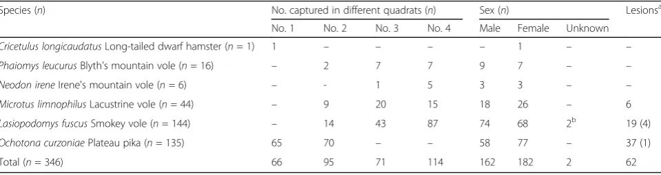
Table 2Statistics of species, gender, and anatomy of captured rodents
Species (n) No. captured in different quadrats (n) Sex (n) Lesionsa
No. 1 No. 2 No. 3 No. 4 Male Female Unknown
Cricetulus longicaudatusLong-tailed dwarf hamster (n= 1) 1 – – – – 1 – –
Phaiomys leucurusBlyth's mountain vole (n= 16) – 2 7 7 9 7 – –
Neodon ireneIrene's mountain vole (n= 6) – - 1 5 3 3 – –
Microtus limnophilusLacustrine vole (n= 44) – 9 20 15 18 26 – 6
Lasiopodomys fuscusSmokey vole (n= 144) – 14 43 87 74 68 2b 19 (4)
Ochotona curzoniaePlateau pika (n= 135) 65 70 – – 58 77 – 37 (1)
Total (n= 346) 66 95 71 114 162 182 2 62
Abbreviation:nnumber of individuals
a
The number of individuals with distinct pathological features/lesions (number of individuals withEchinococcusprotoscoleces) b
Carcasses were partly damaged by raptors
species (L. fuscus, O. curzoniae and M. limnophilus),
both E. multilocularis and E. shiquicus were detected.
There were no mixed Echinococcus spp. infections or
DNA detected in a single host individual (Table 3).
Among the three most-dominant host species, the
prevalence ofE. multilocularisandE. shiquicuswere
sig-nificantly higher in M. limnophilus, whereas the
preva-lence of these twoEchinococcusspp. inO. curzoniaewas
significantly lower. Lasiopodomys fuscus had an
inter-mediate Echinococcus prevalence (Table 3). The
max-imum prevalence of E. multilocularis and the overall
Echinococcus prevalence in L. fuscus were significantly
higher than in O. curzoniae (Table 3). No significant
prevalence bias was detected between male and female
mammals, except forM. limnophilus, in which the
max-imum prevalence of Echinococcus was significantly
higher in females than in males (detected individuals,
fe-male/male, 9/1;χ2= 4.189,df= 1,P= 0.041).
Regression model simulation revealed that Echinococcus
infection was detected in individual hosts with significantly longer RHBL (log odds ratio, 19.134), while their RBWs
were significantly lighter than those without infection (log
odds, -16.010) (Table4). Moreover, although typical lesions
were useful signs of infection (with a log odds ratio of -0. 556 relative to the molecular results), atypical lesions were misleading and caused significantly more misidentification
of infections (log odds ratio of -21.982, Table4).
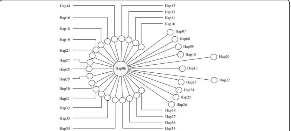
Phylogenetic relationships betweenE. multilocularis cox1 gene haplotypes
In total, 45 haplotypes of a 768 bp longE. multilocularis cox1
gene fragment were acquired from 23 small mammals (76 quences), six human AEs (6 sequences), four dog feces (4 se-quences), and two Tibetan fox fecal samples (5 sequences) in this study, with an additional 17 sequences from online sources. Among the 76 sequences from Shiqu County, 33
haplotypes ofE. multilocularis(i.e. Hap06-Hap38) were
con-firmed (Additional file1: Table S1), of which Hap06 was the
dominant haplotype, with 58 sequences covering all the hu-man AE, dog, and Tibetan fox fecal samples, and 21 small
mammal samples fromL. fuscus,M. limnophilusandO.
cur-zoniae(Fig.1, Additional file1: Table S1). The haplotype and
Fig. 1Network of 33Echinococcus multilocularis cox1 gene haplotypes collected from samples in this study. The size of the circle represents the
number of species of hosts with theE. multilocularisgene haplotype (Hap06 isolated from six species including humans, dogs, Tibetan foxes, two
species of voles and plateau pikas, while each of the other haplotypes has only one host species, see Additional file1: Table S1 for details). The
[image:9.595.58.542.111.165.2]distance between the circle centers shows the variation between two haplotypes (i.e. 1 bp mutation between Hap06 and Hap36)
Table 4Variables of host body condition influencing the general maximum prevalence ofEchinococcusspecies as revealed by the best logistic regression model
Log odds of significant variables ± SE Model evaluation
Relative body weight
Relative head-body length
Lesionsa AICc GeneralizedR2of
the best model
1vs0 2vs0 Best model Null model
-16.010 ± 4.558 19.134 ± 8.521 -21.982 -0.556 49.677 108.473 0.572
a
Categorical data, no SE presented
Abbreviations: 0, individuals without visible lesions; 1, individuals with atypical lesions; 2, individuals with typical lesions;SEstandard error
[image:9.595.57.541.465.683.2]nucleotide diversities for theE. multilocularis cox1 gene from the small mammal community in this study were 0.655 ± 0. 064 and 0.0014 ± 0.0002, respectively. Significant negative
values in both Tajima’sD(D= -2.83311,P< 0.001) and Fu’s
Fs(Fs= -46.942,P< 0.001) tests indicated that theE.
multilo-cularispopulation in the small mammal community of Shiqu
County is currently expanding, and the dominant Hap06 haplotype may be under strong purifying selection.
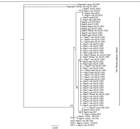
Both ML and Bayesian trees were constructed based
on 56 sequences, including 54 E. multilocularis
se-quences and twoE. granulosus(s.s.) and E. shiquicus
se-quences as outgroups (Additional file 1: Table S1). Both
trees demonstrated identical topological relationships
between haplotypes, but only the Bayesian tree is
in-cluded here (Fig. 2) because of its more concise
struc-ture. The Tibetan Plateau cluster was distinctive from other geographical regions of the world, and mainly comprised the Shiqu haplotypes Hap06-Hap38, the Qinghai Province haplotype Hap05 (also from the Ti-betan Plateau), and two other sequences (Hap04 and
Hap06-Vole-NO.RUS, Additional file 1: Table S1) from
neighboring geographical regions (Fig.2).
Discussion
Transmission ofE. multilocularis and E. shiquicusrelies
on small mammal species as intermediate hosts [3].
Fig. 2Phylogenetic tree comparing the geographical distribution between mtDNAcox1 gene haplotypes ofEchinococcus multilocularis. The
Bayesian phylogenetic analysis was used by setting the“TIM3+I”substitution model, 2,000,000-generation MCMC posterior probability estimation
[image:10.595.58.537.254.694.2]Although monographs of the comprehensive taxonomy of small mammal species with coarse resolution
distribu-tion maps have been published [25,43,44], small
mam-mal assemblages in western China still pose great challenges. Knowledge of the distribution, ecology, and even the basic taxonomy of these small mammals, espe-cially rodent species, is lacking. Consequently, epidemio-logical research focused on small mammal host species based on basic taxonomic and ecological methodologies
in specific E. multilocularis-endemic areas of China is
limited [8,10,14]. Thus, our understanding of the
trans-mission ecology of echinococcosis in the Tibetan Plateau ecosystem is far from complete.
Small mammal host species community composition Although all the six small mammal species reported in this study have been recorded previously on the Tibetan Plateau, information about small rodent species such as arvicolids and cricetids was largely lacking from Shiqu
before the 21st century [45, 46]. He et al. [20] reported
alveolar echinococcosis in the lagomorphs Ochotona
curzoniae and Lepus oiostolus, and also in the
com-mensal rodents N. irene, and Mus musculus in Shiqu.
Raoul et al. [19] studied the habitat ecology of small
mammal communities in Shiqu, reporting for the first
time P. leucurus and C. kamensis in Sichuan Province.
However, neither of these two studies reportedL. fuscus
in Shiqu. The distribution area of L. fuscus was judged
to be restricted to southern Qinghai Province, and this was not considered to be a species distributed to
Si-chuan [24, 25,43, 45]. However, in local plague
surveil-lance studies [47,48] and pest control schemes [49,50],
L. fuscuswas judged to be a dominant rodent species in
the high plateau pasture areas of northwest Sichuan.
Lasiopodomys fuscus is morphologically similar to other
vole species, such as P. leucurus, and thus could be
in-cluded in reports in error if an accurate morphological
identification is lacking [51]. Shiqu is located at the
northwestern point of Sichuan Province, on the southern border of Qinghai; therefore, it is possible that what is
thought to be the southern limit of the range ofL. fuscus
is incorrect [46]. The current study confirmed the
pres-ence ofL. fuscusin Shiqu County, where it was the most
abundant species of the five trapped rodent species and
is likely to have a larger population than that of O.
cur-zoniaein this region (Table2).
Importance of rodent species in the transmission of echinococcosis
Although the potential importance of rodent species as
intermediate hosts ofE. multilocularison the eastern
Ti-betan Plateau has been mentioned previously elsewhere
[10, 14, 51, 52], O. curzoniae were the most frequently
examined small mammal host species with large sample
sizes [8,17,18,20,21,52–54]. For example, when
evalu-ating the prevalence of E. multilocularis, Zhao [21]
re-ported a prevalence of 15.2% in O. curzoniae (34/224)
and 20% in L. fuscus (1/5 individuals). Similarly, Zhang
& Wang [18] reported a prevalence of 5.3% inO.
curzo-niae(62/1177), but only 0.4% in L. fuscus(1/269). Thus,
much smaller sampling sizes might be an important
rea-son for the reported low prevalence ofE. multilocularis
in rodent species. In terms of E. shiquicus, O. curzoniae
was previously the only confirmed intermediate host
species [12, 54]. By contrast, our data revealed that,
among the three most-abundant small mammal species,
both M. limnophilus and L. fuscus had a significantly
higher molecular prevalence of E. multilocularis and E.
shiquicus than did O. curzoniae (Table 3). Moreover, E.
shiquicusDNA was detected not only in the three
most-abundant small mammal species, but also in the other
two rodent species sampled (P. leucurus and C.
longi-caudatus) (Table 3). Although trapping data might not
reflect the exact abundance of each species, the higher
molecular prevalence of both E. multilocularis and E.
shiquicus(Table2) in rodents than in pikas suggests that
rodent species are probably more important intermedi-ate host species than pikas during the transmission of
bothE. multilocularisandE. shiquicus.
Traditionally, epidemiological studies of echinococco-sis in western China mainly focused on human and do-mestic animal populations because of their obvious direct interactions especially regarding transmission of human CE and a role for dogs in risk of human AE.
Phylogenetic analyses revealed that all 33cox1 gene
hap-lotypes from Shiqu County were closely related (Fig.1),
and clustered with haplotypes from other studies to form the Tibetan Plateau group, which is distinct from
haplotypes from other geographical regions (Fig. 2).
Hap06 was the dominant haplotype of E. multilocularis
discovered in humans, domestic dogs, and almost all the wildlife host species tested in this study, including the
three most dominant small mammal species (i.e.O.
cur-zoniae, M .limnophilusand L. fuscus) (Fig.2, Additional
file 1: Table S1). These results confirm that small
mam-mal species and dogs together comprise a wildlife and
peri-domestic ecosystem for transmission of E.
multilo-cularis. Thus, understanding the epidemiology in wildlife
host species is pivotal to understanding the life-cycles and transmission ecology of these parasites.
For a parasite species such asE. multilocularis, which
can utilize multiple host species, understanding the in-terspecific dynamics between host species is essential for
understanding its transmission [55]. Both Tajima’sDand
Fu’s Fs tests revealed the population expansion of E.
multilocularis in the small mammal community in the
study region, as supported by Nakao et al. [28]. Could
this expansion be the result of the frequently reported
increases in small mammal populations and their
inter-specific dynamics in western China?Ochotona curzoniae
and several vole species are the main prey of the Tibetan
fox (V. ferrilata) on grasslands of the eastern Tibetan
Plateau [16]. The significant differences in prevalence of
E. multilocularisamong voles andO. curzoniae(Table3)
suggest that interspecific dynamics among these small mammal species could be essential factors influencing the predator-prey food chain, which would affect the
epidemiology ofE. multilocularisacross all pasture areas
on the Tibetan Plateau. For example, Wang et al. [56]
re-ported a high density of both O. curzoniae and voles in
open grassland areas within 2 km of villages. Ochotona
curzoniae is usually blamed for degrading the grassland
ecosystem of the eastern Tibetan Plateau and, conse-quently, has been a main target for poisoning to protect
the grasslands [49, 50, 57]. Areas around villages are
usually where poisoning programs are located. The fact
that there was a significantly higher prevalence of E.
multilocularis in vole species compared with O.
curzo-niae (Table 3) suggests that pika may have a lesser role
compared to voles in the transmission of E.
multilocu-laris. Furthermore the deliberate poisoning ofO.
curzo-niae could result in increased densities of vole species,
accompanied by a higher prevalence of E. multilocularis
in wildlife, dogs and humans in local areas.
Small mammal body condition and detection of
EchinococcusDNA
The regression model revealed that individuals with
lar-ger RHBL were more likely to be detected with
Echino-coccus, based on the molecular tests (Table 4). Linear
body dimensions are more or less correlated with age,
especially in animals with short lifespans [35]. Thus, the
results of the current study showed that older small
mammals were more likely to have E. multilocularis or
E. shiquicus infections. Similarly, Burlet et al. [34]
re-ported that olderArvicola terrestris had a higher
preva-lence ofE. multilocularisthan younger animals. In small
mammal hosts,E. multilocularisrequires several months
to grow from an oncosphere to a fertile metacestode
[58]. Consequently, the development of metacestode
le-sions can be synchronized with the aging process of its hosts, such as rodents and pikas, further confirming the importance of understanding the host population struc-ture and dynamics when evaluating the present infection burden and predicting the future trend of a specific parasitic species.
Although typical lesions were indicative of infection with
specificEchinococcusspecies, as revealed by the regression
model (Table4), they were harder to find than were the
fre-quently detected atypical lesions (Additional file 1: Table
S2). However, low values of atypical lesions for determining
Echinococcusinfection (Table4, Additional file1: Table S2)
required the use of molecular analyses. Nevertheless, such analyses provide the molecular-positive rate of the parasite, which is not equivalent to its true prevalence. For example,
E. shiquicus DNA was detected in several rodent species;
however, if no typical lesions with metacestodes were
ob-served (Additional file1: Table S2), then it was not possible
to conclude that there was an established parasitic infec-tion. Thus, the function of rodent species as natural
inter-mediate hosts of E. shiquicus still requires further study.
Moreover, many primers have been designed to test various
nuclear [59,60] and mitochondrial [15,26,27,61] genes of
Echinococcus species. In this study, the cox1 and nad1
genes were used. However, the significantly inconsistent
re-sults achieved with both genes (Additional file1: Table S2)
suggested that multiple parasite genes should be tested in the same epidemiological study. In addition, a prevalence interval that is based on the different levels of detection as determined by the available genes (e.g. the maximum and conservative prevalence defined in this study) would be more objective than using a single prevalence.
Conclusions
Our observations suggest that rodent (vole) species are probably more important natural intermediate hosts of
both E. multilocularisand E. shiquicus in Shiqu County
on the eastern Tibetan Plateau. In addition to O.
curzo-niae, the small mammal community sustains
echinococ-cosis transmission in the Tibetan ecosystem. Moreover, small mammal communities usually have complex intra-and interspecific relationships, which influence the population and spatial dynamics of each host species and, thus, the transmission patterns of alveolar
echino-coccosis and E. shiquicus in local areas. Consequently,
we recommend that future studies on the epidemiology of human AE must consider the basic transmission ecol-ogy of the small mammal community as an essential component of research and for control purposes.
Additional file
Additional file 1:Table S1.Information forcox1 sequences used in the
Bayesian phylogenic tree in this study.Table S2.Echinococcusdetection
results of each suspected small mammal sample based on necropsy and molecular analyses. (DOCX 35 kb)
Abbreviations
AE:alveolar echinococcosis; CDC: center for disease control; CE: cystic
echinococcosis; CWL: cross effect of relative weight and head-body length; EB: ethidium bromide; ML: tree maximum likelihood tree; RHBL: relative
head-body length; RW: relative body weight;s.s.:sensu stricto
Acknowledgements
Funding
National Science Foundation of China (NSFC #31071944 and #31470488): supporting the study from the design, field trips and allowance for Chinese authors, data collection in field, laboratory analysis, and data analysis, writing and language polishing. The National Key Program of Research and Development Ministry Science and Technology of China (2016YFC0503200): supporting field trips for Chinese authors, writing, and publishing. NSF EID (US) program (#TW001565) and the Wellcome Trust (#094325/Z/10/Z): supporting trips, data collection in field, laboratory analysis of the European authors.
Availability of data and materials
All data generated or analyzed during this study are included in this
published article and its Additional file1.
Authors’contributions
XW: major writer of the manuscript, molecular analysis and data processing of the echinococcosis materials. JL: collecting echinococcosis materials in Shiqu, small mammal species identification and data processing. QZ, ZM and XW: molecular analysis of small mammal materials. XS and JW: small mammal species identification. BB: dog fecal and human AE sample processing and molecular data analysis. PSC, PG and FR: designing the study and contributing in the writing of the manuscript. ZW: main investigator of the study, designing the study, data analysis and writing the manuscript. All authors read and approved the final manuscript.
Ethics approval and consent to participate
The protocol used to collect the small mammals was approved by the East China Normal University Animal Care and Use Committee (identification number: Q20170501).
Competing interests
The authors declare that they have no competing interests
Publisher’s Note
Springer Nature remains neutral with regard to jurisdictional claims in published maps and institutional affiliations.
Author details
1School of Life Sciences, East China Normal University, Shanghai, China. 2Department of Infectious, Parasitic and Immuno-Mediated Diseases, Istituto Superiore di Sanità, Rome, Italy.3School of Environment and Life Sciences, University of Salford, Greater Manchester, UK.4Chrono-Environment Lab, University of Bourgogne-Franche-Comté and CNRS, Besançon, France.
Received: 27 November 2017 Accepted: 25 April 2018
References
1. Nakao M, Lavikainen A, Yanagida T, Ito A. Phylogenetic systematics of the
genusEchinococcus(Cestoda: Taeniidae). Int J Parasitol. 2013;43:1017–29.
2. Thompson RCA. The taxonomy, phylogeny and transmission of
Echinococcus. Exp Parasitol. 2008;119:439–46.
3. Wang ZH, Wang XM, Liu XQ. Echinococcosis in China, a review of the
epidemiology ofEchinococcusspp. EcoHealth. 2008;5:115–26.
4. Torgerson PR, Keller K, Magnotta M, Ragland N. The global burden of
alveolar echinococcosis. PLoS Negl Trop Dis. 2010;4:e722.
5. Li TY, Qiu JM, Yang W, Craig PS, Chen XW, Xiao N, et al. Echinococcosis in
Tibetan populations, western Sichuan Province, China. Emegr Infect Dis.
2005;11:1866–73.
6. Li TY, Chen XW, Zhen R, Qiu JM, Qiu DC, Xiao N, et al. Widespread
co-endemicity of human cystic and alveolar echinococcosis on the eastern Tibetan Plateau, northwest Sichuan/southeast Qinghai, China. Acta Trop.
2010;113:248–56.
7. National Health and Family Planning Commission of the People’s Republic
of China. Echinococcosis prevention action plan (2010-2015). 2010.http://
www.gov.cn/zwgk/2010-12/14/content_1765485.htm. Accessed 1 Dec 2010.
8. Qiu JM, Chen XW, Ren M, Luo CX, Liu DL, Liu HT, et al. Epidemiological
study on alveolar hydatid disease in Qinghai-Xizang plateau. J Pract Parasit
Dis. 1995;3:106–9.
9. Xiao N, Qui JM, Nakao M, Li TY, Yang W, Chen XW, et al.Echinococcus
shiquicusn. sp., a taeniid cestode from Tibetan fox and plateau pika in
China. Int J Parasitol. 2005;35:693–701.
10. Giraudoux P, Pleydell D, Raoul F, Quéré J-P, Wang Q, Yang YR, et al.
Transmission ecology ofEchinococcus multilocularis: What are the ranges of
parasite stability among various host communities in China? Parasitol Int.
2006;55:S237–46.
11. Craig PS. Epidemiology of human alveolar echinococcosis in China. Parasitol
Int. 2006;55:S221–5.
12. Xiao N, Nakao M, Qiu JM, Budke CM, Giraudoux P, Craig PS, et al. Short
report: dual infection of animal hosts with differentEchinococcusspecies in
the eastern Qinghai-Tibet plateau region of China. Am J Trop Med Hyg.
2006;75:292–4.
13. Boufana B, Qiu JM, Chen XW, Budke CM, Campos-Ponced M, Craig PS. First
report ofEchinococcus shiquicusin dogs from eastern Qinghai-Tibet plateau
region. China. Acta Trop. 2013;127:21–4.
14. Giraudoux P, Raoul F, Afonso E, Ziadinov I, Yang YR, Li L, et al. Transmission
ecosystems ofEchinococcus multilocularisin China and Central Asia.
Parasitology. 2013;140:1655–66.
15. Jiang WB, Liu N, Zhang GT, Renqing PC, Xie F, Li TY, et al. Specific detection
ofEchinococcusspp. from the Tibetan fox (Vulpes ferrilata) and the red fox (V. vulpes) using copro-DNA PCR analysis. Parasitol Res. 2012;111:1531–9.
16. Liu QX, Harris RB, Wang XM. Food habits of the Tibetan fox (Vulpes ferrilata)
in the Kunlun Mountains, Qinghai Province. China. Mamm Biol.
2010;75:283–6.
17. Wang H, Zhao HL, Ma SM, Cao DP, Chai JJ. Investigation on infections of
Echinococcusin animals in Qinghai Plateau. Endem Dis Bull. 2000;15:29–33.
18. Zhang JX, Wang H. Epidemiology ofEchinococcusin animal host species of
Qinghai province. Chin J Parasitol Parasit Dis. 2007;25:350–2.
19. Raoul F, Quéré JP, Rieffel D, Bernard N, Takahashi K, Scheifler R, et al.
Distribution of small mammals in a pastoral landscape of the Tibetan plateaus (Western Sichuan, China) and relationship with grazing practices.
Mammalia. 2006;70:214–25.
20. He JG, Qiu JM, Liu FJ, Chen XW, Liu DL, Chen WD, et al. Epidemiological
survey on hydatidosis in Tibetan region of western Sichuan. II. Infection
situation among domestic and wild animals. Chin J Zoonoses. 2000;16:62–5.
21. Zhao HL. Investigation on infections of alveolar hydatid in small mammals
at south Qinghai Plateau. J Qingh Med Colle. 2002;23:12–4.
22. Wang ZH, Wang XM, Lu QB. Selection of land cover by the Tibetan fox
Vulpes ferrilataon the eastern Tibetan Plateau, western Sichuan Province.
China. Acta Theriol. 2007;52:215–23.
23. Ma B, Wang XM, Liu XQ, Wang ZH. GIS analysis of the spatial relationship
between plateau burrow distribution and vegetation distributional patterns.
Biodivers Sci. 2011;19:71–8.
24. Luo ZX, Chen W, Gao W. Fauna Sinica Mammalia Vol. 6 Rodentia Part III:
Cricetidae. Beijing: Science Press; 2000.
25. Smith AT, Xie Y, editors. A guide to the mammals of China. Princeton:
Princeton University Press; 2008. p. 55–184.
26. Nakao M, Sako Y, Yokoyama N, Fukunaga M, Ito A. Mitochondrial genetic
code in cestodes. Mol Biochem Parasit. 2000;111:415–24.
27. Boufana B, Umhang G, Qiu JM, Chen XW, Lahmar S, Boué F, et al.
Development of three PCR assays for the differentiation between Echinococcus shiquicus,E. granulosus(G1 genotype), andE. multilocularis DNA in the co-endemic region of Qinghai-Tibet plateau, China. Am J Trop
Med Hyg. 2013;88:795–802.
28. Nakao M, Li TY, Han XM, Ma XM, Xiao N, Qiu JM, et al. Genetic
polymorphisms ofEchinococcustapeworms in China as determined by
mitochondrial and nuclear DNA sequences. Int J Parasitol. 2010;40:379–85.
29. Nagelkerke NJD. A note on a general definition of the coefficient of
determination. Biometrika. 1991;78:691–2.
30. Burnhan KP, Anderson DR. Model selection and multimodel inference: a practical
information-theoretic approach. 2nd ed. New York: Springer-Verlag; 2002.
31. Delattre P, Pascal M, LePesteur MH, Giraudoux P, Damange JP.
Caractéristiques éologiques et épidémiologiques de 1'Echinococcus
multilocularisau cours d'un cycle complet des populations d'un hôte
intermédiaire (Microtus arvalis). Can J Zool. 1988;66:2740–50.
32. LePesteur MH, Giraudoux P, Delattre P, Damange JP, Quéré J-P. Spatiotemporal
distribution of four species of cestodes in a landscape of mid-altitude
mountains (Jura, France). Ann Parasitol Hum Comp. 1992;67:155–60.
33. Giraudoux P, Delattre P, Takahashi K, Raoul F, Quéré J-P, Craig P, et al.
Transmission ecology ofEchinococcus multilocularisin wildlife: what can be
learned from comparative studies and multiscale approaches? In: Craig P, Pawlowski Z, editors. Cestode zoonoses: echinococcosis and cysticercosis:
an emergent and global problem. Amsterdam: IOS Press; 2002. p. 251–66.
34. Burlet P, Deplazes P, Hegglin D. Age, season and spatio-temporal factors
affecting the prevalence ofEchinococcus multilocularisandTaenia
taeniaeformisinArvicola terrestris. Parasit Vectors. 2011;4:6.
35. Morris P. A review of mammalian age determination methods. Mammal
Rev. 1972;2:69–104.
36. Burlet P, Deplazes P, Hegglin D. Efficient age determination: how freezing
affects eye lens weight of the small rodent speciesArvicola terrestris. Eur J
Wildlife Res. 2010;56:685–8.
37. Hall T. BioEdit: An important software for molecular biology. GERF Bull
Biosci. 2011;2:60–1.
38. Larkin MA, Blackshields G, Brown NP, Chenna R, Mcgettigan PA, Mcwilliam
H, et al. Clustal W and Clustal X version 2.0. Bioinformatics. 2007;23:2947–8.
39. Librado P, Rozas J. DnaSP v5: a software for comprehensive analysis of DNA
polymorphism data. Bioinformatics. 2009;25:1451–2.
40. Xia XH. DAMBE5: A comprehensive software package for data analysis in
molecular biology and evolution. Mol Biol Evo. 2013;30:1720–8.
41. Darriba D, Taboada GL, Doallo R, Posada D. jModelTest 2: more models,
new heuristics and parallel computing. Nat Methods. 2012;9:772–2.
42. Ronquist F, Huelsenbeck JP. MrBayes 3: Bayesian phylogenetic inference
under mixed models. Bioinformatics. 2003;19:1572–4.
43. Zhang RZ, Jin SK, Quan GQ, Li SH, Ye ZY, Wang FG, et al. Distribution of mammalian
species in China. Beijing: China Forestry Publishing House; 1997. p. 240–1.
44. Jiang ZG, Ma Y, Wu Y, Wang YX, Zhou KY, Liu SY, et al. China’s mammal
diversity and geographic distribution. Beijing: Science Press; 2015. p. 253–
373.
45. Hu QC, Wang YX. Sichuan fauna economica Vol. 2. Chengdu: Sichuan
Science and Technology Press; 1984.
46. Wang YZ. Comparison of the mammal community composition based on
surveys conducted in 1959 and 1987 in Shiqu County. In: Xia WP, Zhang J, editors. The successional changes of mammals in China under the influences of human activities. Beijing: China Science and Technology Press;
1993. p. 155–8.
47. Wang LM, Yang K, Xie F, Tan WM, Li GJ, Wu CX, et al. Surveillance result of
natural foci ofMicrotus fuscusin Sichuan Province from 2000 to 2012. Chin J
Control Endem Dis. 2013;28:335–7.
48. Qi T, Yang K, Wang LM, Duan YJ, Xie F, Yang J, et al. Epidemiology in
plague foci of Shiqu County, China, 2001–2013. Chin J Zoonoses. 2015;31:
485–8.
49. Zhao L, Zhou S, Yan DH, Zhang TY, Tang ZY, Su J. Developing trend and
controlling countermeasures of pests and mice of Sichuan grassland in
2015. Pratac Anim Husb. 2015;220:49–53.
50. Zhao L, Yan DH, Zhang XX, Zhou S. Actuality and control technology of
prairie rodent pests in Sichuan. Pratac Anim Husb. 2015;221:1–7.
51. Giraudoux P, Raoul F, Pleydell D, Li TY, Han XM, Qiu JM, et al. Drivers of
Echinococcus multilocularistransmission in China: small mammal diversity, landscape or climate? PLoS Negl Trop Dis. 2013;7:e2045.
52. Wang H, Zhang JX, Schantz PM, Ito A, Craig PS. Epidemiologic survey and
analysis on echinococcosis in humans and animals from 1995 to 2005 in
Qinghai province. Chin J Zoonoses. 2006;22:1129–34.
53. He DL, Wang H. A report on the epidemiological evaluation of hydatid
disease in Zeku County, Qinghai Province. Endem Dis Bull. 2001;16:36–8.
54. Han XM, Wang H, Ma X, Cai HX, Liu YF, Wei BH, et al. Epidemiological
survey on echinococcosis in Darlag County of Qinghai Province. Chin J
Parasit Dis. 2009;27:22–6.
55. Gandon S. Evolution of multihost parasites. Evolution. 2004;58:455–69.
56. Wang Q, Raoul F, Budke C, Craig PS, Xiao YF, Vuitton DA, et al. Grass height
and transmission ecology ofEchinococcus multilocularisin Tibetan
communities. China. Chin Med J. 2010;123:61–7.
57. Smith AT, Zahler P, Hinds AA. Ineffective and unsustainable poisoning of
native small mammals in temperate Asia: aclassic case of the science-policy divide. In: McNeely JA, McCarthy TM, Smith A, Olsvig-Whittaker L, Wikramanayake ED, editors. Conservation biology in Asia. Kathmandu: Society for Conservation Biology Asia Section and Resources Himalaya;
2006. p. 285–93.
58. Thompson RCA, Mcmanus DP. Aetiology: parasites and life-cycles. In: Eckert
J, Gemmell MA, Meslin FX, Pawlowski ZS, editors. WHO/OIE manual on echinococcosis in humans and animals: A public health problem of global
concern. Paris: World Health Organization; 2001. p. 1–17.
59. Saarma U, Jõgisalu I, Moks E, Varcasia A, Lavikainen A, Oksanen A, et al. A
novel phylogeny for the genusEchinococcus, based on nuclear data,
challenges relationships based on mitochondrial evidence. Parasitology.
2009;136:317–28.
60. Knapp J, Nakao M, Yanagida T, Okamoto M, Saarma U, Lavikainen A, et al.
Phylogenetic relationships withinEchinococcusandTaeniatapeworms
(Cestoda: Taeniidae): an inference from nuclear protein-coding genes. Mol
Phylogenet Evol. 2011;61:628–38.
61. Trachsel D, Deplazes P, Mathis A. Identification of taeniid eggs in the faeces
from carnivores bsed on multiplex PCR using targets in mitochondrial DNA.