W
Wh
haatt m
maak
ke
ess p
paatth
ho
ogge
en
nss p
paatth
ho
ogge
en
niicc
Garth D Ehrlich*
†
, N Luisa Hiller* and Fen Ze Hu*
†
Addresses: *Center for Genomic Sciences, Allegheny General Hospital/Allegheny Singer Research Institute, 320 E. North Ave, Pittsburgh, PA 15212, USA. †Department of Microbiology and Immunology, Drexel University College of Medicine, Allegheny Campus, 320 E. North Ave,
Pittsburgh, PA 15212, USA.
Correspondence: Garth D Ehrlich. Email: [email protected]
A
Ab
bssttrraacctt
Metazoans contain multiple complex microbial ecosystems in which the balance between host and microbe can be tipped from commensalism to pathogenicity. This transition is likely to depend both on the prevailing environmental conditions and on specific gene-gene interactions placed within the context of the entire ecosystem.
Published: 24 June 2008
Genome BBiioollooggyy 2008, 99::225 (doi:10.1186/gb-2008-9-6-225) The electronic version of this article is the complete one and can be found online at http://genomebiology.com/2008/9/6/225
© 2008 BioMed Central Ltd
Metazoans and higher plants are not single-species orga-nisms, but are complex ecosystems composed of a multi-cellular eukaryotic host, with its unique genetic complement [1], and a multitude of ‘microbiomes’. Each microbiome is composed of multiple prokaryotic and eukaryotic symbionts, and the microbiomes and the host collectively make up the ‘symbiome’ (Table 1) [2]. Symbiotic relationships within these ecosystems exist between each of the microbial strains and the host, and also between and among the members of each microbiome. These interdependencies run the gamut from mutualism (in which both or all species benefit) to commensalism (where one party benefits and does no appre-ciable harm to the others) to parasitism (where one of the species benefits at the expense of the other(s)). Finally, a pathogenic relationship exists if the parasite produces a morbid condition in the host. These divisions are themselves an oversimplification of what is, in all likelihood, a con-tinuum: where a given strain of microorganism falls within this spectrum depends not only on its genomic complement but also on the makeup of the microbiome as well as the individual host’s genetics and other environmental factors.
Pathogenicity is not only dependent on qualitative issues such as the presence of specific species, strains, or genes, but also on their relative abundances. Thus, the differential growth of one microbe may result in others transitioning into or out of pathogenic status. It is therefore likely that many pathogens did not initially evolve as pathogens, but
simply take on this role as a result of a lack of ability of the host to maintain homeostasis [3]. Interestingly, not all bacteria associated with pathogenic processes cause disease by their presence; some bacteria are pathogenic by their absence, such as the vaginal lactobacilli whose loss results in an increased pH, which permits overgrowth by invasive species [4-6]. What makes a pathogen, therefore, is the addition, or deletion, of metabolic capabilities in the symbiome that results in a disruption of homeostasis.
G
Ge
en
ne
ettiicc h
he
ette
erro
ogge
en
ne
eiittyy aam
mo
on
ngg b
baacctte
erriiaall p
po
op
pu
ullaattiio
on
nss
m
maak
ke
ess ffo
orr cch
haalllle
en
nggiin
ngg ttaax
xo
on
no
om
myy
gut microflora associated with patients with inflammatory bowel disease [18]) in a manner analogous to damaged sites in the environment that have been shown to have reduced microbial complexity [19-21].
For many bacterial pathogens, such as the non-typeable
Haemophilus influenzae (NTHi) [22,23], Pseudomonas aeruginosa [24,25],Staphylococcus aureus (RJ Boissy, un-published data),Streptococcus agalactiae[26], and Strepto-coccus pneumoniae [27,28], whole-genome sequencing has shown that the supragenome is several times larger than the core genome (see Table 1 for definitions). Thus, for these species there are more distributed genes (see Table 1) than core genes. This leads to the realization that bacterial species-level diagnostics are woefully inadequate as prog-nosticators of disease potential. Therefore, it was not surprising that disease phenotyping for multiple indepen-dent isolates of NTHi [29] and pneumococcus ( Strepto-coccus pneumoniae) [30] revealed a spectrum of diseases -from localized chronic infections to universal lethality.
Similarly, species within the Enterobacteriaceae each reveal a broad spectrum of symbiotic relationships with their hosts. The species Escherichia coli contains both mutualistic strains that have a role in host nutrition, and other strains associated with either chronic urinary disease or acute enterohemorrhagic infections [31,32]. Similarly, pathogenic strains of Enterococcus faecium have emerged from a commensal species, as we discuss below. Whole-genome sequencing of the divergent strains in these species has
revealed massive gene loss and gene gain, resulting in intra-species genomes that vary by more than 30% in size [32].
Bacterial species are usually defined by their 16S rRNA gene. Whereas this is useful for determining phylogenetic relation-ships based on vertically acquired genetic traits, it does not account for horizontally acquired traits, that is, genes acquired by transfer from other species, which are the major driving force in bacterial evolution [23]. Thus, 16S-rRNA-based phylogenies lump together strains that have widely divergent gene distributions, metabolic capabilities, and pathogenic characters [23,26,28-32,33]. A species definition based on possession of a core genome has been proposed [7], but even this is too inclusive to be useful in clinical diagnostics. With the increasing availability of whole-genome sequencing and comparative genomic hybridization (CGH), it should be possible to obtain and analyze very large amounts of bacterial genomic data, which could be cross-indexed with strain-specific disease virulence information to develop effective clinical prognostic indicators.
G
Ge
en
ne
ess aan
nd
d gge
en
ne
e cco
om
mb
biin
naattiio
on
nss d
de
ette
errm
miin
ne
e p
paatth
ho
ogge
en
niicciittyy
As discussed above, within-species comparative genomics combined with disease phenotyping can identify classes of virulence genes that are associated with different pathogenic profiles [22-32]. These findings strongly implicate specific distributed genes and gene combinations as the determi-nants of which bacterial strains are likely to act as patho-gens. Both genotypic and phenotypic heterogeneity have
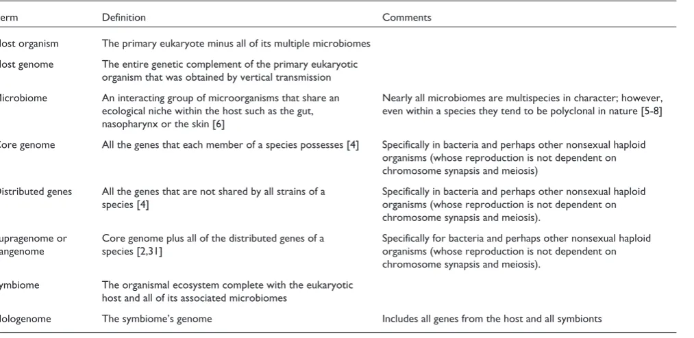
T Taabbllee 11 D
Deeffiinniittiioonnss ooff ssoommee tteerrmmss uusseedd iinn ddiissccuussssiinngg mmiiccrroobbiiaall--hhoosstt ssyymmbbiioossiiss
Term Definition Comments
Host organism The primary eukaryote minus all of its multiple microbiomes Host genome The entire genetic complement of the primary eukaryotic
organism that was obtained by vertical transmission
Microbiome An interacting group of microorganisms that share an Nearly all microbiomes are multispecies in character; however, ecological niche within the host such as the gut, even within a species they tend to be polyclonal in nature [5-8] nasopharynx or the skin [6]
Core genome All the genes that each member of a species possesses [4] Specifically in bacteria and perhaps other nonsexual haploid organisms (whose reproduction is not dependent on chromosome synapsis and meiosis)
Distributed genes All the genes that are not shared by all strains of a Specifically in bacteria and perhaps other nonsexual haploid species [4] organisms (whose reproduction is not dependent on
chromosome synapsis and meiosis).
Supragenome or Core genome plus all of the distributed genes of a Specifically for bacteria and perhaps other nonsexual haploid pangenome species [2,31] organisms (whose reproduction is not dependent on
chromosome synapsis and meiosis). Symbiome The organismal ecosystem complete with the eukaryotic
host and all of its associated microbiomes
[image:2.595.60.567.116.363.2]been demonstrated for the pneumococcus, with some strains associated with chronic indolent infections whereas others are associated with invasive or systemic disease [30]. Similarly, the NTHi display a broad spectrum of phenotypes [29] as well as having a highly plastic genome [22,23], making it likely that correlation studies would find virulence-specific genetic and metabolic pathways.
This view is a departure from classical medical microbiology in which a species-level diagnosis is used to make a prog-nosis. Thus, diagnostics development would profit from large-scale bacterial genotype-phenotype correlation studies designed to provide information on the distributed genes, which are the genes most frequently associated with disease states. Such disease-associated genes may be largely con-fined to a single species, or may be passed among related species, or may be more widely transmitted across broader taxonomic lineages. Examples of species-specific distributed genes include the various heme-acquiring genes found among the NTHi, and the multiple IgA-cleaving proteases isolated among the pneumococci. Within the order Entero-bacteriaceae, the shiga-like toxin genes have been isolated from multiple species, and at higher taxonomic levels, gene cassettes for antibiotic resistance and for natural compe-tence (that is, the ability to take up DNA from the environ-ment) have been passed between negative and Gram-positive bacteria.
The ability to carry out whole-genome sequencing of rela-tively large numbers of bacterial strains using 454-based sequencing technology [34] provides a means of rapidly and inexpensively characterizing the species’ core genomes and supragenomes. Once a relatively complete species supra-genome is available [23,28], microarrays can be constructed containing probes for each distributed gene. These CGH arrays can then be used to interrogate the genomes of large numbers of clinical isolates with different disease pheno-types, providing the information to perform quantitative trait locus-based gene-association studies for the identifica-tion of disease-specific virulence genes. Such a statistical approach to bacterial genetics is new, as until now there have been insufficient sequence data for such an approach. The application of this technology would also provide a comprehensive means of characterizing the functional roles of the plurality of unannotated genes that exist in even the best-studied bacterial species.
H
Ho
ow
w d
do
o p
paatth
ho
ogge
en
nss e
evvo
ollvve
e aan
nd
d w
wh
he
erre
e d
do
o tth
he
eyy cco
om
me
e
ffrro
om
m??
The distributed genome hypothesis [35,36] states that bacterial pathogens arise and acquire virulence traits primarily via horizontal gene transfer (Figure 1). More recently, it has become clear that many bacteria are multi-cellular organisms during part of their life cycle [37], and this has led to the recognition that bacteria possess a
number of virulence traits that are expressed only at the population level and are not operational at the single-cell level [38]. These hypotheses are based on the observation that nearly all classes of pathogenic bacteria maintain highly energy-demanding mechanisms for accessing foreign DNA [39], in spite of the fact that most of these species maintain small genomes. The importance of this observation is that in a background of processes that favor gene deletion [40], the maintenance of multiple horizontal gene transfer mecha-nisms indicates that these traits are highly selected for. The distributed genome hypothesis also posits that chronic pathogens utilize the distribution of non-core genes among strains of a species as a survival strategy, whereby the
F Fiigguurree 11
The distributed genome hypothesis. ((aa)) Schematic showing the distributed (non-core) genes of a species supragenome in a population pool with individual strains below each containing the same set of core genes (green helix). ((bb)) Schematic showing each of the strains of a species with the core genome and a unique distribution of non-core genes.
Distributed genes from the species-level supragenome
Individual strains each with the species core genome
(a)
Individual strains of a species each contain the species core genome as well as a unique distribution
of non-core genes from the supragenome Distributed genes from the species-level
supragenome
continuous recombination of genetic characters between strains serves as a supra-virulence factor that improves popu-lation survival through the generation of new strains with novel combinations of genes. Thus, this population-level gene reassortment acts as a counterpoint to the adaptive immune response of vertebrates, providing a means for pathogens to constantly present the host with novel antigens obtained from any of the constituent species of the symbiome.
Many pathogenic bacteria have complex life cycles that include stages in the environment and passage through multiple hosts. These organisms, therefore, come in contact with many different selective pressures at various stages of their life cycle, and some of the adaptations that provide protection from predation or competition in one stage can induce pathogenicity in another stage. One way in which pathogens evolve is that environmental organisms acquire genes through horizontal transfer that give them an advantage within their non-pathogenic ecosystem. A classic example is the evolution of pathogenic forms of Vibrio cholerae, non-pathogenic progenitor strains of which are principally found in aquatic ecosystems. Pathogenic strains originate from non-pathogenic strains through a multistep process that includes the acquisition of the type IV toxin-co-regulated pilus (TCP). This acquisition is followed by infection with the filamentous phage CTXϕ, which uses the pilus as a point of entry and provides the genes encoding cholera toxin [41]. Studies of cholera epidemics suggest that this general series of genomic rearrangements occurs independently in each epidemic in response to competition among extant environmental strains. These studies led Faruque et al.[41] to hypothesize that “continual emergence of new toxigenic strains and their selective enrichment during cholera outbreaks constitute an essential component of the natural ecosystem for the evolution of epidemic
V. cholerae strains to ensure its continued existence.”
Legionella pneumophila, a bacterium that lives intra-cellularly, also probably evolved its pathogenic characters outside the human host. In humans, L. pneumophila grows and replicates in human alveolar macrophages to cause pneumonia, particularly in immunocompromised hosts. The ability to live within phagocytic cells is the critical virulence factor for this organism and is encoded by the icm/dot secretion system [42], which originally evolved to permit the bacterium’s survival within free-living grazing protozoa. Similarly, E. coliO157, although notorious as a highly viru-lent enterohemorrhagic pathogen of humans, is primarily a commensal microorganism of cattle that also lives in the environment. Although E. coliO157 can be transmitted from person to person, this is not its principal means of propa-gation; thus, it is likely that its virulence in humans is a by-product of other evolutionary forces. Many E. coli strains, including O157, that contain a lambda-like prophage carry-ing the shiga-like toxin genes (stx) have been shown to have a survival advantage in the presence of the ubiquitous
bactivorous protozoan Tetrahymena pyriformis[43]. These investigations showed that most of the survival advantage of the stx-containing strains can be attributed to better survival within the protozoan’s food vacuoles. Thus, for both
L. pneumophila and O157 it would appear that the primary virulence factors associated with human disease actually evolved to play a critical role in the organisms’ survival in other stages of their life cycles. Interestingly, however, the shiga toxin of O157 causes diarrhea in humans, which could lead to increased spread of this strain through fecal contamination. Thus, it is tempting to speculate that acquisition of shiga toxins may be under multiple unrelated evolutionary pressures.
Competition among microorganisms can also generate strains that are pathogenic in their host as a side effect of the intermicrobial arms race. Microorganisms rarely live in isolation, and the myriad interactions amongst co-colonizing species and strains impose a constant selective pressure that ensures the continual evolution of new strains. Thus the same bacterial horizontal gene transfer mechanisms that provide a counterpoint to the host’s adaptive immune response also serve to generate more competitive strains for interspecies competition, with some of these antibacterial mechanisms also resulting in increased virulence towards the host. There is abundant evidence that the numerous bacterial species colonizing the human respiratory mucosa are in competition with each other. Both NTHi and the pneumococcus form biofilms on the middle-ear mucosa that are associated with chronic otitis media but, even when both species are present in the same sample, they do not form mixed biofilms [44]. NTHi can also induce an anti-pneumo-coccal host response during mixed infections that is characterized by increased recruitment of neutrophils into the paranasal spaces [45]. This favors H. influenzae - in spite of the fact that in mixed laboratory culture the pneumo-coccus predominates. Conversely, H. influenzaeis competed against by S. pneumoniae. Both H. influenzaeand Neisseria meningitidis use sialylation of lipooligosaccharides as a mechanism to evade host immune surveillance through mimicry, whereas S. pneumoniae expresses NanA, which desialylates the cell surface of both these bacteria [46]. NanA also alters multiple surface carbohydrates and removes sialic acid residues from human epithelial cells [47]. Disruption of NanA decreases the ability of the pneumococcus to establish a persistent infection, as it can no longer expose the sialylated host-cell receptors needed for attachment [48]. Thus, NanA plays a role in pathogenesis as well as in inter-species competition.
A single molecule is, however, not always advantageous in interactions both with the host and between competing microorganisms. The pore-forming toxin of S. pneumoniae, pneumolysin, increases access of the peptidoglycan of
an advantage to H. influenzae [49]. Thus, the balance between fitness in different environmental settings is critical when considering how pathogens evolve. Mutations that offer a fitness advantage in one environment may confer a disadvantage in another. This is perhaps best understood in respect of microbial drug resistance, where mutations that confer an advantage in the presence of drugs are often deleterious (resulting in slower growth rates) in its absence.
In the monitoring of emerging pathogens it will become increasingly important to recognize the genes and regulatory systems that facilitate transition into a new niche or that balance gene expression within a strain such that it can survive in different environments. In a recent study, Giraud and colleagues [50] created gnotobiotic mice by colonizing germ-free mice with E. coli. In each of eight independent experiments, after habituation, the bacteria were shown to have mutations in the EnvZ-OmpR two-component response regulator, a signal transduction system that controls an entire regulon. This strongly implicates this locus as providing a fitness advantage in this particular environment [50]. This is likely to be the case for many master regulators, and given such an important role in adaptation one might expect these genes to be mostly part of the core genome. In the pneumococcus, however, only a subset of the predicted two-component signal-response systems are core-encoded. Thus, it remains to be determined whether the distributed two-component systems affect pneumococcal fitness under any particular environmental condition, and how the presence, absence, and mutation of these master regulators provides an advantage for one strain over another.
Many pathogens evolve in situ from species that are commensals in the eukaryotic host. This is not surprising, as these organisms are already adapted for survival within the extant symbiome and acquisition of virulence genes can produce a pathogen de novo. Examples of adaptation to a new niche selecting for virulence are commonly observed within the genus Salmonella. Salmonella enterica subspecies I is well adapted to warm-blooded vertebrates. There are more than 1,000 serotypes of this subspecies with different degrees of host adaptation. The level of host specificity among the serotypes correlates with their capacity to cause disease. Mononuclear phagocytes are barriers to the host range of S. enterica, and mechanisms enabling survival of the bacteria within these cells allow adaptation to individual host species [51]. The serotype Typhimurium is successful in mice, and survives well in murine, but not human, macrophages; the reverse is true for the serotype Typhi, which causes disease in humans. In contrast, other subspecies of S. enterica are mainly associated with cold-blooded vertebrates. It is thought that these subspecies survive in the alimentary tract of reptiles, where they are well adapted as commensal organisms [51].
Another example of pathogenic strains evolving from non-pathogenic ones via horizontal gene transfer is the case of Enterococcus faecium. This bacterium has recently evolved from a commensal into a frequently isolated nosocomial (hospital-acquired) pathogen in intensive care units [52]. Comparative genomics has shown that the pathogenic strains have arisen from multiple backgrounds, but all show evidence of having acquired insertion elements (a type of transposable element) that are not present in the commensal strains. Thus, the creation of a new environmental niche, the intensive care unit, has facilitated the evolution of a new subpopulation of this species. The degree of genetic variation among strains in the ‘hospital clade’ of E. faecium
(as assessed by pulsed-field gel electrophoresis and multi-locus sequence typing) was compared with the degree of variation among all other strains. This revealed that the diversity indices (ratio of average genetic similarities) were higher for the hospital clade [52], strongly suggesting increased genomic plasticity within this population that is likely to facilitate its further adaptation.
H
Ho
osstt m
mu
uttaattiio
on
nss aarre
e aasssso
occiiaatte
ed
d w
wiitth
h tth
he
e d
de
evve
ello
op
pm
me
en
ntt
o
off b
baacctte
erriiaall p
paatth
ho
ogge
en
niicciittyy
An example of specific host-bacterium gene combinations resulting in pathogenesis (and the evolution of a pathogen from a commensal) involves the human genetic disease cystic fibrosis. This disease is caused by mutations in the human CFTRgene that lead to the loss of a chloride channel, resulting in highly viscous pulmonary mucus that prevents the normal activity of the ‘mucociliary escalator’, which is designed to sweep bacteria out of the airways. The disease first becomes apparent with colonization and chronic infec-tion by NTHi, which leads inexorably to secondary infecinfec-tion by the opportunistic environmental bacterium P. aerugi-nosa, which establishes a chronic infection involving a biofilm. The pseudomonal infection is ultimately lethal (although modern medical practice can extend life for decades).What is most interesting is that as the P. aeruginosa infection transitions from acute to chronic, there is significant evolution of the bacterial genome [53-56] that makes
P. aeruginosamuch more pathogenic in the lungs of cystic fibrosis patients. Proof of this hypothesis came with the observation that preadolescents with cystic fibrosis who attended the same clinics and summer camps as older adolescents with the disease were experiencing very rapid clinical progression. Molecular typing of the P. aeruginosa
Novel pathogens are constantly emerging from environ-mental and commensal bacterial flora as a result of competi-tive seleccompeti-tive pressures and ubiquitous horizontal gene transfer. Many, perhaps most, virulence traits did not arise originally to damage the host, but rather as a means to compete with other microbes or to prevent predation, or as a means to obtain nutrients from the host. Humans come into contact with a large range of ecological niches through agriculture, aquaculture, and other harvesting, commercial and recreational activities. Given the enormous numbers of microbial species in each of these niches, and the vast size of the accessible supragenomes available to each of these species, novel pathogens are likely to be a permanent feature of human existence.
A
Acck
kn
no
ow
wlle
ed
dgge
em
me
en
nttss
This work was supported by Allegheny General Hospital and Allegheny Singer Research Institute, as well as by grants from the Health Resources and Services Administration and the NIH-NIDCD: DC02148, DC04173, and DC05659. We thank Mary O’Toole for help with the preparation of this manuscript.
R
Re
effe
erre
en
ncce
ess
1. McVean G, Spencer CC, Chaix R: PPeerrssppeeccttiivveess oonn hhuummaann ggeenneettiicc vvaarriiaattiioonn ffrroomm tthhee HHaappMMaapp PPrroojjeecctt..PLoS Genet 2005, 11::e54. 2. Rosenberg E, Koren O, Reshef L, Efrony R, Zilber-Rosenberg I: TThhee
rroollee ooff mmiiccrroooorrggaanniissmmss iinn ccoorraall hheeaalltthh,, ddiisseeaassee aanndd eevvoolluuttiioonn..Nat Rev Microbiol 2007, 55::355-362
3. Goodacre R: MMeettaabboolloommiiccss ooff aa ssuuppeerroorrggaanniissmm.. J Nutr 2007, 113377((11 S
Suuppppll))::259S-266S.
4. Boskey ER, Telsch KM, Whaley KJ, Moench TR, Cone RA: AAcciidd pprro o--d
duuccttiioonn bbyy vvaaggiinnaall fflloorraa iinn vviittrroo iiss ccoonnssiisstteenntt wwiitthh tthhee rraattee aanndd eexxtteenntt o
off vvaaggiinnaall aacciiddiiffiiccaattiioonn..Infect Immun 1999, 6677::5170-175.
5. Sobel JD. IIss tthheerree aa pprrootteeccttiivvee rroollee ffoorr vvaaggiinnaall fflloorraa?? Curr Infect Dis Rep 1999, 11::379-383.
6. Boskey ER, Cone RA, Whaley KJ, Moench TR: OOrriiggiinnss ooff vvaaggiinnaall aacciiddiittyy:: hhiigghh DD//LL llaaccttaattee rraattiioo iiss ccoonnssiisstteenntt wwiitthh bbaacctteerriiaa bbeeiinngg tthhee p
prriimmaarryy ssoouurrccee.. Hum Reprod 2001, 1166::1809-1813.
7. Ehrlich GD, Hu FZ, Shen K, Stoodley P, Post JC: BBaacctteerriiaall pplluurraalliittyy aass aa ggeenneerraall mmeecchhaanniissmm ddrriivviinngg ppeerrssiisstteennccee iinn cchhrroonniicc iinnffeeccttiioonnss.. Clin Orthop Relat Res 2005, 443377::20-24.
8. Murphy TF, Sethi S, Klingman KL, Brueggemann AB, Doern GV: S
Siimmuullttaanneeoouuss rreessppiirraattoorryy ttrraacctt ccoolloonniizzaattiioonn bbyy mmuullttiippllee ssttrraaiinnss ooff n
noonnttyyppeeaabbllee HHaaeemmoopphhiilluuss iinnfflluueennzzaaee iinn cchhrroonniicc oobbssttrruuccttiivvee ppu ull--m
moonnaarryy ddiisseeaassee:: iimmpplliiccaattiioonnss ffoorr aannttiibbiioottiicc tthheerraappyy.. J Infect Dis 1999, 1
18800::404-409.
9. Mukundan D, Ecevit Z, Patel M, Marrs CF, Gilsdorf JR: PPhhaarryynnggeeaall ccoolloonniizzaattiioonn ddyynnaammiiccss ooff HHaaeemmoopphhiilluuss iinnfflluueennzzaaee aanndd HHaaeemmoopphhiilluuss h
haaeemmoollyyttiiccuuss iinn hheeaalltthhyy aadduulltt ccaarrrriieerrss.. J Clin Microbiol 2007, 4
455::3207-3217.
10. Sá-Leão R, Simões AS, Nunes S, Sousa NG, Frazão N, de Lencastre H: IIddeennttiiffiiccaattiioonn,, pprreevvaalleennccee aanndd ppooppuullaattiioonn ssttrruuccttuurree ooff nnoonn--ttyyppaabbllee SSttrreeppttooccooccccuuss ppnneeuummoonniiaaee iinn ccaarrrriiaaggee ssaammpplleess iissoollaatteedd ffrroomm p
prreesscchhoooolleerrss aatttteennddiinngg ddaayy--ccaarree cceennttrreess.. Microbiology 2006, 1
15522::367-376.
11. Sá-Leão R, Nunes S, Brito-Avô A, Alves CR, Carriço JA, Saldanha J, Almeida JS, Santos-Sanches I, de Lencastre H: HHiigghh rraatteess ooff ttrraannssm miiss--ssiioonn ooff aanndd ccoolloonniizzaattiioonn bbyy SSttrreeppttooccooccccuuss ppnneeuummoonniiaaee aanndd H
Haaeemmoopphhiilluuss iinnfflluueennzzaaee wwiitthhiinn aa ddaayy ccaarree cceenntteerr rreevveeaalleedd iinn aa lloon nggii--ttuuddiinnaall ssttuuddyy..J Clin Microbiol 2008, 4466::225-234.
12. Ley RE, Hamady M, Lozupone C, Turnbaugh PJ, Ramey RR, Bircher JS, Schlegel ML, Tucker TA, Schrenzel MD, Knight R, Gordon JI: EEvvo o--lluuttiioonn ooff mmaammmmaallss aanndd tthheeiirr gguutt mmiiccrroobbeess..Science 2008, 33220 0::1647-1651.
13. Lozupone CA, Knight R: SSppeecciieess ddiivveerrggeennccee aanndd tthhee mmeeaassuurreemmeenntt ooff m
miiccrroobbiiaall ddiivveerrssiittyy.. FEMS Microbiol Rev 2008, 3322::557-578.
14. McKenna P, Hoffmann C, Minkah N, Aye PP, Lackner A, Liu Z, Lozupone CA, Hamady M, Knight R, Bushman FD. TThhee mmaaccaaqquuee gguutt m
miiccrroobbiioommee iinn hheeaalltthh,, lleennttiivviirraall iinnffeeccttiioonn,, aanndd cchhrroonniicceenntteerrooccoolliittiiss.. PLoS Pathog 2008, 44::e20.
15. Relman DA: NNeeww tteecchhnnoollooggiieess,, hhuummaann--mmiiccrroobbee iinntteerraaccttiioonnss,, aanndd tthhee sseeaarrcchh ffoorr pprreevviioouussllyy uunnrreeccooggnniizzeedd ppaatthhooggeennss..J Infect Dis 2002, 118866 S
Suuppppll 22::S254-S258.
16. HHuummaann MMiiccrroobbiioommee PPrroojjeecctt [http://nihroadmap.nih.gov/hmp] 17. Job ML, Jacobs NF Jr: DDrruugg--iinndduucceedd ClloCossttrriiddiiuumm ddiiffffiicciillee--aassssoocciiaatteedd
d
diisseeaassee..Drug Safety 1997, 1177::37-46.
18. Manichanh C, Rigottier-Gois L, Bonnaud E, Gloux K, Pelletier E, Frangeul L, Nalin R, Jarrin C, Chardon P, Marteau P, Roca J, Dore J: R
Reedduucceedd ddiivveerrssiittyy ooff ffaaeeccaall mmiiccrroobbiioottaa iinn CCrroohhnn’’ss ddiisseeaassee rreevveeaalleedd bbyy aa mmeettaaggeennoommiicc aapppprrooaacchh..Gut 2006, 5555::205-211
19. Müller AK, Westergaard K, Christensen S, Sørensen SJ: TThhee ddiivveerrssiittyy aanndd ffuunnccttiioonn ooff ssooiill mmiiccrroobbiiaall ccoommmmuunniittiieess eexxppoosseedd ttoo ddiiffffeerreenntt d diiss--ttuurrbbaanncceess.. Microb Ecol 2002, 4444::49-58.
20. Gans J, Wolinsky M, Dunbar J: CCoommppuuttaattiioonnaall iimmpprroovveemmeennttss rreevveeaall ggrreeaatt bbaacctteerriiaall ddiivveerrssiittyy aanndd hhiigghh mmeettaall ttooxxiicciittyy iinn ssooiill..Science 2005, 3
30099::1387-1390.
21. Curtis TP, Sloan WT: MMiiccrroobbiioollooggyy.. EExxpplloorriinngg mmiiccrroobbiiaall ddiivveerrssiittyy --aa vv--aasstt bbeellooww..Science 2005, 330099::1331-1333.
22. Shen K, Antalis P, Gladitz J, Sayeed S, Ahmed A, Yu S, Hayes J, Johnson S, Dice B, Dopico R, Keefe R, Janto B, Chong W, Goodwin J, Wadowsky RW, Erdos G, Post JC, Ehrlich GD, Hu FZ: IIddeen nttiiffiiccaa--ttiioonn,, ddiissttrriibbuuttiioonn,, aanndd eexxpprreessssiioonn ooff nnoovveell ((nnoonnRRdd)) ggeenneess iinn tteenn cclliin nii--ccaall iissoollaatteess ooff nnoonnttyyppeeaabbllee HaaeHemmoopphhiilluuss iinnfflluueennzzaaee.. Infect Immun 2005, 7733::3479-3491.
23. Hogg JS, Hu FZ, Janto B, Boissy R, Gladitz J, Swierczek N, Hayes J, Keefe R, Yu S, Post JC, Hu FZ, Ehrlich GD: CChhaarraacctteerriizzaattiioonn aanndd m
mooddeelliinngg ooff tthhee HHaaeemmoopphhiilluuss iinnfflluueennzzaaee ccoorree aanndd ssuupprraa--ggeennoommee b
baasseedd oonn tthhee ccoommpplleettee ggeennoommiicc sseeqquueenncceess ooff RRdd aanndd 1122 cclliinniiccaall nnoon n--ttyyppeeaabbllee1166 ssttrraaiinnss.. Genome Biol 2007, 88::R103.
24. Shen K, Sayeed S, Antalis P, Gladitz J, Ahmed A, Dice B, Janto B, Dopico R, Keefe R, Hayes J, Johnson S, Yu S, Ehrlich,N, Jocz J, Kropp L, Tadique E, Wong R, Wadowsky RM, Slifkind M, Preston RA, Erdos G, Post JC, Ehrlich GD, Hu FZ: EExxtteennssiivvee ggeennoommiicc ppllaassttiicciittyy iinn P
Psseeuuddoommoonnaass aaeerruuggiinnoossaa rreevveeaalleedd bbyy iiddeennttiiffiiccaattiioonn aanndd ddiissttrriibbuuttiioonn ssttuuddiieess ooff nnoovveell ((nnoonnPPAAOO11)) ggeenneess aammoonngg cclliinniiccaall iissoollaatteess.. Infect Immun 2006, 7744::5272-5283.
25. Mathee K, Narasimhan G, Valdes C, Qiu X, Matewish JM, Koehrsen M, Rokas A, Yandava CN, Engels R, Zeng E, Olavarietta R, Doud M, Smith RS, Montgomery P, White JR, Godfrey PA, Kodira C, Birren B, Galagan JE, Lory S: DDyynnaammiiccss ooff PPsseeuuddoommoonnaass aaeerruuggiinnoossaa ggeennoommee e
evvoolluuttiioonn.. Proc Natl Acad Sci USA 2008, 110055::3100-3105.
26. Tettelin H, Masignani V, Cieslewicz MJ, Donati C, Medini D, Ward NL, Angiuoli SV, Crabtree J, Jones AL, Durkin AS, Deboy RT, David-sen TM, Mora M, Scarselli M, Margarit y Ros I, Peterson JD, Hauser CR, Sundaram JP, Nelson WC, Madupu R, Brinkac LM, Dodson RJ, Rosovitz MJ, Sullivan SA, Daugherty SC, Haft DH, Selengut J, Gwinn ML, Zhou L, Zafar N, et al.: GGeennoommee aannaallyyssiiss ooff mmuullttiippllee ppaatthhooggeenniicc iissoollaatteess ooff SSttrreeppttooccooccccuuss aaggaallaaccttiiaaee:: iimmpplliiccaattiioonnss ffoorr tthhee mmiiccrroobbiiaall ““ppaann--ggeennoommee””.. Proc Natl Acad Sci USA 2005, 110022::13950-13955. 27. Shen K, Antalis P, Gladitz J, Dice B, Janto B, Keefe R, Hayes J, Ahmed
A, Dopico R, Ehrlich N, Jocz J, Kropp J, Yu S, Nistico L, Greenberg D P, Barbadora K, Post JC, Ehrlich GD, Hu FZ: CChhaarraacctteerriizzaattiioonn,, d diissttrrii--b
buuttiioonn aanndd eexxpprreessssiioonn ooff nnoovveell ggeenneess aammoonngg eeiigghhtt cclliinniiccaall iissoollaatteess ooff SSttrreeppttooccooccccuuss ppnneeuummoonniiaaee.. Infect Immun 2006, 7744::321-330. 28. Hiller NL, Janto B, Hogg JS, Boissy R, Yu S, Powell E, Keefe R, Ehrlich
NE, Shen K, Hayes J, Barbadora K, Klimke W, Dernovoy D, Tatusova T, Parkhill J, Bentley SD, Post, JC, Ehrlich GD, Hu FZ: C
Coommppaarraattiivvee ggeennoommiicc aannaallyysseess ooff sseevveenntteeeenn ssttrreeppttooccooccccuuss ppnneeuummo o--n
niiaaee ssttrraaiinnss:: iinnssiigghhttss iinnttoo tthhee ppnneeuummooccooccccaall ssuupprraaggeennoommee..J Bacteriol 2007, 118899::8186-8195.
29. Buchinsky FJ, Forbes M, Hayes J, Hu FZ, Greenberg P, Post JC, Ehrlich GD: PPhheennoottyyppiicc pplluurraalliittyy aammoonngg cclliinniiccaall ssttrraaiinnss ooff nnoon n--ttyyppeeaabbllee HHaaeemmoopphhiilluuss iinnfflluueennzzaaee ddeetteerrmmiinneedd bbyy ssyymmppttoomm sseevveerriittyy iinn tthhee CChhiinncchhiillllaa llaanniiggeerr mmooddeell ooff oottiittiiss mmeeddiiaa.. BMC Microbiol 2007, 7
7::56.
30. Forbes ML, Horsey E, Hiller NL, Buchinsky FJ, Hayes JD, Compli-ment JM, Hillman T, Ezzo S, Shen K, Keefe R, Barbadora K, Post JC, Hu FZ, Ehrlich GD: SSttrraaiinn--ssppeecciiffiicc vviirruulleennccee pphheennoottyyppeess ooff SSttrreepptto o--ccooccccuuss ppnneeuummoonniiaaee aasssseesssseedd uussiinngg tthhee CChhiinncchhiillllaa llaanniiggeerr mmooddeell ooff o
ottiittiiss mmeeddiiaa..PLoS ONE 2008, 33::e1969.
32. Perna NT, Plunkett G 3rd, Burland V, Mau B, Glasner JD, Rose DJ, Mayhew GF, Evans PS, Gregor J, Kirkpatrick HA, Pósfai G, Hackett J, Klink S, Boutin A, ShaoY, Miller L, Grotbeck EJ, Davis NW, Lim A, Dimalanta ET, Potamousis KD, Apodaca J, Anantharaman TS, Lin J, Yen G, Schwartz DC, Welch RA, Blattner FR: GGeennoommee sseeqquueennccee ooff e
enntteerroohhaaeemmoorrrrhhaaggiicc EEsscchheerriicchhiiaa ccoollii OO115577::HH77.. Nature 2001, 440099:: 529-533.
33. Bushman F. Lateral DNA Transfer. Cold Spring Harbor: Cold Spring Harbor Laboratory Press; 2002.
34. Margulies M, Egholm M, Altman WE, Attiya S, Bader JS, Bemben LA, Berka J, Braverman MS, Chen YJ, Chen Z, Dewell SB, Du L, Fierro JM, Gomes XV, Godwin BC, He W, Helgesen S, Ho CH, Irzyk GP, Jando SC, Alenquer ML, Jarvie TP, Jirage KB, Kim JB, Knight JR, Lanza JR, Leamon JH, Lefkowitz SM, Lei M, Li J, et al.: GGeennoommee sseeqquueenncciinngg iinn mmiiccrrooffaabbrriiccaatteedd hhiigghh--ddeennssiittyy ppiiccoolliittrree rreeaaccttoorrss.. Nature 2005, 4
43377::376-380.
35. Ehrlich GD: TThhee bbiiooffiillmm aanndd ddiissttrriibbuutteedd ggeennoommee ppaarraaddiiggmmss pprroovviiddee aa n
neeww tthheeoorreettiiccaall ssttrruuccttuurree ffoorr uunnddeerrssttaannddiinngg cchhrroonniicc bbaacctteerriiaall iinnffe ecc--ttiioonnss.. In 41st Interscience Conference on Antimicrobial Agents and Chemotherapy: December 16-19 2001; Chicago, Illinois. American Society for Microbiology; 2001:524.
36. Shen K, Wang X, Post JC, Ehrlich GD: MMoolleeccuullaarr aanndd ttrraannssllaattiioonnaall rreesseeaarrcchh aapppprrooaacchheess ffoorr tthhee ssttuuddyy ooff bbaacctteerriiaall ppaatthhooggeenneessiiss iinn oottiittiiss m
meeddiiaa.. In Evidence-based Otitis Media, 2nd edition. Edited by Rosen-feld R, Bluestone CD. Hamilton, BC: Decker; 2003: 91-119. 37. Shapiro JA: TThhiinnkkiinngg aabboouutt bbaacctteerriiaall ppooppuullaattiioonnss aass mmuullttiicceelllluullaarr
o
orrggaanniissmmss.. Annu Rev Microbiol 1998; 5522::81-104.
38. Hu FZ, Ehrlich GD: PPooppuullaattiioonn--lleevveell vviirruulleennccee ffaaccttoorrss aammoonnggsstt ppaatth h--o
oggeenniicc bbaacctteerriiaa:: rreellaattiioonn ttoo iinnffeeccttiioonn oouuttccoommee.. Future Microbiol 2008, 33::31-42.
39. Redfield RJ, Findlay WA, Bosse J, Kroll JS, Cameron AD, Nash JH: E
Evvoolluuttiioonn ooff ccoommppeetteennccee aanndd DDNNAA uuppttaakkee ssppeecciiffiicciittyy iinn tthhee P Paass--tteeuurreellllaacceeaaee.. BMC Evol Biol 2006, 66::82
40. Andersson DI: SShhrriinnkkiinngg bbaacctteerriiaall ggeennoommeess.. Microbe 2008, 3 3::124-130.
41. Faruque SM, Sack DA, Sack RB, Colwell RR, Takeda Y, Nair GB: E
Emmeerrggeennccee aanndd eevvoolluuttiioonn ooff VViibbrriioo cchhoolleerraaee OO113399..Proc Natl Acad Sci USA 2003, 110000::1304-1309.
42. Brüggemann H, Cazalet C, Buchrieser C: AAddaappttaattiioonn ooff LLeeggiioonneellllaa p
pnneeuummoopphhiillaa ttoo tthhee hhoosstt eennvviirroonnmmeenntt:: rroollee ooff pprrootteeiinn sseeccrreettiioonn,, e
effffeeccttoorrss aanndd eeuukkaarryyoottiicc--lliikkee pprrootteeiinnss.. Curr Opin Microbiol 2006, 9
9::86-94.
43. Steinberg KM, Levin BR: GGrraazziinngg pprroottoozzooaa aanndd tthhee eevvoolluuttiioonn ooff tthhee E
Esscchheerriicchhiiaa ccoollii OO115577::HH77 SShhiiggaa ttooxxiinn--eennccooddiinngg pprroopphhaaggee..Proc Biol Sci 2007, 227744::1921-1929.
44. Hall-Stoodley L, Hu FZ, Stoodley P, Nistico L, Link TR, Burrows A, Post JC, Ehrlich GD, Kerschner JE: DDiirreecctt ddeetteeccttiioonn ooff bbaacctteerriiaall b
biiooffiillmmss oonn tthhee mmiiddddllee--eeaarr mmuuccoossaa ooff cchhiillddrreenn wwiitthh cchhrroonniicc oottiittiiss m
meeddiiaa..JAMA 2006, 229966::202-211.
45. Lysenko ES, Ratner AJ, Nelson AL, Weiser JN: TThhee rroollee ooff iinnnnaattee iimmmmuunnee rreessppoonnsseess iinn tthhee oouuttccoommee ooff iinntteerrssppeecciieess ccoommppeettiittiioonn ffoorr cco oll--o
onniizzaattiioonn ooff mmuuccoossaall ssuurrffaacceess..PLoS Pathog 2005, 11::e1.
46. Shakhnovich EA, King SJ, Weiser JN: NNeeuurraammiinniiddaassee eexxpprreesssseedd bbyy SSttrreeppttooccooccccuuss ppnneeuummoonniiaaee ddeessiiaallyyllaatteess tthhee lliippooppoollyyssaacccchhaarriiddee ooff N
Neeiisssseerriiaa mmeenniinnggiittiiddiiss aanndd HHaaeemmoopphhiilluuss iinnfflluueennzzaaee:: aa ppaarraaddiiggmm ffoorr iinntteerrbbaacctteerriiaall ccoommppeettiittiioonn aammoonngg ppaatthhooggeennss ooff tthhee hhuummaann rreessp piirraa--ttoorryy ttrraacctt..Infect Immun 2002, 7700::7161-7164.
47. Tong HH, James M, Grants I, Liu X, Shi G, DeMaria TF: CCoommppaarriissoonn o
off ssttrruuccttuurraall cchhaannggeess ooff cceellll ssuurrffaaccee ccaarrbboohhyyddrraatteess iinn tthhee eeuussttaacchhiiaann ttuubbee eeppiitthheelliiuumm ooff cchhiinncchhiillllaass iinnffeecctteedd wwiitthh aa SSttrreeppttooccooccccuuss ppnneeuummo o--n
niiaaee nneeuurraammiinniiddaassee--ddeeffiicciieenntt mmuuttaanntt oorr iittss iissooggeenniicc ppaarreenntt ssttrraaiinn.. Microb Pathog 2001, 3311::309-317.
48. Tong HH, Blue LE, James MA, DeMaria TF: EEvvaalluuaattiioonn ooff tthhee vviirru u--lleennccee ooff aa SSttrreeppttooccooccccuuss ppnneeuummoonniiaaee nneeuurraammiinniiddaassee--ddeeffiicciieenntt m
muuttaanntt iinn nnaassoopphhaarryynnggeeaall ccoolloonniizzaattiioonn aanndd ddeevveellooppmmeenntt ooff oottiittiiss m
meeddiiaa iinn tthhee cchhiinncchhiillllaa mmooddeell..Infect Immun 2000, 6688::921-924. 49. Ratner A J, Aquilar JL, Shchepetov M, Lysenko ES, Weiser JN: NNoodd11
m
meeddiiaatteess ccyyttooppllaassmmiicc sseennssiinngg ooff ccoommbbiinnaattiioonnss ooff eexxttrraacceelllluullaarr bbaacctte e--rriiaa..Cell Microbiol 2007, 99::1343-1351.
50. Giraud A, Arous S, De Paepe M, Gaboriau-Pouthiau V, Bambou JC, Rakotobe S, Lindner AB, Taddei F, Cerf-Bensussan N: DDiisssseeccttiinngg tthhee ggeenneettiicc ccoommppoonneennttss ooff aaddaappttaattiioonn ooff EEsscchheerriicchhiiaa ccoollii ttoo tthhee mmoouussee gguutt..PLoS Genet 2008, 44::e2.
51. Baumler A J, Tsolis RM, Ficht TA, Adams LG: EEvvoolluuttiioonn ooff hhoosstt aaddaap p--ttaattiioonn iinn SSaallmmoonneellllaa eenntteerriiccaa.. Infect Immun 1998, 6666::4579-4587. 52. Leavis H L, Willems RJ, van Wamel WJ, Schuren FH, Caspers MP,
Bonten MJ: IInnsseerrttiioonn sseeqquueennccee--ddrriivveenn ddiivveerrssiiffiiccaattiioonn ccrreeaatteess aa
gglloobbaallllyy ddiissppeerrsseedd eemmeerrggiinngg mmuullttiirreessiissttaanntt ssuubbssppeecciieess ooff EE.. ffaaeecciiuumm.. PLoS Pathog 2007, 33::e7.
53. Jelsbak L, Johansen HK, Frost AL, Thøgersen R, Thomsen LE, Ciofu O, Yang L, Haagensen JA, Høiby N, Molin S: MMoolleeccuullaarr eeppiiddeemmiioollooggyy aanndd ddyynnaammiiccss ooff PPsseeuuddoommoonnaass aaeerruuggiinnoossaa ppooppuullaattiioonnss iinn lluunnggss ooff ccyyssttiicc ffiibbrroossiiss ppaattiieennttss..Infect Immun 2007, 7755::2214-2224.
54. Ciofu O, Riis B, Pressler T, Poulsen HE, Høiby N: OOccccuurrrreennccee ooff h
hyyppeerrmmuuttaabbllee PPsseeuuddoommoonnaass aaeerruuggiinnoossaa iinn ccyyssttiicc ffiibbrroossiiss ppaattiieennttss iiss aassssoocciiaatteedd wwiitthh tthhee ooxxiiddaattiivvee ssttrreessss ccaauusseedd bbyy cchhrroonniicc lluunngg iinnffllaammm maa--ttiioonn..Antimicrob Agents Chemother 2005, 4499::2276-2282.
55. Mathee K, Ciofu O, Sternberg C, Lindum PW, Campbell JI, Jensen P, Johnsen AH, Givskov M, Ohman DE, Molin S, Høiby N, Kharazmi A: M
Muuccooiidd ccoonnvveerrssiioonn ooff PPsseeuuddoommoonnaass aaeerruuggiinnoossaa bbyy hhyyddrrooggeenn ppeerroox x--iiddee:: aa mmeecchhaanniissmm ffoorr vviirruulleennccee aaccttiivvaattiioonn iinn tthhee ccyyssttiicc ffiibbrroossiiss lluunngg.. Microbiology 1999, 114455::1349-1357.