Limitations in using rabbit bladders
in electrophysiological and urodynamic experiments
P. Zerhau
1, Z. Mackerle
1, M. Husar
1, E. Brichtova
1, D. Sochurkova
2,
E. Göpfert
3, M. Faldyna
31Clinic of Paediatric Surgery, Orthopaedics and Traumatology, University Hospital and Masaryk University Brno, Czech Republic
2Clinic of Neurosurgery, University Hospital of St. Anna and Masaryk University Brno, Czech Republic
3Veterinary Research Institute Brno, Czech Republic
ABSTRACT: The aim of this study was to explore the possibility of using rabbit bladder as a model for experimental detrusor electrostimulation research. In a study of urinary bladder activity induced through electrostimulation of the ventral roots, the functional and morphological parameters of the rabbit detrusor were investigated. Under general anaesthesia, open electrostimulation of ventral spinal roots leading towards the detrusor (usually S2, S3) was performed in 20 rabbits. Detrusor response was recorded by repeated electromyography and cystometry in two groups: animals with naturally concentrated urine content (Group A, eight rabbits) and animals after flushing and filling the bladder with saline (Group B, 12 rabbits). Histological examination of bladder wall was performed in both groups. The measured values were compared to one another as well as with data from the veterinary and human literature. The histological specimens were compared with histological specimens of human bladder. The reaction of detrusor fibres was detectable by electromyography in all cases. Elevation of intravesical pressure as a consequence of detrusor contraction was more difficult to detect, as this depends more on the density of the intravesical content. The pressure rise in Group B had a higher amplitude – up to 15 cm H2O versus 5 cm H2O in the first group (P = 0.00046). Histological examination of bladder wall from the two groups of rabbits showed no differences. In comparison with the bladder wall in humans, the only differences found were significantly thinner detrusor layers relative to the overall thickness of bladder wall. It is possible to use rabbit bladder for research into experimentally electrostimulation-induced activity of the detrusor or for experimental detrusor reinnervation research. It is necessary, however, to take into account certain limits – the lower contractility of the bladder wall and the need for qualitative control of bladder content. The present results also suggest that the physiological micturition of rabbits is probably more dependent on abdominal pressure than in humans.
Keywords: electrostimulation; rabbit detrusor; intravesical pressure; micturition; abdominal pressure
Supported by the Ministry of Education, Youth and Sports of the Czech Republic (AdmireVet; Grant No. CZ 1.05/2.1.00/01.0006-ED0006/01/01) and the Internal Grant Agency of the Ministry of Health of the Czech Republic (Grant No. NT 13871-4).
The lower urinary tract consists of the bladder and urethra. The shape, size and position of the bladder depend upon the amount of urine it con-tains. The bladder’s volume increases several fold while filling. The histological structure of the blad-der wall in rabbits and humans has been described as identical. Its external surface is mostly covered
The detrusor muscle is relaxed during the filling phase, is contracted during the voiding phase, and ensures evacuation of the bladder in conjunction with the sphincter mechanism. Abdominal pressure is not involved in micturition in healthy humans. In animals, the function of abdominal pressure is not adequately described in the available literature. Innervation of the bladder is partly sympathetic, facilitated by transmissions from the hypogastric nerves relaxing the bladder muscle, and partly para-sympathetic, mediated by transmissions from the pelvic plexus contracting the detrusor. In humans, sympathetic nerve fibres originate from spinal cord
segments Th10–L2.Parasympathetic innervation
in humans originates from the sacral spinal cord
segments (segments S2–S4) and in rabbits mostly
from segments S2–S3 (Pikov and McCreery 2004).
Urine composition and density is greatly influ-enced by the composition of the diet (especially by the calcium and phosphorus contents), type of housing and overall condition (Harcourt-Brown 2002). If calcium carbonate deposits are present, so-called “sludgy urine” is formed and urination becomes difficult. This situation is commonly en-countered in laboratory animals kept in cages. In humans this phenomenon does not occur under physiological circumstances.
The aim of the present work is to summarise the experience to date with experimentally induced electrophysiological and contractile activity of the rabbit detrusor and simultaneously to examine the suitability of a rabbit model at two qualitatively different bladder loads.
MATERIAL AND METHODS
Between February 2013 and January 2014, un-der general anaesthesia (introduction of ketamine
25 mg/kg i.m. + xylazine 0.4mg/kg i.m., analgesic
butorphanol 0.3 mg/kg i.m. in a single injection at
the start of the surgery and anaesthetic propofol
15mg/kg i.v. continuously throughout the
opera-tion), a group of 20 rabbits (17 males, three females) weighing between 2.8 and 4.9 kg was operated on. First, the bladder was entered through a lower midline laparotomy with the rabbits in a supine position. A bipolar stripped electrode (AU1X4P, Cardion, Brno, Czech Republic) was positioned in the bladder and a CH 10 catheter (Porges, AH 5010, Paris, France) was inserted through the urethra to
measure intravesical pressure (Pves). At the same
time, a small specimen of the bladder wall was re-sected for histological examination (H&E staining, zoom 2.5×). After the abdominal wall was closed, a sagittal incision over the lumbosacral trunk ex-posed the spine with the rabbits in a prone position. Laminectomy and identification of anterior roots
in L4–S3 segments were performed.
After EMG detection of the ventral roots leading towards the detrusor, electrostimulation with a bi-polar hook electrode (Renix International, Sialkot, Pakistan) was performed (amperage 0.5–3.0 mA, 0.05–0.200 ms pulse width, 5 or 15 Hz frequency of repetitive stimulation, 4 s duration of stimulation).
In each animal, all the ventral (motor) roots that provide innervation to the detrusor were probed. The root whose stimulation evoked the largest re-sponse was selected for recordings (usually S2).
Two groups of animals were stimulated: animals with naturally concentrated urine content (30–40 ml of “sludgy urine”, Group A, eight rabbits) and animals after flushing and filling the bladder with 30–40 ml of saline heated to 35 °C (Group B, 12 rabbits). The urinary bladder volume was chosen partly based on the determined mean amount of natural content and partly based on data in the literature (Kontani et al. 2006; Chou et al. 2007; Gomez et al. 2011). No substance was used that could affect the muscle re-sponse during root stimulation.
[image:2.595.325.500.548.740.2]The response of the detrusor was monitored by electromyography (TruTrace EMG, Alien tech-nic, Hronov, Czech Republic), using a bipolar stripped electrode (AU1X4P, Cardion, Brno, Czech Republic), and by cystometric (CTM) measure-ment of intravesical pressure (UD 5500 unit from Dantec, Skovlunde, Denmark) (Figure 1). The total
duration of the experiment (anaesthesia + surgery) ranged from 75–155 min (mean 110 min). Detrusor stimulation was performed repeatedly for 4 s. The measured EMG and CTM values were compared to
one another (using mean, median, Student’s t-test)
and compared with data in the literature on experi-ments performed in animal and human subjects. The histological specimens of rabbit bladder (four from each group) were compared with histological biopsy specimens of human bladder (Institute of Pathology and Anatomy, the University Hospital Brno).
Animal experiments complied with Act No. 246/92 Sb. and were approved by the Branch Commission for Animal Welfare of the Ministry of Agriculture of the Czech Republic (No. 22-2012; Reg. No. 1400).
RESULTS
The reaction of detrusor fibres was detectable by electromyography in both animal groups with
com-parable values. However, intravesical pressure values differed between the two groups. During stimula-tion, significantly higher Pves values were measured in group B, which contained physiological saline. In animals with natural bladder content (“sludgy urine”), the reaction was absent or only minimal.
Group A
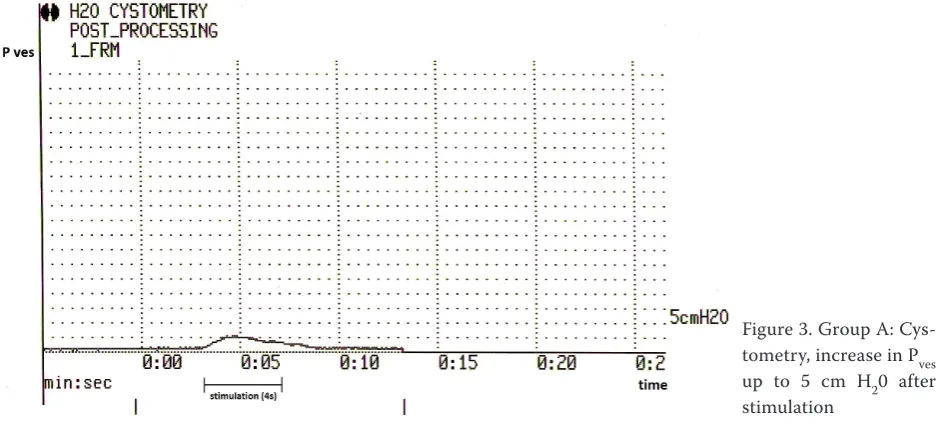
Figure 2 presents EMG records under stimulation for all eight animals (filled with 30–40 ml of sludgy urine) with amplitude of evoked potentials up to 20 uV. Figure 3 and Table 1 show the CTM record in stimulation without elevation of Pves in five cases (62.5%) and with minimal elevation of Pves up to 5 cm
H2O (mean 1.5, median 0) in three cases (37.5%).
Group B
Figure 4 shows EMG records under stimulation for all 12 rabbits (filled with 30–40 ml of
[image:3.595.63.425.84.219.2]physiologi-Figure 2. Group A: Electro-myography of detrusor after anterior spinal root (S2) stim-ulation (0.5 mA current inten-sity, 0.05 ms pulse width, 5 Hz frequency of repetitive stimu-lation, 4 s duration of stimula-tion, response to amplitude up to 20 uV)
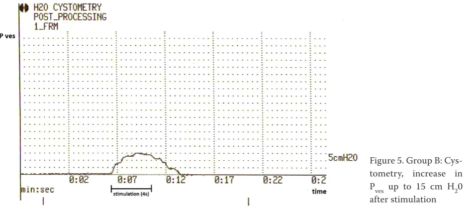
[image:3.595.63.534.549.765.2]cal saline) under conditions comparable with those for Group A (amplitude up to 20 uV). Figure 5 and Table 1 present the CTM record under stimulation
without elevation of Pves in two cases (17%) and
with a rise of Pves to as high as 15 cm H2O (mean
9.9, median 12) in 10 cases (83%). The measured
values of Pves in groups A and B differed with a
significance level of P = 0.00046.
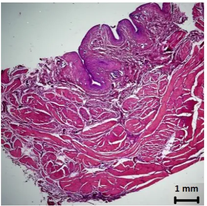
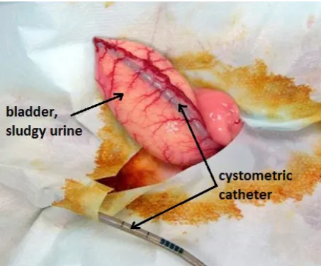
Histological examination of bladder wall (Figure 6) taken from the two groups of rabbits showed no dif-ferences. In comparison with the bladder wall in humans (Figure 7), the only differences found were significantly thinner detrusor layers relative to the overall thickness of the bladder wall. The macro-scopic “membranous” pattern of the rabbit bladder wall is documented in Figure 8.
DISCUSSION
For the experimental study of induced detrusor activity in rabbit bladder via the ventral roots – pel-vic plexus – the detrusor is essential for
determin-ing how realistic it is to monitor this activity. As is clear from our experience, monitoring using elec-tromyography is easier and the pressure response depends on several factors.
The reaction of detrusor fibres (EMG monitor-ing) was detectable in all cases, whether the blad-ders were naturally filled with denser urine as in Group A (having sludgy urine as is often found in rabbits kept in hutches) or after the bladders were flushed and filled with saline, as in Group B.
Elevation of intravesical pressure as a con-sequence of detrusor contraction (cystometric monitoring of Pves) is more difficult to detect. This depends more on the density of the intravesical content. A minimal increase in intravesical pres-sure was observed in only three of eight (38%) ani-mals in Group A (with sludgy urine), while pressure elevation was observed in 10 of 12 (83%) rabbits in Group B (with saline). Furthermore, the pressure
rise had a higher amplitude – up to 15 cm H2O
versus 5 cm H2O in the first group (P = 0.00046).
[image:4.595.62.405.84.206.2]Nevertheless, none of the rabbits in either group released urine during the experiments.
[image:4.595.66.542.550.764.2]Figure 4. Group B: Electromy-ography of detrusor muscle after anterior spinal root (S2) stimulation (0.5 mA current intensity, 0.05 ms pulse width, 5 Hz frequency of repeti-tive stimulation, 4 s duration of stimulation, response to amplitude up to 20 uV)
Our experience has shown that in experimental studies it is necessary to account for the different absolute value of intravesical pressure in rabbits and humans. The strength of human detrusor contraction as evaluated by Pves can be as high as 50 cm H2O (Dai
and Xiao 2005; Xiao 2006) or even 80 to 90 cm H2O
(Dolezel et al. 2002; Martens and Heesakkers 2011; Creasey and Craggs 2012). The values of Pves in rabbits (with comparable parameters of stimulating poten-tials) are significantly lower – in our experiments up
to 5 cm H2O for Group A and up to 15 cm H2O for
Group B. There are no data in the available literature of comparable experiments concerning these animals. Pharmacologically modified Pves values in rabbits have
been described to reach up to 20 cm H2O (Kontani
et al. 2006; Chou et al. 2007), although no parameter for comparison at different bladder filling densities is described. Lin (Lin et al. 2005) also described Pves
values of under 20 cm H2O for rabbits in a study of
the influence of ischaemia on the bladder, although the study focused on pressure in overfilled bladders achieved under entirely different circumstances.
The different Pves values in rabbits and humans
(including children of comparable weight) raise an associated question: to what extent does detrusor
contraction itself influence micturition intravesi-cal pressure and what role does abdominal pres-sure play? Although the voiding phase and the role of individual muscle systems have been described in detail for human medicine (Krhut et al. 2005; Yoshimura and Chancellor 2012), this issue has not yet been addressed within the available literature for veterinary medicine. One of the reasons for this is certainly an absence of open cooperation in monitoring the subject, which is essential for proper urodynamic examination.
The lower detrusor pressure during micturition in rabbits compared to that in humans can be in-directly inferred, too, from the macroscopic and histological structures of the bladder wall. Rabbit bladder is thin-walled, and when filled it resembles the gall bladder in humans. The muscle width in the histological examination supports this obser-vation. For purposes of studying evoked potentials and detrusor pressure responses, abdominal pres-sure nevertheless plays no essential role.
[image:5.595.65.528.102.160.2]We conclude that the rabbit model can be used in experimental studies of detrusor muscle activity by electrostimulation of the spinal roots. It is nev-ertheless necessary to recognise certain limitations,
Figure 6. Histogram of rabbit bladder wall; H&E
[image:5.595.313.525.512.722.2]stain-ing, magnification 2.5× Figure 7. Histogram of human bladder wall, biopsy; H&E staining, magnification 2.5× Table 1. Intravesical pressure (Pves) values for both groups of experimental animals
Group A 1 2 3 4 5 6 7 8 Mean Median
Pves H₂O 0 0 0 0 0 5 3 4 1.5 0
Group B 1 2 3 4 5 6 7 8 9 10 11 12
[image:5.595.63.291.512.722.2]namely, the lower contractility of the bladder wall and the need for qualitative control of the bladder content.
REFERENCES
Chou ECL, Whitbeck C, Herz J, Demopulos G, Levin RM (2007): The effect of intravesical ketoprofen on acetylcholine-evoked urinary bladder contractility and detrusor overactivity in the anesthetized rabbit model. International Urology and Nephrology 39, 1055–1059. Creasey GH, Craggs MD (2012): Functional electrical
stimulation for bladder, bowel, and sexual function. Handbook of Clinical Neurology 109, 247–257. Dai CF, Xiao CG (2005): Electrophysiological
monitor-ing and identification of neural roots durmonitor-ing somatic-autonomic reflex pathway procedure for neurogenic bladder. Chinese Journal of Traumatology 8, 74–76. Dolezel J, Cejpek P, Miklanek D (2002): Sacral
deaffer-entation and neurostimulation of the anterior spinal roots in the treatment of neuropathic urinary bladder in patients with complete transverse spinal lesions – initial clinical experience (in Czech). Rozhledy v Chi-rurgii, 81, 203–209.
Gomez MM, Mendoza-Martinez G, Corona-Quintanilla DL, Fajardo V, Rodriquez-Antolin J, Castelan F (2011):
Multiparity causes uncoordinated activity of pelvic- and perineal-striated muscles and urodynamic changes in rabbits. Reproductive Sciences 18, 1246-1252. Hartcourt-Brown F (2002): Lower urinary tract disease.
In: Textbook of Rabbit Medicine. 1st ed.
Butterworth-Heinemann, Oxford. 338–340.
Konig HE, Liebich HG (2005): Veterinary Anatomy of Domestic Mammals. 4th ed. Schattauer, Stuttgart.
403–404.
Kontani H, Hamamoto T, Takeuchi S, Nomura Y, Sawan-ishi H, Saito H (2006): Comparison of the effects of percutaneous and intraduodenal administration of oxybutynin on bladder contraction and salivation in rabbits. International Journal of Urology 13, 977–984. Krhut J et al. (2005): Physiology of micturition. In:
Neu-rology (in Czech). 1st ed. Galen, Prague. 11–14.
Lin AD, Mannikarottu A, Chaudhry A, Whitbeck C, Kogan BA, Chichester P, Levin RM (2005): Protective effects of grape suspension on in vivo ischaemia/reperfusion of the rabbit bladder. BJU International 96, 1397–1402.
Martens FMJ, Heesakkers JPFA (2011): Clinical results of a Brindley procedure: sacral anterior root stimula-tion in combinastimula-tion with a rhizotomy of the dorsal roots. Advances in Urology, http://www.hindawi.com/ journals/au/2011/709708/.
Maxie MG (2008): Lower urinary tract. In: Pathology of Domestic Animals. 5th ed. Elsevier Saunders,
Edin-burgh. 503–504.
Pikov V, McCreery DB (2004): Mapping of spinal cord circuits controlling the bladder and external urethral sphincter functions in the rabbit. Neurourology and Urodynamics 23, 172–179
Xiao CG (2006): Reinnervation for neurogenic bladder: historic review and introduction of a somatic-auto-nomic reflex pathway procedure for patients with spinal cord injury or spina bifida. European Urology 49, 22–29. Yoshimura N, Chancellor MB (2012): Physiology and
pharmacology of the bladder and urethra. In: Wein AJ, Kavousii LR, Novick AC, Partin AW (eds.): Camp-bell-Walsh Urology. Vol. 3, 10th ed. Elsevier Saunders,
Philadelphia. 1786–1834.
[image:6.595.63.290.84.272.2]Received: 2014–06–19 Accepted after corrections: 2014–09–05
Figure 8. Intraoperative photo of rabbit bladder, “sludgy urine” and catheter for cystometry inside
Corresponding Author:
Pavel Zerhau, University Hospital and Masaryk University Brno, Clinic of Pediatric surgery, orthopedics and traumatology, Cernopolni 9, 625 00 Brno, Czech Republic