Copyright © 1998, American Society for Microbiology. All Rights Reserved.
Direct Susceptibility Testing with Positive BacT/Alert Blood Cultures
by Using MicroScan Overnight and Rapid Panels
KEN B. WAITES,1* E. S. BROOKINGS,1S. A. MOSER,1ANDB. L. ZIMMER2 Department of Pathology, University of Alabama at Birmingham, Birmingham, Alabama,1
and Dade MicroScan, Inc., West Sacramento, California2
Received 4 December 1997/Returned for modification 16 March 1998/Accepted 24 April 1998
Studies were conducted on a method of direct inoculation of MicroScan dried overnight and of rapid panels with positive aerobic blood cultures obtained from the BacT/Alert to determine antimicrobial susceptibilities. Inocula were limited to specimens that appeared unimicrobic on Gram stain. Results were compared to those obtained from panels inoculated following subculture. For 133 gram-negative bacilli, there were 94.7 and 93.5% categorical agreements between direct and standard methods for all drugs tested with overnight and rapid panels, respectively. For 104 gram-positive cocci, there were 93.2 and 93.1% categorical agreements for over-night and rapid panels, respectively. The major error (false resistance) rate for gram negatives was 1.4% for overnight versus 0.7% for rapid panels. The very major error (false susceptibility) rate was 2.7% for overnight versus 8.1% for rapid panels. The total error rates were 1.6% for overnight panels and 1.5% for rapid panels. The major error rates for gram-positive direct susceptibility tests were 2.6% for overnight and 2.5% for rapid panels. The very major error rates were 8.8 and 7.2% for overnight and rapid panels, respectively. Total error rates were 3.6% for overnight and rapid gram-positive panels. These findings suggest that susceptibility results obtained from directly inoculated gram-negative overnight panels have the greatest correlation to those ob-tained by standard methods. When discrepant results occur with direct-susceptibility testing, they are more likely to show false susceptibility than false resistance.
Results of antimicrobial susceptibility tests are essential to guide clinicians in the selection of the most appropriate and cost-effective treatment for persons with bacteremia or other serious infections. Availability of these data as soon as possible after infection is confirmed may result in reduced costs for the pharmacy, laboratory, and other general charges. These data may also lead to timely changes to more effective therapy in the event the infecting organism is resistant to coverage provided by empiric antimicrobial choices (3, 14). Continuously monitor-ing automated blood culture systems such as the BacT/Alert (Organon Teknika Corporation, Durham, N.C.) minimize the length of time required for detection of positive blood cultures from bacteremic patients, often indicating a positive culture within 24 h following the initial incubation, and have emerged as the new standard in blood culture technology (16). Conven-tional methodology requires instrument-positive bottles to be subcultured to solid media and incubated overnight to pro-duce bacterial colonies, which are then used to prepare stan-dardized suspensions for species identification and suscep-tibility testing (8). Direct inoculation from positive blood culture bottles into MicroScan panels (Dade MicroScan, Inc., West Sacramento, Calif.) is a potential alternative to de-crease further the length of time from initial inoculation of blood culture media to reporting of susceptibility results. We performed a prospective study in which fluid from positive blood culture bottles from the BacT/Alert were inoculated di-rectly into MicroScan rapid and overnight panels for gram-negative and gram-positive bacteria and compared susceptibil-ity results with those obtained in standard fashion with each panel type.
MATERIALS AND METHODS
Specimens tested.Positive aerobic blood cultures collected between Septem-ber 1995 and April 1996 from patients suspected of having bacteremia at the University of Alabama at Birmingham Hospital were examined by Gram stain. Specimens that appeared to contain a single organism were included in the study. Specimens that were unimicrobic on Gram stain but yielded more than one isolate after subculture were excluded from analysis. Only aerobic and facultative bacteria were included. Haemophilus spp., Neisseria spp., Streptococcus
pneu-moniae, yeasts, and anaerobes were excluded.
MicroScan instrumentation and panels.The WalkAway/40 instrument with Version 20.30 of the Data Management System was used for all reading and interpretation of panel results. MicroScan Rapid Neg Combo Type 2 panels and MicroScan Dried Overnight Neg Combo Type 15 panels were used for gram-negative isolates. MicroScan Rapid Pos Combo Type 1 panels and MicroScan Overnight Pos Combo Type 6 panels were used for gram-positive isolates.
Blood culture instrumentation and media. Blood culture testing was per-formed on the Organon Teknika BacT/Alert instrument with standard aerobic media.
Direct susceptibility testing.Preliminary studies were performed to determine the optimum method of direct inoculation of MicroScan panels by using seeded cultures (1) and actual patient specimens (18). Serum separator tubes (Becton-Dickinson Vacutainer Systems, Rutherford, N.J.) containing 0.2 ml of Triton X-100 (Sigma, St. Louis, Mo.) were inoculated with 9.5 ml of each blood spec-imen from positive BacT/Alert bottles with a 22-gauge needle. The tubes were centrifuged at 1,4003g for 10 min at room temperature. Bacteria were
har-vested from the surface of the silicon layer by using a cotton swab to make an inoculum suspension equivalent to a 0.5 McFarland standard, confirmed by a MicroScan turbidity meter. This technique has been shown previously to be an acceptable means for preparation of bacterial suspensions for direct susceptibil-ity testing (16). All blood culture specimens were tested the same day that the instrument flagged them as positive. MicroScan panels were inoculated and incubated in the MicroScan WalkAway/40 according to the manufacturer’s in-structions, by using the suspensions prepared as described above.
Standard susceptibility testing.A small amount of the blood culture fluid was inoculated onto Trypticase soy agar with 5% sheep blood (BBL, Cockeysville, Md.) and a MacConkey agar plate (BBL) with crystal violet and lactose. The blood agar plate was incubated in 5% CO2, and the MacConkey agar plate was incubated in air for 18 to 24 h at 35°C to produce bacterial colonies for inocu-lation. Rapid gram-negative panels were inoculated from MacConkey agar. All other panels were inoculated from blood agar plates according to the manufac-turer’s instructions. Panels were then incubated in the same manner as in the direct susceptibility test panels.
Quality control.Appropriate identification and susceptibility quality control organisms for the MicroScan rapid and overnight panels as defined by the
* Corresponding author. Mailing address: Department of Pathology, WP 230, University of Alabama at Birmingham, Birmingham, AL 35233. Phone: (205) 934-6421. Fax: (205) 975-4468. E-mail: [email protected] .edu.
2052
on May 15, 2020 by guest
http://jcm.asm.org/
manufacturer were tested weekly, and all results were acceptable. All other daily maintenance for the MicroScan WalkAway/40 and the BacT/Alert was per-formed in accordance with each manufacturer’s instructions.
Data analysis.Direct testing on MicroScan rapid panels was compared to standardized testing on rapid panels. Direct testing on overnight panels was compared to standardized testing on overnight panels. Drug-organism combina-tions which have not been approved by the Food and Drug Administration for testing with MicroScan were not included in data analysis. Only antimicrobial agents considered appropriate by the National Committee for Clinical Labora-tory Standards guidelines were evaluated by using their published susceptibility breakpoints (11). Gram-positive isolates resistant to oxacillin were considered resistant to other beta-lactam antimicrobials. All drug-organism contraindica-tions were based on the direct test method identification. Organism identification used for summary purposes was that obtained with the standard method.
Categorical agreement for the susceptibility interpretations by direct versus standard methods was determined for each antimicrobial agent. Categorical agreement was defined as complete agreement of the direct and standard inter-pretive results. Since several of the antimicrobials were present in three or fewer dilutions on some panels, no attempt was made to compare directly the actual MICs obtained with direct and standard methods. Major errors (false resistance) occurred when the test method categorized the isolate as resistant when the standard method result was susceptible. Very major errors (false susceptibility) occurred when the direct method characterized the isolate as susceptible while the standard method characterized it as resistant. Minor errors occurred when the direct method characterized the isolate as intermediate while the standard method characterized is as susceptible or resistant, or if the direct method
characterized the isolate as susceptible or resistant while the standard method characterized it as intermediate. The standard method was repeated on isolates with five or more very major or major errors in comparison with the direct method to control for problems which may have occurred with performance of the standardized tests. The repeated standard results were then substituted for original testing results for analytical purposes. Isolates showing only minor errors were not considered in data analysis. The study design involved testing sequential bacterial isolates without regard for duplicate isolates of the same organism in multiple blood culture bottles.
RESULTS
A total of 253 specimens appeared unimicrobic based on ini-tial Gram-stained smears. However, 16 (6.3%) specimens were eventually shown to be polymicrobic when subcultured and ex-cluded from analysis, leaving 237 evaluable specimens. Among these, there were 133 negative bacilli and 104 gram-positive cocci, broken down into species in Table 1.
Standardized susceptibility results were available for all 133 gram-negative bacteria tested on overnight panels. For 114 of 133 (85.7%) gram-negative isolates there were rapid panel MIC results. For 102 of 104 (98.1%) gram-positive cocci there were standardized MIC results on overnight panels, whereas for 81 of 104 (77.9%) there were rapid panel results. Two gram-posi-tive cocci tested by overnight panels were excluded from anal-ysis because of contamination and erroneous readings by the standard method, which were not detected until after the sam-ples had been evaluated. All gram-negative and gram-positive isolates that did not give rapid panel results were due to in-sufficient growth. Six of 133 (4.5%) gram-negative isolates were retested due to five or more very major or major errors on ini-tial testing. Four were due to errors in the overnight panel, and one was due to errors in the rapid panel. Five of 104 (4.8%) of the gram-positive cocci were retested due to five or more major or very major errors on initial testing. Four were retested due to errors in the overnight panel and one was retested because of errors in the rapid panel. After repeat testing, one coagu-lase-negative Staphylococcus sp. (CNS), two Escherichia coli spp., and one Pseudomonas aeruginosa sp. still had five major errors.
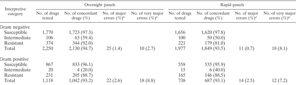
[image:2.612.49.290.79.298.2]One hundred fifteen of 133 (86.4%) gram-negative isolates showed no major or very major errors for any drugs on the overnight panel, compared to 98 of 114 (85.9%) on the rapid panel. Complete categorical agreement, i.e., no very major, major, or minor errors for any drug tested, was observed with 94.7 and 93.5% of drugs tested on overnight and rapid gram-negative panels, respectively (Table 2). Seventy-seven of 102 (75.5%) gram-positive cocci showed no major or very major TABLE 1. Isolates tested
Organism type testedNo. overnight MICNo. providing results
No. providing rapid MIC
results
Gram negative
Acinetobacter spp. 6 6 5 Enterobacter spp. 13 13 12 Escherichia coli 48 48 48 Klebsiella spp. 30 30 30 Pseudomonas aeruginosa 13 13 1 Proteus and Morganella spp. 8 8 8
Salmonella spp. 2 2 2
Serratia spp. 3 3 3
Other 10 10 5
Total 133 133 114
Gram positive
Beta-hemolytic
Strepto-coccus spp. 8 8 8
Enterococcus spp. 7 7 7
Micrococcus spp. 2 2 0
Staphylococcus aureus 37 36 35 Coagulase-negative
Staphylo-coccus spp. 50 49 31
Total 104 102 81
TABLE 2. Categorical agreement of individual susceptibility results for direct versus standard methods
Interpretive category
Overnight panels Rapid panels
No. of drugs
tested No. of concordantdrugs (%) No. of majorerrors (%)a No. of very majorerrors (%)a No. of drugstested No. of concordantdrugs (%) No. of majorerrors (%)a No. of very majorerrors (%)a
Gram negative
Susceptible 1,770 1,723 (97.3) 1,656 1,620 (97.8)
Intermediate 106 63 (59.4) 100 50 (50.0)
Resistant 374 344 (92.0) 221 179 (81.0)
Total 2,250 2,130 (94.7) 25 (1.4) 10 (2.7) 1,977 1,849 (93.5) 11 (0.7) 18 (8.1)
Gram positive
Susceptible 867 833 (96.1) 558 535 (95.9)
Intermediate 20 4 (20.0) 15 6 (40.0)
Resistant 231 205 (88.7) 165 146 (88.5)
Total 1,118 1,042 (93.2) 22 (2.6) 18 (8.8) 738 687 (93.1) 14 (2.5) 12 (7.2)
aDenominator for major error rate is total number of MICs indicating bacterium susceptibility by standard method, while that for very major errors is total number
of MICs indicating bacterium resistance by standard method.
on May 15, 2020 by guest
http://jcm.asm.org/
[image:2.612.51.548.565.708.2]errors on overnight panels, versus 64 of 81 (79.0%) on rapid panels. Complete concordance was observed with 93.2 and 93.1% of drugs tested on overnight and rapid gram-positive panels, respectively.
The major error rate for all gram-negative direct suscepti-bility tests was 1.4% for overnight panels versus 0.7% for rapid panels. The very major error rate was 2.7% for overnight ver-sus 8.1% for rapid panels. The total error rates for gram-negative bacteria, obtained by combining very major and major errors and dividing by the total number of those bacteria for which MICs indicated susceptibility and resistance, were 1.6% for overnight panels and 1.5% for rapid panels.
The major error rates for gram-positive direct susceptibility tests were 2.6% for overnight panels and 2.5% for rapid panels. The very major error rates were 8.8 and 7.2% for overnight and rapid panels, respectively. Total error rates were 3.6% for both overnight and rapid gram-positive panels.
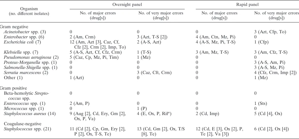
Analysis of errors detected by direct susceptibility testing with overnight and rapid panels according to individual anti-microbials and bacterial species is shown in Table 3. Among drugs on the gram-negative panels, only amikacin and genta-micin had no major or very major errors. Among those on gram-positive panels, narrow-spectrum cephalosporins and gentamicin synergy had no major or very major errors. Aztreo-nam had the most errors (seven on overnight and one on rapid panels), followed by mezlocillin (two on overnight and five on rapid panels), and cefuroxime (five on overnight and one on rapid panels) among agents tested on gram-negative panels. Drugs with the most errors on gram-positive panels were clindamycin (four on overnight and eight on rapid pan-els), oxacillin (four on overnight and seven on rapid panpan-els), and trimethoprim-sulfamethoxazole (nine on overnight pan-els).
One P. aeruginosa and two E. coli isolates produced 15 of 25 major errors on overnight gram-negative panels. One CNS
iso-late gave 5 of 12 major errors on rapid panels. Overall, CNS gave 42 of 66 errors on both panel types combined.
DISCUSSION
Direct susceptibility testing on positive blood cultures has been recommended to facilitate early confirmation of antimi-crobial susceptibility (2, 4, 6–10, 12, 13). Automated blood cul-ture systems such as BacT/Alert and rapid MIC panels provid-ed by automatprovid-ed bacterial identification and susceptibility testing systems such as MicroScan can potentially make direct suscep-tibility testing even more clinically valuable, allowing prelim-inary results in a matter of hours after growth is first detected in the blood culture bottle. Although the concept of direct in-oculation of fluid from positive blood culture bottles into MIC panels was investigated in the early 1980s (6), no prospective evaluations by using the current MicroScan instrumentation, panels, software, and database, newer antimicrobials, or with more recently developed continuously monitoring blood cul-ture instruments such as the BacT/Alert have been published. This study demonstrated that MicroScan overnight and rap-id panels can be used to provrap-ide direct susceptibility test results for unimicrobic gram-negative blood culture isolates detected by the BacT/Alert with 94.7 and 93.1% overall categorical agreement for MICs in comparison to standardized method-ology by using the same instrumentation. For gram-positive bacteria, direct and standard testing with overnight and rapid panels provided 93.2 and 93.1% categorical agreement.
The definitive evaluation of direct susceptibility testing would involve a third reference test method for arbitration of discrepancies between the standardized result and the direct result. However, this was not attempted in the present evalu-ation, mainly because the objective of the study was to deter-mine whether direct susceptibility testing was an acceptable alternative for use with an automated instrument already pres-TABLE 3. Analysis of errors by species and antimicrobial
Organism (no. different isolates)
Overnight panel Rapid panel
No. of major errors
(drug[s]) No. of very major errors(drug[s]) No. of major errors(drug[s]) No. of very major errors(drug[s])
Gram negative
Acinetobacter spp. (3) 0 0 0 3 (Azt, Cfp, To)
Enterobacter spp. (6) 2 (Am, Crm) 3 (Azt, T-S [2]) 4 (Am, Ctn, Mz, Pi) 0 Escherichia coli (7) 12 (Am, Azt [3], Caz, Cf,
Cfz [2], Crm [2], Imp, To) 2 (A-S, Azt) 4 (A-S, Mz, Pi, T-S) 1 (Cfp) Klebsiella spp. (7) 5 (A-S, Azt, Cf, Cfz, Crm) 1 (T-S) 3 (Am, Mz, T-S) 3 (Am, Cfz, T-S) Pseudomonas aeruginosa (2) 5 (Caz, Cp, Mz, Pi, Tim) 1 (Mz) 0 0
Proteus-Morganella spp. (1) 0 0 0 3 (A-S, Am, Pi)
Salmonella-Shigella spp. (1) 0 0 0 3 (A-S, Mz, Pi)
Serratia marcescens (2) 0 3 (Caz, Cft, Crm) 0 4 (Cfz, Crm, Imp [2])
Other (1) 1 (Azt) 0 0 1 (Mz)
Gram positive
Beta-hemolytic
Strepto-coccus spp. 0 0 0 0
Enterococcus spp. (1) 2 (Am, P) 0 0 1 (Sts)
Micrococcus spp. (1) 0 1 (P) 0 0
Staphylococcus aureus (14) 9 (Aug [2], Cd, Ery, Gm [2],
Ox, P, Va) 4 (E, Ox, P, Rif
a) 2 (Cd, Imp) 5 (Cd [4], Ox)
Coagulase-negative
Staphylococcus spp. (21) 11 (Cd [2], Cp, Gm, Ery [2],
P [2], Ox, T-S, Te) 13 (Cd, Gm [2], Ox, T/S[8], Te) 12 (Cd, E [3], Ox [2], P,Te [2], Va [3]) 6 (Cd [2], Ox [4]) aRifampin was not present on rapid gram-positive panels. Am, ampicillin; A-S, ampicillin-sulbactam; Azt, aztreonam; Cd, clindamycin; Cfz, cefazolin; Cfp,
cefo-perazone; Cft, cefotaxime; Ctn, cefotetan; Caz, ceftazidime; Crm, cefuroxime; Cf, cephalothin; Cp, ciprofloxacin; Ery, erythromycin; Gm, gentamicin; Imp, imipenem; Mz, mezlocillin; Ox, oxacillin; P, penicillin; Pi, piperacillin; Rif, rifampin; Sts, streptomycin synergy; Tim, ticarcillin-clavulanate; To, tobramycin; T-S, trimethoprim-sulfamethoxazole; Va, vancomycin.
on May 15, 2020 by guest
http://jcm.asm.org/
[image:3.612.51.548.81.313.2]ent in our laboratory. MicroScan susceptibility test capability has already been compared with reference methods numerous times (7, 17).
Only 6.3% of the specimens tested that appeared unimicro-bic were later found to be polymicrounimicro-bic and unsuitable for direct susceptibility testing by this method, numbers which are in agreement with previously published rates for polymicrobic bacteremia (10). Verification of direct inoculum purity could be made following incubation of overnight panels and prior to reporting results if a subculture to agar is performed at the time of direct inoculation of susceptibility panels. This would not be possible for rapid panels for which MIC data would be available prior to verification of culture purity if rapid direct susceptibility data are to be reported as soon as results are available.
False susceptibility occurred with direct susceptibility tests with a greater frequency than false resistance in overnight and rapid panels for gram-negative as well as gram-positive bacte-ria. When considering the total error rates, there was minimal difference between overnight versus rapid gram-negative pan-els and none between overnight versus rapid gram-positive panels. Total error rates for overnight gram-negative panels were less than the corresponding values for overnight gram-positive panels.
Among gram-positive cocci, CNS were responsible for the most errors. Of major concern, however, was false susceptibil-ity in directly inoculated panels for oxacillin in two isolates of Staphylococcus aureus, one with an overnight panel and the other with a rapid panel. This finding was not surprising since oxacillin-resistant S. aureus is often present in heterogeneous populations and tends to grow more slowly.
In comparative evaluations of susceptibility testing proce-dures, very major errors should occur in,1.5% of all tests, and the overall agreement between tests and the reference method should be$95% (12). Rigorous application of these criteria to data from the present evaluation would mean that direct sus-ceptibility testing with either overnight or rapid panels would be inappropriate, mainly because of the high false-susceptibil-ity rates. However, it has been suggested that these criteria may be too restrictive (12).
Even though automated systems have reduced the time re-quired to detect microorganisms in bloodstream infections, the relatively low number of bacteria in culture bottles may com-promise the inoculum concentration, if not standardized, for susceptibility tests. This may partially account for the relatively high error rates for direct susceptibility testing with MIC meth-ods reported by some studies evaluating blood cultures per-formed in automated instruments (5, 12), but others (2, 6, 13) yielded more favorable comparisons.
Kiehn et al. (6) found only 0.7% categorical discrepancies between MicroScan MIC panels inoculated directly and by standard methodology from nonautomated blood culture sys-tems. More recently, Pettigrew et al. (13) compared results of direct inoculation from positive BacT/Alert bottles to Micro-Scan gram-positive and gram-negative rapid panels. Complete agreements for 1,403 antimicrobial agent-organism combina-tions were 96.9% for gram-negative bacilli and 96.8% for 1,083 gram-positive coccus combinations, slightly more favorable re-sults than obtained in our investigation, which was performed in a similar manner.
Zimmer et al. (18) also evaluated direct susceptibility testing by using MicroScan rapid and dried overnight panels, testing three different inoculum preparation methods and employing multiple media from three different blood culture systems. Due to a small number of positive cultures, results from all media and blood culture systems were combined in data analysis. Overall
categorical agreement rates similar to those reported in the present study were described. They also detected false suscep-tibility by using direct testing more often than they detected false resistance, consistent with our findings.
Direct susceptibility testing of bacteria from positive blood culture bottles by agar disk diffusion is a relatively inexpensive procedure, not tied to bacterial identification, in contrast to direct testing with an automated system such as MicroScan. Agar disk diffusion has been the most widely studied technique of direct susceptibility testing and has the greatest correlation with standardized methods, according to most studies (9).
Most laboratories which invest in MicroScan technology use the instrument for both bacterial identification and suscepti-bility testing, typically employing the same biochemical-antimi-crobial panel for each organism tested. Due to the costs of panels, it is not practical to perform direct susceptibility testing with this technology if results must be repeated with a standardized inoculum. Likewise, it would be impractical to perform direct susceptibility testing unless direct bacterial iden-tification also proves to be accurate so that confirmation fol-lowing subculture is unnecessary.
Organism identification evaluated as part of this investiga-tion has been preliminarily described (15). Ninety-six percent of overnight and 72% of rapid panels showed complete agree-ment between direct and standard methods for identification of gram-negative bacteria, with the highest concordance (99%) occurringwithEnterobacteriaceae.Asmightbeexpected,nonfer-mentative gram-negative bacilli proved more difficult to iden-tify by direct inoculation. Significant problems occurred with gram-positive cocci, for both overnight and rapid panels inoc-ulated directly. Only 82% concordance with standard identifi-cation occurred for overnight and 52% for rapid panels directly inoculated with gram-positive cocci. These discrepancies were predominantly due to the inability to distinguish among CNS, but problems also occurred with identification of S. aureus in di-rectly inoculated panels. Even though Pettigrew and colleagues (13) had a better correlation in their study with respect to di-rect versus standardized susceptibility testing by using Micro-Scan rapid panels, their identification agreement was only 89% for gram-negative bacilli and 70% for gram-positive cocci.
Our practice has been to perform direct susceptibility testing on all blood culture isolates by agar disk diffusion and confirm results for reporting purposes by using MicroScan overnight panels which provide both susceptibility and organism identi-fication. We did not change this practice after performance of this evaluation. If MicroScan panels are to be used for direct susceptibility testing, results should be considered preliminary, mainly because of potential false susceptibility. However, di-rect susceptibility results obtained from gram-negative over-night panels indicating resistance are likely to be in agreement with standard methods and could be considered for immediate reporting if laboratories choose to use such a direct method to improve turnaround time.
Given the many potential advantages of rapid turnaround times for antimicrobial susceptibility results, periodic evalua-tions of automated microbiological instrumentation are ex-tremely important. It is particularly relevant to the study of emerging resistant organisms and newer antimicrobials. Reli-ability of direct susceptibility testing by using any automated method should be tested by individual laboratories before the method is considered for routine use.
ACKNOWLEDGMENTS
Financial support for this project was provided by Dade MicroScan, Inc., West Sacramento, Calif., and Organon Teknika Corporation, Durham, N.C.
on May 15, 2020 by guest
http://jcm.asm.org/
The technical assistance of Linda Van Pelt and Mary McKinnon of Dade Microscan, Inc., is appreciated.
REFERENCES
1. Devitt, K. A., J. M. O’Sullivan, M. L. McKinnon, and B. L. Zimmer. 1995. Feasibility of direct susceptibility testing from various seeded blood culture media into MicroScan rapid and dried overnight susceptibility panels, abstr. C-345, p. 60. In Abstracts of the 95th General Meeting of the American Society for Microbiology 1995. American Society for Microbiology, Wash-ington, D.C.
2. Dipersio, J. R., S. M. Ficorilli, and F. J. Varga. 1984. Direct identification and susceptibility testing of gram-negative bacilli from Bactec bottles by use of MS-2 System with updated bacterial identification software. J. Clin. Mi-crobiol. 20:1202–1204.
3. Doern, G. V., R. Vautour, M. Gaudet, and B. Levy. 1994. Clinical impact of rapid in vitro susceptibility and bacterial identification. J. Clin. Microbiol. 32: 1757–1762.
4. Fay, D., and J. E. Oldfather. 1979. Standardization of direct susceptibility tests for blood cultures. J. Clin. Microbiol. 9:347–350.
5. Howard, W. J., B. J. Buschelman, M. J. Bale, M. A. Pfaller, F. P. Koontz, and
R. N. Jones.1996. Vitek GPS card susceptibility testing accuracy using direct inoculation from Bactec 9240 blood culture bottles, abstr. C-311, p. 56. In Abstracts of the 96th General Meeting of the American Society for Micro-biology 1996. American Society for MicroMicro-biology, Washington, D.C. 6. Kiehn, T. E., C. Capitelo, and D. Armstrong. 1982. Comparison of direct and
standard microtiter broth dilution of susceptibility testing of blood culture isolates. J. Clin. Microbiol. 16:96–98.
7. McGregor, A., F. Schio, S. Beaton, V. Boulton, M. Perman, and G. Gilbert. 1995. The MicroScan WalkAway diagnostic microbiology system—an eval-uation. Pathology 27:172–176.
8. Mirrett, S. 1994. Antimicrobial susceptibility testing and blood cultures. Clin. Lab. Med. 14:171–179.
9. Mirrett, S., and L. B. Reller. 1979. Comparison of direct and standard antimicrobial disk susceptibility testing for bacteria isolated from blood. J. Clin. Microbiol. 10:482–487.
10. Moore, D. F., S. S. Hamada, E. Marso, and W. J. Martin. 1981. Rapid identification and antimicrobial susceptibility testing of gram-negative bacilli from blood cultures by the automicrobic system. J. Clin. Microbiol. 13: 934–939.
11. National Committee for Clinical Laboratory Standards. 1994. Methods for dilution antimicrobial susceptibility tests for bacteria that grow aerobically. Publication M7-A3, 3rd ed. National Committee for Clinical Laboratory Standards, Villanova, Pa.
12. Nolte, F. S., P. B. Contestable, D. Lincaslis, and A. Punsalang, Jr. 1986. Rapid, direct antibiotic susceptibility testing of blood culture isolates using the Abbott Advantage System. Am. J. Clin. Pathol. 86:665–669.
13. Pettigrew, J., D. Ballou, and J. Salyer. 1993. Direct susceptibility testing of positive BacT/Alert blood culture systems with AutoSCAN-W/A rapid combo panels, abstr. C-66, p. 457. In Abstracts of the 93rd General Meeting of the American Society for Microbiology 1993. American Society for Mi-crobiology, Washington, D.C.
14. Trenholme, G. M., R. L. Kaplan, P. H. Karakusis, T. Stine, J. Fuhrer, W.
Landau, and S. Levin.1989. Clinical impact of rapid identification in sus-ceptibility testing of bacterial blood culture isolates. J. Clin. Microbiol. 27: 1342–1345.
15. Waites, K. B., E. S. Brookings, S. A. Moser, M. L. McKinnon, L. Van Pelt,
and B. L. Zimmer.Direct bacterial identification from positive BacT/Alert blood culture specimens using MicroScan overnight and rapid panels. Diagn. Microbiol. Infect. Dis., in press.
16. Wilson, M. L., M. P. Weinstein, and L. B. Reller. 1994. Automated blood culture systems. Clin. Lab. Med. 14:149–169.
17. York, M. K., C. F. Brooks, and E. H. Fiss. 1992. Evaluation of the autoScan W/A rapid system for identification and susceptibility testing of gram-nega-tive fermentagram-nega-tive bacilli. J. Clin. Microbiol. 30:2901–2910.
18. Zimmer, B. L., K. Devitt, M. McKinnon, S. Mirrett, and L. B. Reller. 1995. Direct susceptibility testing from positive blood culture specimens into Mi-croScan rapid and dried overnight susceptibility panels, abstr. C-346, p. 60.
In Abstracts of the 95th General Meeting of the American Society for
Microbiology 1995. American Society for Microbiology, Washington, D.C.