Copyright © 1998, American Society for Microbiology
Development of PCR Assays for Species- and Type-Specific
Identification of Pasteurella multocida Isolates
KIRSTY M. TOWNSEND,1* ALAN J. FROST,1CHIANG W. LEE,1 JOHN M. PAPADIMITRIOU,2ANDHUGH J. S. DAWKINS3
University of Western Australia Department of Pathology2and Urological Research Centre,3
Queen Elizabeth II Medical Centre, Nedlands 6009 Western Australia, and Division of Veterinary Pathobiology, University of Queensland,
Brisbane 4072 Queensland,1Australia
Received 4 August 1997/Returned for modification 19 November 1997/Accepted 14 January 1998
Genomic subtractive hybridization of closely related Pasteurella multocida isolates has generated clones use-ful in distinguishing hemorrhagic septicemia-causing type B strains from other P. multocida serotypes. Oligo-nucleotide primers designed during the sequencing of these clones have proved valuable in the development of PCR assays for rapid species- and type-specific detection of P. multocida and of type B:2 in particular. This study demonstrated that the primer pair designed from the sequence of the clone 6b (KTT72 and KTSP61) specifically amplified a DNA fragment from types B:2, B:5, and B:2,5 P. multocida and that the primers KMT1T7 and KMT1SP6 produced an amplification product unique to all P. multocida isolates analyzed. It was also shown that PCR amplification performed directly on bacterial colonies or cultures represents an extremely rapid, sensitive method of P. multocida identification.
Hemorrhagic septicemia (HS) is a peracute disease of cattle and buffalo, and recently swine, that is endemic in most parts of tropical Asia, Africa, and India (5, 6). Definitive diagnosis of HS is presently made by laboratory identification of the caus-ative agent, Pasteurella multocida serotype B:2 or E:2, although some isolates demonstrate cross-reactivity with type 5 antisera. In recent years, group B isolates possessing somatic antigens other than serotype 2 or 5 have been implicated in causing HS-like disease (or septicemic pasteurellosis) in wild rumi-nants (17, 18). In addition, reexamination of P. multocida strains isolated from outbreaks of HS in North America dem-onstrated that certain strains presumed to be serotype B:2 were in fact serotype B:3,4 (20). These findings emphasize the necessity of employing both capsular and somatic typing meth-ods for definitive serological characterization of P. multocida. The identification of serotypes other than B:2 and E:2 from reported HS outbreaks clearly indicates that the definition of HS and its distinction, if any, from septicemic pasteurellosis require reevaluation.
Accurate laboratory detection of P. multocida depends on the isolation and identification of suspect bacterial colonies by microscopy and biochemical tests. Samples taken immediately from animals that died of suspected pasteurellosis yield almost pure cultures of P. multocida from, e.g., heart blood, liver, spleen, bone marrow, or lung. However, isolation of P.
multo-cida can prove difficult during field surveys of carrier status
when samples are taken from a contaminated site on the ani-mal, such as the nose or throat. Extensive subculturing is then required to obtain a pure culture of the causative organism. In addition, difficulties experienced in the preparation of antisera and the time required for current P. multocida serotyping pro-cedures have meant that definitive serological determination is impractical for most laboratories in countries where HS is endemic (19). This may lead to an increased lag between the
collection of animal material and serotype identification if lengthy transportation is required for the material to reach a laboratory able to perform definitive serotyping procedures.
In recent years, genotypic methods of bacterial identification have proved beneficial in overcoming some limitations of tra-ditional phenotypic procedures. Nucleic acid-based assays al-low the detection of organisms directly from clinical samples or from small amounts of cultured bacterial cells, thus dramati-cally improving the sensitivity and decreasing the time required for bacterial identification. PCR has been particularly useful in this regard, with the use of primer sequences designed to facilitate identification at any level of specificity: strain, species, genus, or all members of a domain (16).
Genomic subtractive hybridization has been of great value in the identification of unique DNA sequences, with its recent application to the identification of differences between closely related bacterial genomes (3, 7, 24). The original subtractive hybridization method described was designed to isolate and clone differentially expressed mRNA sequences (21). Modifi-cations to include the use of genomic DNA have expanded the application of the technique in molecular biology. In recent years, there have been an increasing number of reports of differential cloning of genomic DNA, particularly from pro-karyotic genomes. Genomic subtraction has proved effective in isolating DNA fragments for direct use as probes for strain identification (3, 4, 7, 8).
The incorporation of streptavidin-coated paramagnetic par-ticles and a low-background cloning strategy (9, 24) has expo-nentially increased the efficiency of the subtraction procedure and remains applicable to the employment of competitive re-association of DNA fragments of any cell types to identify unique DNA sequences. This report details the replacement of Streptavidin Magnesphere Paramagnetic Particles (Promega, Sydney, Australia) with Dynabeads M-280 streptavidin (Dy-nal), allowing the addition of solid-phase driver fragments to ensure the enrichment of unique tester DNA sequences fol-lowing magnetic separation.
Oligonucleotide primers designed from cloned subtracted fragments have contributed to the development of PCR-based * Corresponding author. Mailing address: Division of Veterinary
Pathobiology, School of Veterinary Science, The University of Queensland, Brisbane Qld 4072, Australia. Phone: 61 7 3365 2667. Fax: 61 7 3365 1355. E-mail: [email protected].
1096
on May 15, 2020 by guest
http://jcm.asm.org/
assays for species- and type-specific identification of P.
multo-cida and of P. multomulto-cida type B, the causal agent of HS. Rapid
identification of P. multocida and presumptive confirmation of the HS-causing serotype have the potential to reform HS di-agnosis in Southeast Asia, as this technique could be imple-mented in regional laboratories that are currently not able to perform serological determination.
MATERIALS AND METHODS
Bacterial strains.The bacterial strains used in the genomic subtraction and determination of species specificity are listed in Table 1. All bacteria were grown overnight at 37°C on sheep blood agar plates, except for Actinobacillus
pleuro-pneumoniae and Haemophilus influenzae, which were grown on 8% sheep blood
chocolate agar with a Vitox supplement (Oxoid) overnight at 37°C in 5% CO2.
Subtractive hybridization and nucleotide sequence analysis.Genomic subtrac-tive hybridization with Dynabead magnetic separation was performed essentially as described previously (24) with minor modifications. Genomic DNA of tester and driver P. multocida strains was prepared as described by Townsend et al. (23). The tester DNA was from isolate 0113 (type I), while the cocktail driver mix was comprised of 20mg of sonicated (two 5-min bursts), biotinylated DNA from
13B&W buffer (5 mM Tris-HCl [pH 7.5], 0.5 mM EDTA [pH 8.0], 1 M NaCl).
Sau3AI-digested tester DNA was denatured by boiling, cooled on ice, and then
added to the biotinylated DNA-coated beads. Hybridization of driver and tester DNA was performed in a hybridization buffer containing 40 mM
piperazine-N,N9-bis(2-ethanesulfonic acid) (PIPES) (pH 6.4), 1 mM EDTA (pH 8.0), 0.4 M NaCl, and 80% deionized formamide, at 42°C for 24 to 48 h with constant rolling in a Hybaid hybridization oven (Hybaid Limited, Teddington, United Kingdom). Following hybridization, the magnetic beads were captured, and the hybrid-ization mixture was transferred to a new Eppendorf tube. The hybridhybrid-ization mixture was then denatured by heating at 95°C for 5 min and stored on ice until required. The magnetic beads were regenerated by alkali denaturation with immediate magnetic separation. The beads were washed three times, resus-pended in the denatured hybridization mixture, and incubated for a further 24 to 48 h at 42°C. Following the second round of subtraction, the magnetic beads were captured, and enriched subtracted DNA was purified with the BRESA-CLEAN kit (Bresatec Ltd., Thebarton, Australia) and resuspended in 10ml of nuclease-free water (Promega). All subsequent steps were performed as de-scribed previously (24) with additional purification of partially end-filled vector and enriched DNA with the BRESA-CLEAN kit prior to ligation to remove unincorporated nucleotides. Isolated clones successfully amplified by PCR with SP6-T7 promoter primers were examined by Southern blot hybridization with membrane-bound PstI-digested P. multocida DNA, and nucleotide sequence analysis was performed.
Amplification by PCR.Oligonucleotide primers used to sequence the clones 6b (24) and KMT1 were synthesized by the Centre for Cell and Molecular Biology, Queen Elizabeth II Medical Centre, Nedlands, Western Australia, Aus-tralia. The primer sequences are as follows: SP6 promoter primer, 59-TATTTA GGTGACACTATAG-39; T7 promoter primer, 59-d(TAATACGACTCACTAT AGGG)-39; KTSP61, 59-ATCCGCTAACACACTCTC-39(internal sequencing primer for 6b); KTT72, 59-AGGCTCGTTTGGATTATGAAG-39(internal se-quencing primer for 6b); KMT1SP6, 59-GCTGTAAACGAACTCGCCAC-39
(internal sequencing primer for KMT1); and KMT1T7, 59-ATCCGCTATTTA CCCAGTGG-39(internal sequencing primer for KMT1).
Specificity of the PCR assays.In order to determine the specificities of the primers KMT1SP6-KMT1T7 and KTSP61-KTT72, a broad range of bacterial species and P. multocida serotypes (Table 1) were examined. For ease and rapidity, PCR was performed directly from single colonies grown on agar plates. A pipette tip was lightly touched onto a colony, and this sample was then resuspended in PCR amplification mixture containing 10 ng of each primer per
ml, 200mM concentrations of each dNTP, 13Expand High Fidelity buffer with 1.5 mM MgCl2, and 1 U of Expand High Fidelity PCR System enzyme mix
(Boehringer Mannheim). The PCR was performed on an FTS-320 thermal sequencer (Corbett Research), with an initial denaturation at 95°C for 4 min, followed by 30 cycles of denaturation at 95°C for 1 min, annealing at 55°C for 1 min, extension at 72°C for 1 min, and a final extension at 72°C for 9 min. Amplification products were separated by agarose gel electrophoresis (2% aga-rose in 13TAE) at 4 V/cm for 1 h and stained with ethidium bromide. DNA fragments were viewed by UV illumination and photographed.
Nucleotide sequence accession number.The GenBank accession numbers for the subtracted clones KMT1 and 6b are AF016259 and AF016260, respectively.
RESULTS
Genomic subtraction utilizing Dynabead magnetic separa-tion produced three candidate clones, of which one clone (KMT1) was amplified successfully with SP6-T7 promoter primers. The amplified product was radioactively labelled and used to probe membrane-bound PstI-digested P. multocida DNA. Hybridization of the clone KMT1 revealed binding to all serotypes of P. multocida; however, type B and type E isolates could be distinguished from other strains on the basis of frag-ment size (data not shown). In addition, the clone KMT1 was able to distinguish HS-causing P. multocida B:2 from type B strains possessing other somatic serotypes.
Nucleotide sequence analysis of the clone KMT1 was per-formed, and the size of the subtracted fragment was deter-mined to be 866 nucleotides (nt) after allowances were made for the partial end-fill of both the fragment and the vector (Fig. 1). Analysis of open reading frame (ORF) location demon-strated a large ORF of.260 amino acids with a termination at 1778 in reading frame 2 of the sequence obtained with the T7 promoter primer. Multiple terminations were demonstrated in all reading frames of the sequence by means of the SP6 pro-P. multocida
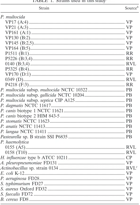
VP17 (A:4) ... VP VP21 (A:3) ... VP VP161 (A:1) ... VP VP130 (B:2)... VP VP145 (B:2,5)... VP VP164 (B:5)... VP P1511 (B:1)... RR P5226 (B:3,4)... RR 0140 (B:3,4) ... RVL P5325 (B:4)... RR VP170 (D:1) ... VP 0349 (D)... RVL P4218 (F:3)... RR P. multocida subsp. multocida NCTC 10322... PB P. multocida subsp. gallicida NCTC 10204... PB P. multocida subsp. septica CIP A125... PB P. dagmatis NCTC 11617... PB P. canis biotype 1 NCTC 11621... PB P. canis biotype 2 HIM 843-5 ... PB P. stomatis NCTC 11623... PB P. anatis NCTC 11413... PB P. langaa NCTC 11411 ... PB Pasteurella sp. B strain SSI P6835 ... PB P. haemolytica
0155 (A5)... RVL 0158 (T10) ... RVL H. influenzae type b ATCC 10211... CP A. pleuropneumoniae FD131 ... VP Actinobacillus sp. strain 0134 ... RVLc
E. coli K-12... VP P. aeruginosa FD28... VP S. typhimurium FD27 ... VP S. aureus Oxford FD32 ... VP S. faecalis FD72 ... VP B. cereus FD8 ... VP
aKnown serotypes of P. multocida and P. haemolytica are given in parentheses
following each isolate identification number.
bVP, Department of Veterinary Pathology, The University of Queensland,
Brisbane, Queensland, Australia. RR, R. Rimler, U.S. Department of Agricul-ture, National Animal Disease Center, Ames, Iowa. RVL, Regional Veterinary Laboratory, Benalla, Victoria, Australia. PB, P. Blackall, Queensland Depart-ment of Primary Industries, Animal Research Institute, Yeerongpilly, Queens-land, Australia. CP, J. J. Sullivan, N. J. Nicolaides, and Partners, Consulting Pathologists, Brisbane, Queensland, Australia.
cOriginally obtained from RVL as P. multocida, this strain, upon subculture,
was determined to be an Actinobacillus species by the Medvet Microbact 24E system (Medvet, Adelaide, Australia).
on May 15, 2020 by guest
http://jcm.asm.org/
[image:2.612.49.292.72.426.2]moter primer. Therefore, it was assumed that the strand ob-tained with the T7 promoter sequence was more likely to be the coding strand, and this primer was used for subsequent database similarity searches. While a search of the
Haemophi-lus influenzae Rd genome (http://www.tigr.org/) did not
dem-onstrate significant identity between the latter and KMT1, a GenBank database search (November, 1995) revealed a degree of identity (59.1% of 115-nt overlap with the T7 sequence) with
bexB of the Haemophilus influenzae type b capsulation locus
(11). Identity (56.0%) in 243 overlapping nt was also observed with an ORF adjacent to the Escherichia coli crp divergent RNA (1). However, recent analysis (November, 1997) of the nucleotide and partial amino acid sequences did not reveal any significant homology to published DNA or protein sequences in either the GenBank or the Swiss-Prot database.
Specificities of PCR primers. In order to determine the
specificities of the regions encoded by clones 6b and KMT1, the internal sequencing primers from each fragment were used to amplify DNA sequences from a broad range of P. multocida isolates, other members of the Pasteurellaceae family, and un-related bacteria. The primer pair KMT1SP6-KMT1T7 ampli-fied a product of approximately 460 bp from all strains of
P. multocida, from the three P. multocida subspecies reference
strains (subsp. multocida, subsp. gallicida, and subsp. septica), and from Pasteurella canis biotype 2 (Fig. 2). No product was detected from any of the remaining cultures. Some variation in the intensity of the amplified product was observed, illustrating
the inconsistency of the DNA concentration used in each PCR by the pipette tip method. However, a positive result is still easily determined. PCR amplification with the primer pair designed during the sequencing of clone 6b (KTSP61-KTT72) specifically produced a product of approximately 590 bp from HS-causing type B isolates of P. multocida (Fig. 3). These primers were unable to amplify DNA from other P. multocida serotypes, other Pasteurella species, other members of the
[image:3.612.52.292.67.361.2]Pas-teurellaceae family, or unrelated bacteria. It was also clearly FIG. 1. Predicted nucleotide sequence of the clone KMT1. Predicted
[image:3.612.309.545.69.237.2]nucle-otide sequence of the clone KMT1 from the T7 promoter primer (GenBank accession number AF016259). The sequence contains an ORF of.260 amino acids in frame 2 before reaching a termination codon at1781 (marked in bold and underlined). The oligonucleotide primers used for sequencing and the PM-PCR assay are underlined and marked KMT1T7 and KMT1SP6.
FIG. 2. P. multocida-specific PCR assay. This figure illustrates fragments specifically amplified by PCR in all P. multocida subspecies and serotypes by means of the primers KMT1SP6 and KMT1T7. The upper panel shows the following: lane 1, negative control; lane 2, P. multocida subsp. multocida; lane 3,
P. multocida subsp. gallicida; lane 4, P. multocida subsp. septica; lane 5, Pasteu-rella dagmatis; lane 6, P. canis biotype 1; lane 7, P. canis biotype 2; lane 8, Pasteurella stomatis; lane 9, Pasteurella anatis; lane 10, Pasteurella langaa; lane 11, Pasteurella species B; lane 12, Pasteurella haemolytica A5; lane 13, Pasteurella haemolytica T10; lane 14, Actinobacillus species 0134; and lane 15, 100-bp DNA
marker (Promega). The lower panel shows the following: lane 1, negative con-trol; lane 2, P. multocida Carter type A; lane 3, type B; lane 4, type D; lane 5, type E; lane 6, type F; lane 7, H. influenzae type b; lane 8, A. pleuropneumoniae; lane 9, E. coli; lane 10, Pseudomonas aeruginosa; lane 11, Salmonella typhimurium; lane 12, Staphylococcus aureus; lane 13, Streptococcus faecalis; lane 14, Bacillus
cereus; and lane M, 100-bp DNA marker. Samples were electrophoresed at 2
V/cm for 2 h in a 2% agarose gel (13TAE), stained with ethidium bromide, visualized by UV illumination, and photographed.
FIG. 3. HS-causing type B P. multocida-specific PCR assay. This figure illus-trates fragments specifically amplified by PCR from type B P. multocida organ-isms that cause hemorrhagic septicemia by means of the primers KTSP61 and KTT72. It can be seen that only P. multocida B:2, B:5, and B:2,5 produced amplification products. This gel shows a negative control (lane 1), P. multocida strain VP161, serotype A:1 (lane 2), VP21, A:3 (lane 3), VP17, A:4 (lane 4), P1511, B:1 (lane 5), 0332, B:2 (lane 6), VP164, B:5 (lane 7), VP145, B:2,5 (lane 8), P5226, B:3,4 (lane 9), P5325, B:4 (lane 10), 0349, D (lane 11), VP170, D:1 (lane 12), 0350, E (lane 13), P4218, F:3 (lane 14), and a 100-bp DNA marker (Promega) (lane M). Samples were electrophoresed at 2 V/cm for 2 h in a 2% agarose gel (13TAE), stained with ethidium bromide, visualized by UV illumi-nation, and photographed.
on May 15, 2020 by guest
http://jcm.asm.org/
[image:3.612.332.525.511.627.2]DISCUSSION
The development of genomic subtractive hybridization has revolutionized the search for virulence genes in pathogenic bacteria with the use of virulent and related avirulent strains to enhance the isolation of DNA fragments related to pathoge-nicity. In addition, this technique is capable of isolating species-specific sequences useful for identification of bacterial species. A modified magnetic cloning strategy incorporating the use of Dynabeads has produced a cloned fragment (KMT1) that, with subsequent hybridization analysis, is capable of distinguishing type B:2 P. multocida from other serotypes. Oligonucleotide primers designed from the nucleotide sequence of this clone and a previously isolated subtracted DNA fragment arbitrarily named 6b (24) have formed the basis for two PCR assays that specifically identify P. multocida, and in particular type B iso-lates that cause HS.
Knowledge of the identity and function of the gene partially encoded by KMT1 would enhance our understanding of the distinction between HS and septicemic-pasteurellosis-caus-ing isolates. However, recent analysis (November, 1997) of sequences in the GenBank database did not reveal any signif-icant identity. While the initial search of the GenBank data-base (November, 1995) demonstrated a degree of identity be-tween clone KMT1 and bexB from H. influenzae type b and also with crp divergent RNA from E. coli, the failure of primers KMT1SP6 and KMT1T7 to produce an amplification product with either species suggests that either this fragment is unique to P. multocida or the primer sequences are not conserved.
The positive amplification of DNA from P. canis biotype 2 was of some interest, as this strain was originally classified as a
P. multocida-like strain, designated Taxon 13, isolated from a
pneumonic calf lung (13). DNA-DNA hybridization studies by Mutters et al. (14) indicated high homology of this strain to isolates now designated as P. canis biotype 1 (previously known as P. multocida biovar 6). At the time of submission of this report, there had not been any published studies documenting the use of specific primers for the detection of P. multocida. Therefore, it is not known whether other laboratories have also observed false-positive amplification of P. canis biotype 2 DNA when testing the specificity of PCR assays for the detection of
P. multocida. These results may, however, indicate a higher
degree of genomic relatedness of P. canis biotype 2 to P.
mul-tocida than was previously seen by DNA-DNA hybridization
analysis. Alternatively, the distinction of P. canis biotype 2 (Orn2) from P. multocida (Orn1) by DNA-DNA hybridization could reflect the findings of Bisgaard et al. (2), in which orni-thine-positive and -negative strains of P. multocida subsp.
sep-tica showed only 44% DNA binding. Comparison of the 16S
rRNA sequences from P. multocida and P. canis biotype 2 could provide clarification of the phylogenetic relationship be-tween these two strains and determine whether these strains represent two species or ornithine variants of P. multocida.
In order to assess accurately the impact of pasteurellosis on the poultry and livestock industries, a rapid diagnostic method specific for the detection of P. multocida is essential. The development of a P. multocida-specific PCR assay will provide rapid species identification without relying on phenotypic dif-ferentiation, which could require up to 2 weeks before defin-itive biotype results are obtained. This assay will also assist in the rapid detection of P. multocida from mixed cultures, a common activity when the clinical sample is obtained from a
atrophic rhinitis (10, 12, 25), with one report detailing the use of arbitrary primers to differentiate P. multocida subsp.
multo-cida (2).
The present study describes the development of a PCR assay that will detect all subspecies of P. multocida, a technique useful for the identification of P. multocida directly from bac-terial cultures without extraction and purification of genomic DNA. As isolation of P. canis biotype 2 has only been reported with pneumonic calves and swine (15, 22), it is unlikely that a false-positive reaction due to this species will hinder field trials aimed at ascertaining the level of carriage or infection with
P. multocida in poultry. Therefore, protocols to detect P. mul-tocida in chicken blood and feces by means of P. mulmul-tocida-
multocida-specific PCR (PM-PCR) are currently being developed, with the aim of providing a rapid, sensitive method for the detection of clinically infected birds. It is hoped that future optimization of this protocol will either eliminate false-positive amplifica-tion from P. canis biotype 2 or clarify the phylogenetic rela-tionship between these two species, thus permitting the use of this technique in field studies of cattle and swine.
Discrimination of the B:2 serotype with the clone KMT1 requires additional hybridization analysis. However, this study has shown that oligonucleotide primers designed during nucle-otide sequencing analysis of the clone 6b (24) can be used to identify type B P. multocida that causes HS (types B:2, B:5, and B:2,5). It is understood that this assay will not identify all HS-causing strains of P. multocida, as these primers do not amplify DNA from type E:2 strains that cause HS in Africa. Nor will this assay identify type B strains of other somatic serotypes that have been implicated in septicemic pasteurello-sis of wild ruminants. However, the ability of the PCR assays described in this study to provide rapid identification of P.
mul-tocida and confirmation of the HS-causing serotype has the
potential to reform HS diagnosis in Southeast Asia. This tech-nique could be implemented in regional laboratories that are currently not able to perform serological determination and be used to rapidly confirm a field diagnosis of HS without the need to obtain pure cultures and perform extensive biochem-ical tests.
ACKNOWLEDGMENT
This work was supported in part by the Australian Centre for Inter-national Agricultural Research.
REFERENCES
1. Bhasin, R., and M. Freundlich. 1991. The nucleotide sequence of the
Esch-erichia coli crp divergent RNA and an overlapping ORF. Biochim. Biophys.
Acta 1129:109–111.
2. Bisgaard, M., S. B. Houghton, R. Mutters, and A. Stenzel. 1991. Reclassifi-cation of German, British and Dutch isolates of so-called Pasteurella
multo-cida obtained from pneumonic calf lungs. Vet. Microbiol. 26:115–124.
3. Bjourson, A. J., C. E. Stone, and J. E. Cooper. 1992. Combined subtraction hybridization and polymerase chain reaction amplification procedure for isolation of strain-specific Rhizobium DNA sequences. Appl. Environ. Mi-crobiol. 58:2296–2301.
4. Bjourson, A. J., and J. E. Cooper. 1988. Isolation of Rhizobium loti strain-specific DNA sequences by subtraction hybridization. Appl. Environ. Micro-biol. 54:2852–2855.
5. Carter, G. R., and M. C. L. De Alwis. 1989. Haemorrhagic septicaemia, p. 131–160. In C. Adlam and J. M. Rutter (ed.), Pasteurella and pasteurellosis. Academic Press Limited, London, England.
6. Chanter, N., and J. M. Rutter. 1989. Pasteurellosis in pigs and the determi-nants of virulence of toxigenic Pasteurella multocida, p. 161–195. In C. Adlam and J. M. Rutter (ed.), Pasteurella and pasteurellosis. Academic Press Lim-ited, London, England.
7. Chen, J., R. Brosch, and J. B. Luchansky. 1993. Isolation and characteriza-tion of Listeria monocytogenes-specific nucleotide sequences. Appl. Environ. Microbiol. 59:4367–4370.
on May 15, 2020 by guest
http://jcm.asm.org/
8. Darrasse, A., A. Kotoujansky, and Y. Bertheau. 1994. Isolation by genomic subtraction of DNA probes specific for Erwinia carotovora subsp. atroseptica. Appl. Environ. Microbiol. 60:298–306.
9. Fletcher, S., D. Darragh, Y. Fan, M. D. Grounds, C. J. Fisher, and M. W. Beilharz.1993. Specific cloning of DNA fragments unique to the dog Y chromosome. Genet. Anal. Tech. Appl. 10:77–83.
10. Kamp, E. M., G. C. Bokken, T. M. Vermeulen, M. F. de Jong, H. E. Buys, F. H. Reek, and M. A. Smits. 1996. A specific and sensitive PCR assay suitable for large-scale detection of toxigenic Pasteurella multocida in nasal and tonsillar swab specimens of pigs. J. Vet. Diagn. Invest. 8:304–309. 11. Kroll, J. S., B. Loynds, L. N. Brophy, and E. R. Moxon. 1990. The bex locus
in encapsulated Haemophilus influenzae: a chromosomal region involved in capsule polysaccharide export. Mol. Microbiol. 4:1853–1862.
12. Lichtensteiger, C. A., S. M. Steenbergen, R. M. Lee, D. D. Polson, and E. R. Vimr.1996. Direct PCR analysis for toxigenic Pasteurella multocida. J. Clin. Microbiol. 34:3035–3039.
13. Madsen, E. B., M. Bisgaard, R. Mutters, and K. B. Pedersen. 1985. Char-acterization of Pasteurella species isolated from lungs of calves with pneu-monia. Can. J. Comp. Med. 49:63–67.
14. Mutters, R., P. Ihm, S. Pohl, W. Fredericksen, and W. Mannheim. 1985. Reclassification of the genus Pasteurella Trevisan 1887 on the basis of de-oxyribonucleic acid homology, with proposals for the new species Pasteurella
dagmatis, Pasteurella canis, Pasteurella stomatis, Pasteurella anatis, and Pas-teurella langaa. Int. J. Syst. Bacteriol. 35:309–322.
15. Nagai, S., S. Someno, and T. Yagihashi. 1994. Differentiation of toxigenic from nontoxigenic isolates of Pasteurella multocida by PCR. J. Clin. Micro-biol. 32:1004–1010.
16. Relman, D. A., and D. H. Persing. 1996. Genotypic methods for microbial identification, p. 3–31. In D. H. Persing (ed.), PCR protocols for emerging infectious diseases: a supplement to Diagnostic Molecular Microbiology:
Prin-ciples and Applications. ASM Press, Washington, D.C.
17. Rhoades, K. R., and R. B. Rimler. 1992. Serological characterisation of
Pasteurella multocida strains isolated from wild ruminants as capsular
sero-group B. Vet. Rec. 130:331–332.
18. Rimler, R. B. 1996. Passive immune cross-protection in mice produced by rabbit antisera against different serotypes of Pasteurella multocida. J. Comp. Pathol. 114:347–360.
19. Rimler, R. B., and K. R. Rhoades. 1989. Pasteurella multocida, p. 37–73. In C. Adlam and J. M. Rutter (ed.), Pasteurella and pasteurellosis. Academic Press Limited, London, England.
20. Rimler, R. B., and M. A. Wilson. 1994. Re-examination of Pasteurella
mul-tocida serotypes that caused haemorrhagic septicaemia in North America.
Vet. Rec. 134:256.
21. Sargent, T. D., and I. B. Dawid. 1983. Differential gene expression in the gastrula of Xenopus laevis. Science 222:135–139.
22. Schimmel, D., and K. Sachse. 1993. Classification of Pasteurella field strains isolated from farms in Germany using traditional methods and DNA-DNA hybridization. Zentbl. Bakteriol. 279:125–130.
23. Townsend, K. M., H. J. S. Dawkins, and J. M. Papadimitriou. 1997. Analysis of haemorrhagic septicaemia-causing isolates of Pasteurella multocida by ribotyping and field alteration gel electrophoresis (FAGE). Vet. Microbiol. 57:383–395.
24. Townsend, K. M., H. J. S. Dawkins, B. J. Zeng, M. W. Watson, and J. M. Papadimitriou.1996. Cloning of a unique sequence specific to type B:2
Pasteurella multocida isolates. Res. Vet. Sci. 61:199–205.
25. Zucker, B., M. Kruger, and F. Horsch. 1996. Differentiation of Pasteurella
multocida subspecies multocida isolates from the respiratory system of pigs
using polymerase chain reaction fingerprinting techniques. Zentbl. Vetmed. Reihe B 43:585–591.