stability
Rajiv Kumar
J Clin Invest.
2009;
119(10)
:2887-2891.
https://doi.org/10.1172/JCI40784
.
Secondary hyperparathyroidism often occurs in chronic kidney disease (CKD) and vitamin
D deficiency, resulting in increased fractures and mortality. Understanding factors that
stimulate parathyroid hormone (PTH) synthesis is important for devising methods to treat
this condition. Previous work has demonstrated that murine
Pth
mRNA levels are regulated
by proteins that bind AU-rich elements (AREs) within the 3
¢
UTR region of
Pth
mRNA and
influence
Pth
mRNA stability. In this issue of the
JCI
, Nechama et al. demonstrate that in
murine secondary hyperparathyroidism associated with CKD or Ca deficiency, the activity of
Pin1, a peptidyl-prolyl isomerase, is reduced (see the related article beginning on page
3102). Reduced Pin1 activity resulted in the phosphorylation and degradation of an
ARE-binding protein, K-homology splicing regulator protein (KSRP), which normally enhances
the degradation of
Pth
mRNA. The activity of other ARE-binding proteins, such as AU-rich
binding factor 1 (AUF1), that increase
Pth
mRNA stability, was increased, thereby
increasing PTH synthesis. This work suggests new ways by which to regulate PTH
synthesis in secondary hyperparathyroidism.
Commentary
Find the latest version:
setbacks recently (21), the overwhelming clinical need for these interventions is clear. A definition of affordable, safe, and effective therapeutic intervention that can enhance T cell renewal is clearly a challenge worth pursuing.
Address correspondence to: Steven G. Deeks, University of California, Ward 84, Building 80, 995 Potero Avenue, San Fran-cisco, California 94110, USA. Phone: (415) 476-4082 ext. 404; Fax: (415) 476-6953; E-mail: [email protected].
1. Sauce, D., et al. 2009. Evidence of premature immune aging in patients thymectomized during early childhood. J. Clin. Invest. 119:3070–3078. 2. Prelog, M., et al. 2009. Thymectomy in early
childhood: significant alterations of the CD4(+) CD45RA(+)CD62L(+) T cell compartment in later life. Clin. Immunol. 130:123–132.
3. Hadrup, S.R., et al. 2006. Longitudinal studies of clonally expanded CD8 T cells reveal a repertoire shrinkage predicting mortality and an increased number of dysfunctional cytomegalovirus-specific T cells in the very elderly. J. Immunol.
176:2645–2653.
4. Wikby, A., et al. 2006. The immune risk phenotype
is associated with IL-6 in the terminal decline stage: findings from the Swedish NONA immune longi-tudinal study of very late life functioning. Mech. Ageing Dev. 127:695–704.
5. Strindhall, J., et al. 2007. No Immune Risk Profile among individuals who reach 100 years of age: findings from the Swedish NONA immune longi-tudinal study. Exp. Gerontol. 42:753–761.
6. Deayton, J.R., et al. 2004. Importance of cyto- megalovirus viraemia in risk of disease progres- sion and death in HIV-infected patients receiv-ing highly active antiretroviral therapy. Lancet.
363:2116–2121.
7. Hsue, P.Y., et al. 2006. Increased carotid intima-media thickness in HIV patients is associated with increased cytomegalovirus-specific T-cell responses. AIDS. 20:2275–2283.
8. Peggs, K.S., et al. 2003. Reconstitution of T-cell rep-ertoire after autologous stem cell transplantation: influence of CD34 selection and cytomegalovirus infection. Biol. Blood Marrow Transplant. 9:198–205. 9. Sylwester, A.W., et al. 2005. Broadly targeted human
cytomegalovirus-specific CD4+ and CD8+ T cells dominate the memory compartments of exposed subjects. J. Exp. Med. 202:673–685.
10. Bourgeois, C., Hao, Z., Rajewsky, K., Potocnik, A.J., and Stockinger, B. 2008. Ablation of thymic export causes accelerated decay of naive CD4 T cells in the periphery because of activation by environmental antigen. Proc. Natl. Acad. Sci. U. S. A.
105:8691–8696.
11. Napolitano, L.A., et al. 2008. Growth hormone
enhances thymic function in HIV-1–infected adults. J. Clin. Invest. 118:1085–1098.
12. Barton, E.S., et al. 2007. Herpesvirus latency con-fers symbiotic protection from bacterial infection. Nature. 447:326–329.
13. Nikolich-Zugich, J. 2008. Ageing and life-long maintenance of T-cell subsets in the face of latent persistent infections. Nat. Rev. Immunol. 8:512–522.
14. Pawelec, G., et al. 2004. Is immunosenescence infec-tious? Trends Immunol. 25:406–410.
15. Hakim, F.T., et al. 2005. Age-dependent incidence, time course, and consequences of thymic renewal in adults. J. Clin. Invest. 115:930–939.
16. Chu, Y.W., et al. 2008. Exogenous insulin-like growth factor 1 enhances thymopoiesis predomi-nantly through thymic epithelial cell expansion. Blood. 112:2836–2846.
17. Williams, K.M., et al. 2008. CCL25 increases thy-mopoiesis after androgen withdrawal. Blood.
112:3255–3263.
18. Sutherland, J.S., et al. 2005. Activation of thymic regeneration in mice and humans following andro-gen blockade. J. Immunol. 175:2741–2753. 19. Sportes, C., et al. 2008. Administration of rhIL-7 in
humans increases in vivo TCR repertoire diversity by preferential expansion of naive T cell subsets. J. Exp. Med. 205:1701–1714.
20. Levy, Y., et al. 2009. Enhanced T cell recovery in HIV-1-infected adults through IL-7 treatment. J. Clin. Invest. 119:997–1007.
21. Kuritzkes, D.R. 2009. Interleukin-2: trials and trib-ulations. J. Infect. Dis. 200:164–165.
Pin1 regulates parathyroid hormone
mRNA stability
Rajiv Kumar
Division of Nephrology and Hypertension, Departments of Medicine, Biochemistry, and Molecular Biology, Mayo Clinic and Foundation, Rochester, Minnesota, USA.
Secondary hyperparathyroidism often occurs in chronic kidney disease
(CKD) and vitamin D deficiency, resulting in increased fractures and
mor-tality. Understanding factors that stimulate parathyroid hormone (PTH)
synthesis is important for devising methods to treat this condition.
Pre-vious work has demonstrated that murine
Pth
mRNA levels are regulated
by proteins that bind AU-rich elements (AREs) within the 3
′
UTR region
of
Pth
mRNA and influence
Pth
mRNA stability. In this issue of the
JCI
,
Nechama et al. demonstrate that in murine secondary hyperparathyroidism
associated with CKD or Ca deficiency, the activity of Pin1, a
peptidyl-pro-lyl isomerase, is reduced (see the related article beginning on page 3102).
Reduced Pin1 activity resulted in the phosphorylation and degradation of
an ARE-binding protein, K-homology splicing regulator protein (KSRP),
which normally enhances the degradation of
Pth
mRNA. The activity of
other ARE-binding proteins, such as AU-rich binding factor 1 (AUF1), that
increase
Pth
mRNA stability, was increased, thereby increasing PTH
syn-thesis. This work suggests new ways by which to regulate PTH synthesis in
secondary hyperparathyroidism.
Conflict of interest: The author has declared that no conflict of interest exists.
Citation for this article: J. Clin. Invest. 119:2887–2891 (2009). doi:10.1172/JCI40784.
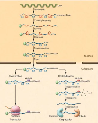
RNA processing in the cytoplasm regulates RNA concentrations
Following transcription, nascent RNA is processed by 5′ methyl capping, splicing,
several copies of the AUUUA motif dispersed within U-rich regions; class II AREs possess at least two overlapping UUAUUUA(U/A) nonamers; and class III AREs are less well defined and generally do not contain an AUUUA sequence (1, 10, 13). Whether an mRNA species containing an ARE bound to ARE-binding proteins (ARE-BPs) is degraded or stabilized is partly dependent upon the cellular milieu, physiological cir-cumstances, and the relative amounts of different bound stabilizing or destabilizing ARE-BPs. Following binding of ARE-BPs to an ARE, RNAs are targeted for translation or degradation. RNAs targeted for degra-dation undergo deadenylation, decapping, and degradation in a large multiprotein complex, the exosome, or in cytoplasmic compartments known as GW bodies or pro-cessing bodies (P-bodies) (14–16).
Parathyroid hormone serum concentrations are dependent on parathyroid hormone secretion and synthesis
The parathyroid (PT) glands play a central role in Ca homeostasis by regulating bone resorption and formation, the synthesis of 1a , 25-dihydroxyvitamin D in the renal prox-imal tubule, and the reabsorption of Ca2+ in
the distal nephron of the kidney (17–21). Changes in serum Ca2+ concentrations are
sensed by PT chief cells via a cell-surface, G protein–coupled receptor, the Ca2+-sensing
receptor, and result in rapid (within min-utes) alterations in parathyroid hormone (PTH) secretion (22, 23). More long-term changes in serum Ca2+ concentrations (over
several hours) result in increases or decreases in PTH synthesis and PTH mRNA concen-trations in the PT gland (24, 25).
Pathogenesis of secondary hyperparathyroidism
Secondary hyperparathyroidism occurs in the clinical context of vitamin D deficiency, Ca deficiency, and chronic kidney disease (CKD) (26, 27). The pathogenesis of second- ary hyperparathyroidism in CKD is multi-factorial and includes phosphate retention and hyperphosphatemia, hypocalcemia, 1a, 25-dihydroxyvitamin D deficiency, intestinal Ca malabsorption, the reduction in vitamin D receptor concentrations with-in the PT gland, and reduced Ca2+-sensing
receptor amounts in PT tissue (28–30). Not only is PTH synthesis increased with con- comitant increases in serum PTH concen-trations, but PT hyperplasia often occurs as well (29, 30). In CKD and dialysis patients, uncontrolled secondary hyperparathyroid- ism is associated with an increased inci-dence of fractures and increased mortality (31–34). Numerous methods, including the control of serum phosphate concen-trations, the administration of vitamin D analogs, Ca supplementation, and the
administration of calcimimetics, have been developed to control PTH levels in CKD and dialysis patients (35–38), but second-ary hyperparathyroidism in CKD remains a significant problem. Additional methods for the treatment of this condition would therefore be of value.
Pth mRNA amounts are regulated by Ca2+ and Pi by posttranscriptional mechanisms
In murine PT glands, changes in Pth mRNA concentrations following altera-tions in serum Ca2+
[image:3.585.44.372.83.494.2](or inorganic phos- phate, Pi) concentrations are due to alter-ations in Pth mRNA stability rather than changes in Pth mRNA transcription (39, 40). Such changes are brought about by the binding of proteins to the terminal
Figure 1
portion of the 3′ UTR of Pth mRNA (Fig-ure 2). A 63-nt ARE in the 3′ UTR regulates
Pth mRNA stability in response to changes in Ca2+ and Pi concentrations (39, 41, 42).
The 63-nt element consists of a core 26-nt minimal binding sequence and adjacent
flanking regions. The Pth RNA ARE does not contain AUUUA sequences and falls into the class III category of AREs (42, 43). Two proteins, AU-rich binding factor 1 (AUF1) and K-homology splicing regula-tor protein (KSRP), bind the ARE in the 3′
UTR of Pth mRNA (43, 44). AUF1 increas-es Pth mRNA half-life, whereas KSRP has the opposite effect (43, 44). Both proteins are regulated by changes in serum Ca2+
[image:4.585.50.527.109.381.2]and Pi concentrations and are altered by CKD (43, 45).
Table 1
Effect of ARE-BPs on mRNA stability
ARE-BP Effect on stability of mRNA of various genes
Increase Decrease
AUF1 (HNRNPD, heterogenous nuclear ribonucleoprotein D0) c-myc, c-fos, PTH, GMCSF, TNFA c-myc, c-fos, p21, cyclin D1,
GMCSF, IL3
HuR (Hu-antigen R, ELAV-like protein 1) c-myc, c-fos, MyoD, cyclin A, cyclin B1, –
cyclin D1, NOS2, GMCSF, TNFA, COX2,
IL3, VEGF, myogenin
Hel-N1 (Hu-antigen B, ELAV-like protein 2) TNFA, GLUT1 – HuD (Hu-antigen D, ELAV-like protein 4) GAP43 –
KSRP (far upstream element-binding protein 2, FUSE-binding – c-fos, NOS2, TNFA, IL2, c-Jun, PTH
protein 2)
TTP (tristetraproline) – c-fos, GMCSF, TNFA, COX2, IL3, IL2
BRF-1 (B-related factor 1, transcription factor IIIB – TNFA, IL3
90 kDa subunit)
CUG-BP2 (CUG triplet repeat RNA-binding protein 2, CUG-BP– COX2 – and ETR-3–like factor 2, Bruno-like protein 3, RNA-binding
protein BRUNOL-3, ELAV-type RNA-binding protein 3, neuroblastoma apoptosis-related RNA-binding protein)
Nucleolin Bcl-2 –
TINO (RNA-binding protein MEX3D, RING finger- and – Bcl-2
KH domain–containing protein 1, RING finger protein 193)
PAIP2 (Polyadenylate-binding protein–interacting protein 2) VEGF –
[image:4.585.52.531.111.379.2]Modified with permission from Nucleic Acids Research (9).
Figure 2
[image:4.585.44.372.502.739.2]Pin1, a peptidyl-prolyl isomerase, alters KSRP phosphorylation and binding of KSRP to the Pth ARE
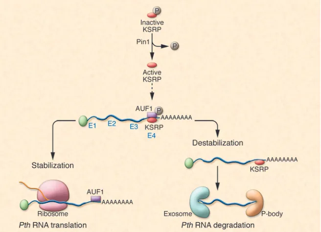
In the accompanying article, Nechama et al. have elegantly dissected the man-ner in which Pth mRNA is degraded via AREs and cognate ARE-BPs in secondary hyperparathyroidism in rodents and have shown a role for the enzyme peptidyl-pro-lyl cis/trans isomerase NIMA-interacting 1 (Pin1) in this process (46) (Figure 2). The authors demonstrate that the secondary hyperparathyroidism associated with CKD or Ca deficiency is due in part to reduced Pin1 activity in the PT glands. The reduc-tion in Pin1 activity reduced the ratio of the ARE-BPs, KSRP, and AUF1, which nor-mally exert opposite effects on Pth mRNA stability. As a result, Pth mRNA half-life and stability were increased due to unop-posed AUF1 activity. The data suggest that it is possible to modulate Pth mRNA half-life and stability by altering the activity of Pin1 and by changing KSRP concentra-tions within the PT cell.
Pin1 is a peptidyl-prolyl isomerase that specifically binds serine/threonine-pro-tein motifs and catalyzes the cis-trans isomerization of peptide bonds, thereby changing the activity of proteins (47, 48). Previous work from other laboratories has shown that Pin1 interacts with AUF1 and stabilizes GMCSF and TGFB mRNAs (49, 50). Nechama et al. (46) hypothesized that Pin1 might also alter Pth mRNA stability and play a role in the pathogenesis of sec-ondary hyperparathyroidism seen in CKD. They showed the presence of Pin1 epitopes and Pin1 enzymatic activity in PT glands and PT extracts. Induction of secondary hyperparathyroidism by feeding rats a diet low in Ca or by inducing CKD with adenine reduced Pin1 activity. Reduced Pin1 activ-ity correlated with increased Pth mRNA levels in the PT glands of such animals. Inhibition of Pin1 activity with the inhibi-tor juglone increased Pth mRNA levels. The increase in Pth mRNA levels in juglone-treated PT was posttranscriptional, since nuclear run-on experiments revealed no changes in the transcription rate of Pth. In a surrogate cell line, HEK293 (a PT cell line is not available for transfection experi-ments), Pin1 overexpression accelerated Pth mRNA decay, whereas Pin1 knockdown with siRNA decreased Pth mRNA decay. Also, Pin1 overexpression was without effect on the half-life of a Pth transcript lacking the ARE-containing Pth 3′ UTR. Both the protein-interaction domain and
the peptidyl-prolyl cis-trans isomeriza-tion domains of Pin1 were necessary for the effect of this protein on Pth mRNA stability. Interestingly, when the 63-nt Pth ARE was introduced into the 3′ UTR of a growth hormone reporter gene (GH63nt), Pin1 overexpression decreased chimeric GH mRNA levels. Treatment with juglone of cells transfected with such a construct decreased Pin1 activity and increased GH63-nt mRNA levels. Pin1 bound KSRP, an ARE-BP that increases the degradation of Pth mRNA. In cells transfected with a chi-meric GH63-nt reporter containing the Pth 63-nt ARE, KSRP overexpression decreased GH63-nt mRNA levels. Conversely, KSRP depletion increased GH63-nt mRNA levels in the presence of Pin1 expression. In such cells, Pin1 inhibition prevented KSRP-mediated decreases in GH63-nt mRNA. The authors also demonstrated that inhi-bition of Pin1 by juglone in PT glands in vivo reduced KSRP binding to Pth mRNA, thus increasing Pth mRNA half-life. Necha-ma et al. showed that Pin1 prevents the phosphorylation of KSRP at serine residue 181 and that a mutant KSRP (S181A) that is incapable of being phosphorylated had increased activity. Pin1-knockout mice had increased PT gland PTH levels and circu-lating serum PTH concentrations without changes in serum Ca and Pi levels. In summary, the data reported in this issue by Nechama et al. (46) are consistent with a biological role for Pin1 in the patho-genesis of secondary hyperparathyroidism in rat PT via regulation of the amount of active, nonphosphorylated KSRP in PT cells. Several additional areas of investiga-tion remain to be explored. For example, it is unclear what triggers the reduction in Pin1 activity in the parathyroids in chronic renal failure and Ca deficiency. Future studies might be directed at identifying fac- tors that regulate Pin1 activity and expres- sion in the PT gland. Precise quantifica-tion of KSRP/AUF1 ratios in the PT gland in different conditions would also be of value. The development of PT gland–spe-cific modulators of ARE-BPs might result in drugs that are effective for the control of secondary hyperparathyroidism and PT hyperplasia. Stay tuned for developments in this area. Acknowledgments The author’s laboratory is supported by NIH grants DK76829 and DK77669 and a grant from Genzyme (Genzyme Renal Innovations Program [GRIP]). Address correspondence to: Rajiv Kumar, Division of Nephrology and Hyperten- sion, Departments of Medicine, Biochem-istry and Molecular Biology, Mayo Clinic and Foundation, 200 1st St. SW, Roches- ter, Minnesota 55905, USA. Phone: 507-284-0020; Fax: 507-538-9536; E-mail: [email protected].
1. McKee, A.E., and Silver, P.A. 2007. Systems perspec-tives on mRNA processing. Cell Res. 17:581–590.
2. Bentley, D.L. 2005. Rules of engagement: co-tran-scriptional recruitment of pre-mRNA processing factors. Curr. Opin. Cell Biol. 17:251–256. 3. Blencowe, B.J. 2006. Alternative splicing: new
insights from global analyses. Cell. 126:37–47. 4. Kornblihtt, A.R., de la Mata, M., Fededa, J.P., Munoz,
M.J., and Nogues, G. 2004. Multiple links between transcription and splicing. RNA. 10:1489–1498. 5. Hieronymus, H., and Silver, P.A. 2004. A systems
view of mRNP biology. Genes Dev. 18:2845–2860. 6. Moore, M.J. 2005. From birth to death: the complex
lives of eukaryotic mRNAs. Science. 309:1514–1518.
7. Sanchez-Diaz, P., and Penalva, L.O. 2006. Post-transcription meets post-genomic: the saga of RNA binding proteins in a new era. RNA Biol. 3:101–109. 8. Singh, R., and Valcarcel, J. 2005. Building specific-ity with nonspecific RNA-binding proteins. Nat. Struct. Mol. Biol. 12:645–653.
9. Barreau, C., Paillard, L., and Osborne, H.B. 2005. AU- rich elements and associated factors: are there unify-ing principles? Nucleic Acids Res. 33:7138–7150. 10. Misquitta, C.M., Chen, T., and Grover, A.K. 2006.
Control of protein expression through mRNA stabil-ity in calcium signalling. Cell Calcium. 40:329–346. 11. Schiavi, S.C., Belasco, J.G., and Greenberg, M.E.
1992. Regulation of proto-oncogene mRNA stabil-ity. Biochim. Biophys. Acta. 1114:95–106.
12. Schiavi, S.C., et al. 1994. Multiple elements in the c-fos protein-coding region facilitate mRNA dead-enylation and decay by a mechanism coupled to translation. J. Biol. Chem. 269:3441–3448. 13. Chen, C.Y., and Shyu, A.B. 1995. AU-rich elements:
characterization and importance in mRNA degra-dation. Trends Biochem. Sci. 20:465–470.
14. Lebreton, A., Tomecki, R., Dziembowski, A., and Seraphin, B. 2008. Endonucleolytic RNA cleavage by a eukaryotic exosome. Nature. 456:993–996. 15. McPheeters, D.S., et al. 2009. A complex gene
regulatory mechanism that operates at the nexus of multiple RNA processing decisions. Nat. Struct. Mol. Biol. 16:255–264.
16. Schilders, G., and Pruijn, G.J. 2008. Biochemical studies of the mammalian exosome with intact cells. Methods Enzymol. 448:211–226.
17. Habener, J.F., Rosenblatt, M., and Potts, J.T., Jr. 1984. Parathyroid hormone: biochemical aspects of biosynthesis, secretion, action, and metabolism. Physiol. Rev. 64:985–1053.
18. Potts, J.T. 2005. Parathyroid hormone: past and present. J. Endocrinol. 187:311–325.
19. Kumar, R. 1984. Metabolism of 1,25-dihydroxyvi-tamin D3. Physiol. Rev. 64:478–504.
20. DeLuca, H.F., and Schnoes, H.K. 1983. Vitamin D: recent advances. Annu. Rev. Biochem. 52:411–439.
21. Gesek, F.A., and Friedman, P.A. 1992. On the mech- anism of parathyroid hormone stimulation of cal-cium uptake by mouse distal convoluted tubule cells. J. Clin. Invest. 90:749–758.
22. Hofer, A.M., and Brown, E.M. 2003. Extracellular calcium sensing and signalling. Nat. Rev. Mol. Cell Biol. 4:530–538.
parathyroid hormone gene expression by hypocal-cemia, hypercalcemia, and vitamin D in the rat. J. Clin. Invest. 86:1313–1319.
25. Naveh-Many, T., Friedlaender, M.M., Mayer, H., and Silver, J. 1989. Calcium regulates parathyroid hormone messenger ribonucleic acid (mRNA), but not calcitonin mRNA in vivo in the rat. Dominant role of 1,25-dihydroxyvitamin D. Endocrinology.
125:275–280.
26. Audran, M., Gross, M., and Kumar, R. 1986. The physiology of the vitamin D endocrine system. Semin. Nephrol. 6:4–20.
27. Audran, M., and Kumar, R. 1985. The physiology and pathophysiology of vitamin D. Mayo Clin. Proc.
60:851–866.
28. Cozzolino, M., et al. 2005. Pathogenesis of parathy-roid hyperplasia in renal failure. J. Nephrol. 18:5–8.
29. Dusso, A.S., et al. 2007. Molecular basis of parathy-roid hyperplasia. J. Ren. Nutr. 17:45–47.
30. Dusso, A.S., et al. 2006. Pathogenic mechanisms for parathyroid hyperplasia. Kidney Int. Suppl.
102:S8–S11.
31. Danese, M.D., et al. 2006. PTH and the risks for hip, vertebral, and pelvic fractures among patients on dialysis. Am. J. Kidney Dis. 47:149–156. 32. Jadoul, M., et al. 2006. Incidence and risk factors
for hip or other bone fractures among hemodialy-sis patients in the Dialysis Outcomes and Practice Patterns Study. Kidney Int. 70:1358–1366. 33. Rudser, K.D., de Boer, I.H., Dooley, A., Young,
B., and Kestenbaum, B. 2007. Fracture risk after parathyroidectomy among chronic hemodialysis patients. J. Am. Soc. Nephrol. 18:2401–2407.
34. Block, G.A., et al. 2004. Mineral metabolism, mor-tality, and morbidity in maintenance hemodialysis.
J. Am. Soc. Nephrol. 15:2208–2218.
35. Block, G.A., et al. 2004. Cinacalcet for secondary hyperparathyroidism in patients receiving hemo-dialysis. N. Engl. J. Med. 350:1516–1525.
36. Drueke, T.B. 2004. Calcimimetics versus vitamin D: what are their relative roles? Blood Purif. 22:38–43.
37. Locatelli, F., et al. 2002. Management of distur-bances of calcium and phosphate metabolism in chronic renal insufficiency, with emphasis on the control of hyperphosphataemia. Nephrol. Dial. Transplant. 17:723–731.
38. Moe, S.M., and Drueke, T.B. 2003. Management of secondary hyperparathyroidism: the impor-tance and the challenge of controlling parathyroid hormone levels without elevating calcium, phos-phorus, and calcium-phosphorus product. Am. J. Nephrol. 23:369–379.
39. Moallem, E., Kilav, R., Silver, J., and Naveh-Many, T. 1998. RNA-Protein binding and post-transcrip-tional regulation of parathyroid hormone gene expression by calcium and phosphate. J. Biol. Chem.
273:5253–5259.
40. Levi, R., et al. 2006. Increased parathyroid hormone gene expression in secondary hyperparathyroidism of experimental uremia is reversed by calcimimet-ics: correlation with posttranslational modification of the trans acting factor AUF1. J. Am. Soc. Nephrol.
17:107–112.
41. Naveh-Many, T., Sela-Brown, A., and Silver, J. 1999. Protein-RNA interactions in the regulation of PTH gene expression by calcium and phosphate. Nephrol. Dial. Transplant. 14:811–813.
42. Kilav, R., Bell, O., Le, S.Y., Silver, J., and Naveh-Many, T. 2004. The parathyroid hormone mRNA 3’-untrans-
lated region AU-rich element is an unstructured func-tional element. J. Biol. Chem. 279:2109–2116. 43. Naveh-Many, T., Bell, O., Silver, J., and Kilav, R.
2002. Cis and trans acting factors in the regulation of parathyroid hormone (PTH) mRNA stability by calcium and phosphate. FEBS Lett. 529:60–64. 44. Nechama, M., Ben-Dov, I.Z., Briata, P., Gherzi, R.,
and Naveh-Many, T. 2008. The mRNA decay pro- moting factor K-homology splicing regulator pro-tein post-transcriptionally determines parathyroid hormone mRNA levels. FASEB J. 22:3458–3468.
45. Nechama, M., Ben-Dov, I.Z., Silver, J., and Naveh- Many, T. 2009. Regulation of PTH mRNA stabil-ity by the calcimimetic R568 and the phosphorus binder lanthanum carbonate in CKD. Am. J. Physiol. Renal Physiol. 296:F795–F800.
46. Nechama, M., Uchida, T., Mor Yosef-Levi, I., Silver, J., and Naveh-Many, T. 2009. The peptidyl-prolyl isom-erase Pin1 determines parathyroid hormone mRNA levels and stability in rat models of secondary hyper-parathyroidism. J. Clin. Invest. 119:3102–3114. 47. Lu, K.P., Liou, Y.C., and Zhou, X.Z. 2002. Pinning
down proline-directed phosphorylation signaling. Trends Cell Biol. 12:164–172.
48. Lu, K.P., and Zhou, X.Z. 2007. The prolyl isomerase PIN1: a pivotal new twist in phosphorylation signal-ling and disease. Nat. Rev. Mol. Cell Biol. 8:904–916. 49. Shen, Z.J., Esnault, S., and Malter, J.S. 2005. The
peptidyl-prolyl isomerase Pin1 regulates the stabil-ity of granulocyte-macrophage colony-stimulating factor mRNA in activated eosinophils. Nat. Immu-nol. 6:1280–1287.
50. Shen, Z.J., et al. 2008. Pin1 regulates TGF-beta1 production by activated human and murine eosin-ophils and contributes to allergic lung fibrosis. J. Clin. Invest. 118:479–490.
Resolving lung injury: a new role for Tregs
in controlling the innate immune response
Anthony Pietropaoli and Steve N. Georas
Division of Pulmonary and Critical Care Medicine, University of Rochester Medical Center, Rochester, New York, USA.
Inflammation-associated lung injury is a major cause of morbidity and
mor-tality for patients in intensive care units. Although the cellular and
molec-ular events that initiate lung inflammation are now well understood, the
mechanisms that promote its resolution remain poorly defined. In this issue
of the
JCI
, D’Alessio et al. show in a mouse model that recovery from acute
lung injury is not simply a passive process, but involves Tregs in an active
resolution program (see the related article beginning on page 2898).
Conflict of interest: The authors have declared that no conflict of interest exists.
Citation for this article: J. Clin. Invest. 119:2891–2894 (2009). doi:10.1172/JCI40880.
Overview of lung injury
Acute lung injury (ALI) is a syndrome defined by bilateral pulmonary infiltrates on chest radiography and arterial hypox- emia in the absence of left atrial hyperten- sion. This syndrome has a rapidly progres-sive clinical presentation characterized by
acute onset of dyspnea and respiratory fail-ure requiring mechanical ventilation; when arterial hypoxemia is particularly severe, it is classified as acute respiratory distress syndrome (ARDS; ref. 1). ALI can be caused by direct and indirect pulmonary insults. The initial pathophysiologic events are endothelial and epithelial injury, excessive permeability of the alveolar-capillary unit, and alveolar and interstitial accumulation of proteinaceous fluid and inflammatory cells (1). This inflammatory response dis-rupts alveolar function, impairing fluid clearance, surfactant production, and gas
exchange (2). Neutrophils are a key com-ponent of lung inflammation in ALI and are recruited to the lung by chemotactic signals produced by alveolar macrophages and other lung cell types (3, 4).