ORIGINAL RESEARCH ARTICLE
EFFECTS OF HIGH-FAT DIET ON ADIPOSE TISSUE ANDPANCREATIC ISLET FUNCTION IN
OVARIECTOMIZED RATS
1,2
Aluana Santana Carlos,
1Luciano Pinto Souza,
1Rodrigo de Azeredo Siqueira,
1Adalgiza Mafra
Moreno,
1Joana da Costa Pinto d'Avila,
2Carlos Alberto Soares da Costa,
3Aline de Souza Santos,
3Patrícia Cristina Lisboa and
3Celly Cristina Alves do Nascimento-Saba
1
Preclinical Research Laboratory, Faculdade Ciências Biológicas e da Saúde, Iguaçu University,
Nova Iguaçu, RJ, Brazil
2
Laboratory of Experimental Nutrition, Departament of Nutrition and Dietetics, Fluminense Federal University,
Niteroi, RJ, Brazil
3
Physiological Sciences, Institute of Biology Roberto Alcantara Gomes, State University of Rio de Janeiro,
Rio de Janeiro, RJ, Brazil
ARTICLE INFO ABSTRACT
The increase in life expectancy allows women to live longer, with the deleterious effects of gonadal deficiency, such as the accumulation of abdominal fat; and the new habits of modern society are dedicated to eating foods rich in fats and carbohydrates, which contribute to overweight and obesity.The objective of this study was to evaluate intra-abdominal fat tissue and pancreas in ovariectomized rats. Wistar rats, 90 days-old, after evaluation by vaginal cytology were ovariectomized. The animals were divided into four groups and were fed diets with soybean oil concentrations of 4% or 19% for 30 days.Body weight and food intake was evaluated. At 120 days-old, total fat and lean mass were assessed by densitometry (DEXA) and after the blood was collected. Serum was used for determination of triglyceride, cholesterol, HDL, glucose, estradiol and insulin.Adipose tissue was excised, weighed and used to evaluate the morphology and expressionERα. Pancreas used to evaluate the morphology and immunohistochemistry by insulin.The mass of the uterus, serum estradiol were lower in OVX and OVXHf groups.The mass of intra-abdominal adipose tissue and the total body fat were higher in OVXHf group. Serum insulin triglycerides, cholesterol and glucoseincreased in ShamHf and OVXHf groups. There was an increase on area of adipocytes and islet pancreasOVX, OVXHf groups. ERα expression was lower in OVX and OVXHf.We concluded, the deficiency of estrogen associated the intake of high fat diet can be accelerating the onset of metabolic syndrome.he works in the area metabolic diseases.
Copyright © 2019,Aluana Santana Carlos et al. This is an open access article distributed under the Creative Commons Attribution License, which permits
unrestricted use, distribution, and reproduction in any medium, provided the original work is properly cited.
INTRODUCTION
In recent years, the increase on food intake rich in carbohydrates and specially lipids induces to obesity, a risk factor for many disorders (Paniagua, 2016 and Engin, 2017). Increased adiposity, especially the intra-abdominal adiposity, has a well-known association with an increased risk of insulin resistance and the development of type 2 diabetes that can lead to the metabolic syndrome (Wang, 2017 and Fändriks, 2017).
*Corresponding author: Aluana Santana Carlos
It is well established that high fat diets (HF) are related to metabolic alterations including liver lipid infiltration, accumulation of intra-abdominal adipose tissue and negative changes of lipid profile, glucose metabolism and contributes to insulin resistance (Soares da Costa, 2009; Paquette, 2007; Pataky, 2017). The polyunsaturated fatty acids (PUFA) can induce obesity, acting directly on preadipocytes, increasing the rate of replication and/or differentiation, mainly in early stages of adipose tissue development (Wu, 2017; Sun, 2003; Vishvanath, 2016). Adipose tissue is highly specialized to store lipid and/or release energy from lipid stores in response
ISSN: 2230-9926
International Journal of Development Research
Vol. 09, Issue, 03, pp.26348-26354, March, 2019
Article History:
Received 10th December, 2018 Received in revised form 21st January, 2019
Accepted 16th February, 2019 Published online 31st March, 2019
Key Words:
High fat diet, Adiposity, Pancreas, Ovariectomized.
Citation: Aluana Santana Carlos, Luciano Pinto Souza, Rodrigo de Azeredo Siqueira, Adalgiza Mafra Moreno, Carlos Alberto Soares da Costa,
Aline de Souza Santos, Patrícia Cristina Lisboa, Celly Cristina Alves do Nascimento-Saba. 2019. “Effects of high-fat diet on adipose tissue
andpancreatic islet function in ovariectomized rats”, International Journal of Development Research, 09, (03), 26348-26354.
to a variety of signals (Cinti, 2005 and Cinti, 2001). In addition, to managing the most important energy reserve of the body, it secretes a multitude of soluble proteins called adipokines, which have beneficial or, alternatively, deleterious effects on the homeostasis of the whole body (Freitas Lima, 2015; Jennifer, 2011 and Alvarez-Castro, 2011). The expression of these adipokines is an integrated response to various signals received from many organs, which depends heavily on the integrity and physiological status of the adipose tissue (Singla, 2010; Ouchi, 2011; Myers, 2011). One of the main regulators of gene expression in fat is the transcription factor peroxisome proliferator-activated receptor gamma (PPARgamma), which is a fatty acid- and eicosanoid-dependent nuclear receptor that plays a key role in the development and maintenance of the adipose tissue (Myers, 2011 and de la Rosa Rodriguez, 2017). It has long been recognized that estrogen is the main regulator factor of adipose tissue development and triglycerides deposition in females and its biological activities are performed binding to nuclear estrogen receptors (ERα, ERβ) (Dahlman-Wright, 2006). The deficiency of ovarian steroids induced by ovariectomy or in menopause, is another factor that favors metabolic changes, favoring visceral fat tissue deposition (Santana, 2011) and insulin resistance (Paquette, 2007). Menopause by itself was found as a risk factor for the metabolic syndrome, independently of the influence of estrogen deficiency on central obesity in nondiabetic women (Wang, 2017). The roles of both high fat diet development and estrogen deficiency in obesity have been extensively studied23but the association of these two parameters is poorly explored. So, based on the change on eating and in the increase of life expectancy, meaning that women live longer after menopause, this study was designed to investigate the effects of high-fat diet associated with the deficiency of estrogen on adiposity of female adult rats.
MATERIALS AND METHODS
Animals: A total of 48 female Wistar rats weighing 200-210g, reproduced and maintained in the animal room of the Laboratory of Endocrine Physiology, Institute of Biology Roberto Alcantara Gomes, University of the State of Rio de Janeiro, were used. They were housed, six per cage, receiving food and water ad libitum, under 12h light–dark cycle (lights on from 7:00 AM to 7:00 PM) and temperature-controlled (25 ± 1°C). The protocol to use and handling the experimental animals was approved by the Ethical Committee of the Biology Institute of the State University of Rio de Janeiro, which based their analysis on the principles adopted and promulgated by the Brazilian Law that concerns the rearing and use of animals in teaching and research activities in Brazil (Marques, 2009).
Groups:The stage of the estrous cycle was evaluated by daily
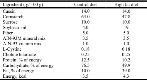
vaginal smears, and only the animals showing two consecutive regular cycles were used in the study. At ninety days, rats were randomly divided into two groups: Sham operation (Sham; n = 24) and Ovariectomy (Ovx; n = 24)21. For surgery, rats were anaesthetized with thiopental (0,1ml/100gPC). After surgery, the rats were fed by 30 days with manufactured diets (table 1), according the suggestion of the American Institute of Nutrition (AIN93-M). In the composition of control diet, it was used 4% soybean oil, and for high fat diet (Hf) was used 19% soybean oil. Rats were divided in Sham (n=12, receiving control diet), ShamHf (n=12, receiving high fat diet), Ovx (n=12, receiving control diet) and OvxHf (n=12, receiving high fat diet). Food
intake (g) was measured each three days and body weight (g) was verified each seven days.
Dual Energy X-ray Absortiometry (DEXA):At the end of the
nutritional period, 120-days-old rats, after 8 h of fasting, were anesthetized with Avertin (Tribromoethanol,300 mg/kg) and subjected to dual-energy X-ray absorptiometry (DEXA), using a Lunar DEXA 200368 GE instrument (Lunar, Wisconsin, USA) with specific software (encore 2008. Version 12.20 GE Healthcare). The evaluation was blind, since the DEXA technician did not know the experimental protocol. Total lean (g), fat mass (g) and trunk fat mass analysis.
Sacrifice: Immediately after DEXA evaluation rats were killed
(between 12:00 and 15:00 h) by exsanguination. Blood samples were centrifuged were centrifuged (1500 g/20 min per 4°C) to obtain serum, which were kept at -20°C until the assay. The abdominal cavity was opened following the median line of the abdomen and tissues were removed in the following order: intra-abdominal adipose tissue (retroperitoneal depot), uterus and pancreas. Adipose tissue was weighed and immediately frozen in liquid nitrogen and stored samples at -70C until performed analyses.
Analytical procedures: All measurements were performed in
one assay; all samples were analyzed in duplicate. Triglycerides, cholesterol, HDL cholesterol and glucose concentrations were determined using colorimetric assay (Bioclin®,Quibasa, MG, Brasil) and evaluated by spectrophotometer. Leptin was measured by a specific RIA kit (Linco Research, Inc., St Louis, MO, USA) that measures both rat and mouse leptin (range of detection, 0.5–50 ng/ml; intraassay variation, 2.9%). Insulin was determined by a commercial RIA kit (Linco Research Inc., St Louis, MO, USA), with an assay sensitivity of was 1 mUl/ml, and intra-assay variation was 8.9%. 17β-estradiol was measured using a RIA kit (Linco Research, Inc., St Louis, MO, USA), with an coefficient of variation was 5.3%.
Morphometry: Samples of tail pancreas and adipose tissue
were fixed in Bouin and buffered formaldehyde, respectively. After 24 h of fixation, samples were dehydrated with increasing concentration of ethanol (1x 80%, 1x 90%, 2x 100%), incubated with xylol (3x) and liquid paraffin (3x), and embedded into paraffin. Sections of 5µm were obtained (microtome Microtec-CUT 4050, SC, USA), placed onto slides and stained with Hematoxylin/Eosin. A light microscope (model BX40 Olympus coupled to a digital camera (Olympus DP71, USA) was used to examine the morphology. A morphometric analysis was realized with captured images of about 10 slides for animal. For each slide, the area of 10 adipocytes or pancreatic islets were randomly selected and analyzed using the IMAGEM-J/NHI software.
Immunohistochemistry: Sections of pancreatic islets were
Western blotting: Western Blot was also perfomed as previously reported (Conceição, 2011). In brief, about 20 µg of total proteins from each sample were loaded onto 12% SDS-PAGE and transferred onto a nitrocellulose membrane (Hybond P ECL membrane; Amersham Pharmacia Biotech). Membranes were incubated with 5%TBS containing of nonfat dry milk for 90 min. Primary antibody (SC- Santa Cruz Biotechnology, Santa Cruz, CA, USA) ERα (1:1000) was incubated with the membrane overnight at 4°C. Further, membranes were washed and incubated with appropriate secondary antibodies conjugated to biotin (Santa Cruz Biotechnology; 0.5 % nonfat dry milk TTBS diluted) for 1 h at room temperature. Membranes were washed and incubated for 1 h at room temperature with streptavidin HRP-conjugated (Zymed, CA, USA) in the same dilution of the secondary antibody. Immunoreactive proteins were visualized by horseradish peroxidase substrate (ECL-plus; Amersham Pharmacia Biotech, NJ, USA) and then exposed to X-ray fi lm. Area and density of protein bands were quantified by Image J 1.34 s software (Wayne Rasband National Institute of Health, MA, USA).
Statistical analysis:Statistical analyses were carried out using
the Graph Pad Prism statistical package version 5.00, 2007 (San Diego, CA, USA). Food intake and body mass were analyzed by two-way ANOVA, followed by Bonferroni post-test. The other data were analyzed by one-way ANOVA, followed by Newman–Keuls post-test. Differences were considered significant at P<0.05.
RESULTS
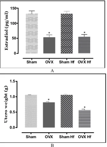
As expected, estradiol concentrations were significantly decreased (P<0.05) in ovarietomized groups (OVX: -60%; OVXHf: - 58%) and was accompanied by a lower uterus mass (OVX: -22%; OVXHf: -46%) (Fig. 1).
A
B
Fig. 1. (A) Serum estradiol concentration; (B) Utero weight. Sham (Sham, n= 12), Ovariectomized (Ovx, n=12), Sham fed with high fat diet with soybean oil 4% during 30 days (ShamHf, n=12) and Ovariectomized fed
with high fat diet with soybean oil 19% during 30 days (OvxHf, n= 12). Values are means. *Significantly different from corresponding OVX Hf,
Ovx, vsSham, ShamHf (one-way ANOVA, P < 0.05)
During experimental period, neither ovariectomy nor the high fat diet changed food intake (Fig. 2A). Nevertheless, the lack of sexual hormones associated with HF diet intake determined an increase in body mass from the nintety day, well evidenced in OvxHf group. Among the other groups (Sham, Ovx and ShamHf) the gain of body mass was similar (Fig. 2B). But, the analysis of body compartments by DEXA showed that ovariectomy alone was capable to induce a significant increase in total fat mass and when associated to HF diet the increase is even higher (ShamHf: +7.2%; Ovx: +14%; OvxHf: +32.3%, P<0.001).
Fig. 3- A. Photomicrography of intra-abdominal adipose tissue staining with HE (original magnification 200x); (B) Size of intra-abdominal adipocytes; (C) Wheight intra-intra-abdominal adipose tissue.Sham (Sham, n= 12), Ovariectomized (Ovx, n=12), Sham fed with high fat diet with soybean oil 4% during 30 days (ShamHf, n=12), Ovariectomized fed with high fat diet with soybean oil 19% during 30 days (OvxHf, n= 12). Values are
means±S.E. #Significantly different from corresponding OVX Hf
vsOVX, Sham, ShamHf (one-way ANOVA, P < 0.05).
*Significantly different from corresponding OVX, ShamHf
vsOVXHf (one-way ANOVA, P < 0.05)
Sham OVX Sham Hf OVX Hf
0 50 100 150
* *
E
s
tr
a
d
io
l (
p
g
/
m
l)
Sham OVX Sham Hf OVX Hf
0.0 0.5 1.0 1.5
*
[image:3.595.319.546.185.676.2] [image:3.595.70.258.475.732.2]By the other hand, the trunk fat mass was higher in OVX and OVX Hf (OVX: +25%; OvxHf: +56.9%,P<0.05, table 2) compared to Sham females. Total lean mass did no differ among groups (Table 2).
[image:4.595.174.425.78.214.2]The weight of intra-abdominal adipose tissue depot (Figure 3) was higher in both ovariectomized groups (OVX: +18%; OvxHf: +74%,P<0.05). And, interestingly, sham rats fed with high fat diet (ShamHf) did not differ from sham fed with
Table 1. Composition of experimental diets
Ingredient ( g/ 100 g) Control diet High fat diet
Casein 14.0 14.0
Cornstarch 63.0 47.8
Sucrose 10.0 10.0
Soybean oil 4.0 19
Fiber 5.0 5.0
AIN-93M mineral mix 3.5 3.5
AIN-93 vitamin mix 1.0 1.0
L-Cystine 0.18 0.18
Choline bitartrate 0.25 0.25
Protein, % of energy 12.5 10.2
Carbohydrate, % of energy 76.5 49.9
Fat, % of energy 10.0 39.0
Energy, kcal 3.5 4.3
C, control diet; HF, high fat diet; AIN, American Institute of Nutrition.
Mineral and Vitamin Mix; L-Cystine; Choline Bitartrate: PragSoluções®; Casein; Cornstarch: FARMOS®; Soybean: Lisa® and Sucrose: União®.
Formulated on recommendations of the AIN-93M for rodent diets.
Table 2. Body composition by DXA at 120 days
Sham OVX ShamHf OVXHf
Mean SEM Mean SEM Mean SEM Mean SEM Total lean mass (g) 160.3 5.6 116.3 10.3* 93.33 3.8# 130.3 8.5* Total fat mass (g) 79.67 5.5 156.0 7.5 165.3 5.6 177.0 8.9 Trunk fat mass (g) 54.0 5.8 68.50 3.5* 58.6 1.3 86.0 4.5*
[image:4.595.118.480.401.481.2]Sham (Sham, n= 12), Ovariectomized (Ovx, n=12), Sham fed with high fat dietwith soybean oil 4% during 30 days (ShamHf, n=12) and Ovariectomized fed with high fat diet with soybean oil 19% during 30 days (OvxHf, n= 12).Values are means. #Significantly different from Sham vs Sham Hf (one-way ANOVA, P < 0.05); *Significantly different from corresponding OVX Hf vs Ovx, ShamHf(one-way ANOVA, P < 0.05).
Table 3. Serum hormonal, lipids and glucose at 120 days
Sham OVX ShamHf OVXHf
Mean SEM Mean SEM Mean SEM Mean SEM Estradiol (pg/mL)
Glucose (mg/dL) Insulin (pg/mL)
131.5 70.2 24.9
9.1 2.5 0.4
52.1 7.8* 116.9 3.8& 40.4 5.6
131.1 8.5 116.8 8.5& 53.6 3.8$
55.6 105.2 51,91
6.5* 5.9& 1,7$ Triglycerides (mg/dL) 70.5 1.9 112.0 8.5 96.11 5.7 191.5 1.5@ HDL-cholesterol(mg/dL) 56.5 9.8 58.5 4.5 60.75.9 58.4 2.4 Cholesterol (mg/dL) 60.1 4.9 67.23.6 79.54.3 78.3 5.4
[image:4.595.191.405.540.720.2]Sam (Sham, n= 12), Ovariectomized (Ovx, n=12), Sham fed with high fat diet with soybean oil 4% during 30 days (ShamHf, n=12) and Ovariectomized fed with high fat diet with soybean oil 19% during 30 days (OvxHf, n=12).Values are means. *Significantly different from corresponding OVX, OVX Hf vs ShamHf(one-way ANOVA, P < 0.05); &Significantly different from OVX, Sham Hf, OVX Hf vs Sham (one-way ANOVA, P < 0.05); $Significantly different from OVX, Sham Hf vsOVX, Sham(one-way ANOVA, P < 0.05); @Significantly different from corresponding OVX Hf vs OVX, Sham, ShamHf(one-way ANOVA, P < 0.05).
Fig. 4-Expression of ERα in intra abdominal adipose tissue. Sham (Sham, n= 12), Ovariectomized (Ovx, n=12), Sham fed with high fat diet with soybean oil 4% during 30 days (ShamHf, n=12), Ovariectomized fed with high fat diet with soybean oil 19% during 30 days (OvxHf, n= 12). Values are means±S.E.
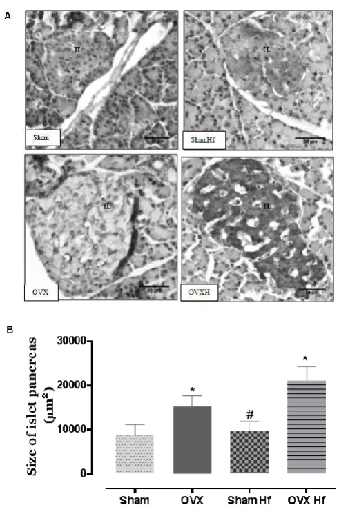
normolipidic diet (Sham). But, the area of intra-abdominal adipocytes was higher in all groups, the increase being more evident in OvxHf (+127%, p< 0.0001) (Figure 3). The analysis of intra-abdominal adipose tissue by protein expression showed a significant decrease of ERα in both ovarietomized groups (OVX: -61%, OVXHf: -67.1%, P<0.05) (Figure 4). Glucose concentrations were significantly increased in all groups, compared to Sham. But, insulin was higher in the groups fed with high fat diet (ShamHf and OVXHf, P<0.05). Triglycerides were increased only in OVXHf group (+27.16%, P<0.05). There was no significant effect of ovariectomy and/or high fat diet on serum HDL-col, cholesterol and leptin. In order to better explain insulin and glucose serum concentrations, pancreatic islets were evaluated. Sham groups maintained morphologic characteristics of exocrine pancreas and homogeneous and rounded islets. But, in the ovariectomized rats the islets were higher (OVX: +48%, OVXHf: +120%, P<0.05, Figure 5) and present amyloid depots. The detection of pancreatic islets insulin content by immunohistochemistry showed an exacerbated marking in OVXHf and ShamHf groups, while OVX group had a lower marking than Sham group (Figure 5).
[image:5.595.39.285.318.691.2]
Fig. 5. Morphologic analysis of pancreatic islets. A) Immunohistochemical staining for insulin in islets of Langerhans (original magnification 400x). B) Morphometric analysis of pancreatic islet.The arrows shown that indicate areas have immunomarker; IL= islets of Langerhans.Sham (Sham, n= 12), Ovariectomized (Ovx, n=12), Sham fed with high fat diet with soybean oil 4% during 30 days (ShamHf, n=12), Ovariectomized fed with high fat diet with soybean oil 19% during 30 days (OvxHf, n= 12).
Values are means±S.E. #Significantly different from corresponding
ShamHf vsOVX, OVXHf (one-way ANOVA, P < 0.05).*Significantly
different from corresponding OVX, OVXHf vsSham, ShamHf (one-way
ANOVA, P < 0.05).
DISCUSSION
The increase of longevity allows greater insight of the deleterious effects of loss of gonadal function; among the many changes it causes central obesity (Conceição, 2011). In addition, the lifestyle of modern society includes high energy consumption with meals rich in lipids, which contributes to adverse effects on health such as obesity (Wang, 2017 and Chandran, 2014). Changes in eating habits, increasing the consumption of high-fat diet, constitute one of the factors determinants of metabolic syndrome in humans and rodents (Costa, 2011 and D'Eon, 2005). In this study, it was observed that the association of ovarian function loss and intake of high fat diet was deleterious in the short experimental period. Deficiency of sex steroids determined by bilateral removal of the ovaries is a very common experimental practice to simulate menopause (Santana, 2011). Deficiency of sex steroids determined by bilateral removal of the ovaries is a very common experimental practice to simulate menopause (21). This was confirmed by the low serum estradiol and decreased uterus mass, an organ that maintains its integrity by stimulation of ovarian sex steroids. In animals, ovariectomy (OVX) leads to increased adiposity (Blaustein, 1976; Stubbins, 2012), that is repressed by estrogen replacement (Lovre, 2017). Although OVX induces a transient rise in food intake in rodents and E2 replacement reduces food intake (Clegg, 2003), hyperphagia does not completely explain the changes in metabolism and development of obesity after OVX. In our study, the experimental period was sufficient to detect the increase of intra-abdominal adipose tissue and total fat content in OVX and OVXHf groups, determined by weighing and DEXA. Nevertheless, the higher body mass, that is commonly seen in ovariectomized animals (Zhang, 2014), was observed only in OVXHf group. In this model, the evaluation by DEXA was more efficient and was able to detect fat depot increase even without changes in body mass. Maybe in a larger trial period, an increase in body mass could be observed in OVX group.
(Mauvais-Jarvis, 2013 and Martínez de Morentin, 2014). There is some evidence that a lack of estrogen changes the gene expression of a variety of ECM proteins culminating in the ECM destabilization and impairing the interaction between ECM proteins and adipocytes, leading to adipose tissue accumulation44. Is also important to mention that the increase of lipoprotein lipase (LPL) activity, accumulating TG is promoted by insulin but it can also be triggered by estrogen deficiency (D'Eon, 2005). The increased deposition of TG is in agreement with the increased adipocyte area determined by the lack of estrogen and its association with high fat diet. As intra-abdominal adipocytes are considered metabolically more active, it requires a higher glucose uptake to provide glycerol-3-phosphate necessary to increase TG synthesis and depot in this tissue that has a high rate of lipolysis (Wang, 2016). The insulin regulation on lipid metabolism seems to be maintained by insulin serum concentrations. The increase in polyunsaturated fat acids intake causes hyperglycemia and hyperinsulinemia and a greater content of insul/in in pancreatic islets, but without alteration of the islets morphology when estrogen is present. By the other hand, when occurs the loss of ovaries, despite no alteration be detected on insulin concentrations and content, probably by the adaptation/augmentation of islet structure, a hyperglycemia state is establishedaccompanied by amyloid depots characterizing a prediabetes state (Aarabi, 2017). Similarly, Zhang et al. Did not observe a difference in fasting insulinemia in oophorectomized animals, however, they noted a higher glucose uptake and lower insulinemia after intraperitoneal glucose administration, compared to the control group (Zhang, 2014). Our results can be explained by the fact that accompanied by amyloid depots characterizing a prediabetes state (Aarabi, 2017 and Guardado-Mendoza, 2009). The amyloids depot are partially ordered, fibrillar, protein aggregates that formation occurs progressive loss of beta cell (Tatsuo Tomita, 2016). Hyperglycemia and islet amyloidosis have been implicated in the cause beta cell death by occupying extracellular space, thereby impairing nutrients and oxygen uptake. In the group submitted to ovariectomy and fed with Hf this association determined the worst response. So, the deficiency of estrogen and the intake of high fat diet rich in polyunsaturated fat acids can alone cause the increase of fat depots, adipocytes size, glycemia and insulinemia. But, when the two conditions are together the set of changes worsening the overall condition of the animal and accelerate the onset of metabolic syndrome.
Conflicts of Interest: None.
REFERENCES
Aarabi MH, Mirhashemi SM. To estimate effective antiamyloidogenic property of melatonin and fisetin and their actions to destabilize amyloid fibrils. Pak J Pharm Sci., 2017, 30, 1589-1593.
Alvarez-Castro P, Sangiao-Alvarellos S, Brandón-Sandá I, Cordido F. Endocrine function in obesity. Endocrinol Nutr., 2011, 58, 422-32.
Blaustein JD, Wade GN. Ovarian influences on the meal patterns of female rats. Physiol Behav., 1976, 17, 201-8. Brown LM, Clegg DJ. Central effects of estradiol in the
regulation of food intake, body weight, and adiposity. J Steroid Biochem Mol Biol., 2010, 122, 65-73.
Chandran U, McCann SE, Zirpoli G, Gong Z, Lin Y, Hong CC, Ciupak G, Pawlish K, Ambrosone CB, Bandera EV.
Intake of energy-dense foods, fast foods, sugary drinks, and breast cancer risk in African American and European American women. Nutr Cancer., 2014, 66, 1187-99. Cinti S. The adipose organ. Prostaglandins Leukot Essent
Fatty Acids., 2005, 73, 9-15.
Cinti S. The adipose organ: morphological perspectives of adipose tissues. Proc Nutr Soc., 2001,60,319-28.
Clegg DJ, Riedy CA, Smith KA, Benoit SC, Woods SC. Differential sensitivity to central leptin and insulin in male and female rats. Diabetes., 2003, 52, 682-7.
Compston J. Obesity and fractures in postmenopausal women.
Curr Opin Rheumatol., 2015,27, 414-9.
Conceição EP, Trevenzoli IH, Oliveira E, Franco JG, Carlos AS. Higher white adipocyte area and lower leptin production in adult rats overfed during lactation. Horm Metab Res., 2011, 43, 513-6.
Costa CA, Carlos AS, Gonzalez GD, Reis RP, Ribeiro MD, et. al. Diet containing low n-6/n-3 polyunsaturated fatty acids ratio, provided by canola oil, alters body composition and bone quality in young rats. Eur J Nutr., 2011, 51, 191-8. Dahlman-Wright K, Cavailles V, Fuqua SA, Jordan VC,
Katzenellenbogen JA, et al. International Union of Pharmacology. LXIV. Estrogen receptors. Pharmacol Rev., 2006, 58, 773-81.
De Groot LJ, Chrousos G, Dungan K, Feingold KR, Grossman A, Hershman JM, Koch C, Korbonits M, McLachlan R, New M, Purnell J, Rebar R, Singer F, Vinik A. The Effect of Endocrine Disorders on Lipids and Lipoproteins.South Dartmouth (MA): MDText.com, Inc,2017.
de la Rosa Rodriguez MA, Kersten S. Regulation of lipid droplet-associated proteins by peroxisome proliferator-activated receptors. Biochim Biophys Acta., 2017, 1862, 1212-1220.
D'Eon TM, Souza SC, Aronovitz M, Obin MS, Fried SK, et. al. Estrogen regulation of adiposity and fuel partitioning. Evidence of genomic and non-genomic regulation of lipogenic and oxidative pathways. J Biol Chem., 2005, 280, 35983-91.
Duarte FO, Gomes-Gatto CDV, Oishi JC, Lino ADS, Stotzer US, Rodrigues MFC, Gatti da Silva GH, Selistre-de-Araújo HS. Physical training improves visceral adipose tissue health by remodelling extracellular matrix in rats with estrogen absence: a gene expression analysis. Int J Exp Pathol., 2017, 8, 203-213.
Engin A. The Definition and Prevalence of Obesity and Metabolic Syndrome. Adv Exp Med Biol., 2017, 960:1-17. Fändriks L. Roles of the gut in the metabolic syndrome: an
overview. Intern Med., 2017, 281, 319-336.
Faulds MH, Zhao C, Dahlman-Wright K, Gustafsson JA. Regulation of metabolism by estrogen signaling. J Endocrinol., 2012, 212, 3-12.
Freitas Lima LC, Braga VA, do Socorro de França Silva M, Cruz JC, Sousa Santos SH, de Oliveira Monteiro MM, Balarini CM. Adipokines, diabetes and atherosclerosis: an inflammatory association. Front Physiol., 2015, 3, 304. Guardado-Mendoza R, Davalli AM, Chavez AO, Hubbard GB,
Dick EJ, Majluf-Cruz. Pancreatic islet amyloidosis, beta-cell apoptosis, and alpha-beta-cell proliferation are determinants of islet remodeling in type-2 diabetic baboons. Proc Natl Acad Sci., 2009, 106, 13992-7.
Hamilton KJ, Hewitt SC, Arao Y, Korach KS.Estrogen Hormone Biology. Curr Top Dev Biol., 2017, 125, 109-146.
body weight, appetite, and lifespan in female rats. Horm Behav., 2018, 97, 25-30.
Jennifer L. Parker, Jesse J. Lugus, Kenneth Walsh, Noriyuki Ouchi. Adipokines in inflammation and metabolic disease.
Nat Rev Immunol., 2011, 11, 85–97.
Lovre D, Lindsey SH, Mauvais-Jarvis F. Effect of menopausal hormone therapy on components of the metabolic syndrome. Ther Adv Cardiovasc Dis., 2017, 11, 33–43. Marques RG, Morales MM, Petroianu A. Brazilian law for
scientific use of animals. Acta Cir Bras., 2009, 24, 69-74. Martínez de Morentin PB, González-García I, Martins L, Lage
R, Fernández-Mallo D, Martínez-Sánchez N, Ruíz-Pino F, Liu J, Morgan DA, Pinilla L, Gallego R, Saha AK, Kalsbeek A, Fliers E, Bisschop PH, Diéguez C, Nogueiras R, Rahmouni K, Tena-Sempere M, López M.Estradiol regulates brown adipose tissue thermogenesis via hypothalamic AMPK. Cell Metab., 2014, 20, 41-53. Mauvais-Jarvis F, Clegg DJ, Hevener AL. The role of
estrogens in control of energy balance and glucose homeostasis. Endocr Rev., 2013, 34, 309–338.
Myers MG Jr, Burant CF. PPAR-γ action: it's all in your head.
Nat Med ., 2011, 17, 544-5.
Ouchi N, Parker JL, Lugus JJ, Walsh K. Adipokines in inflammation and metabolic disease. Nat Rev Immunol., 2011, 11, 85-97.
Palmer BF, Clegg DJ. The sexual dimorphism of obesity. Mol Cell Endocrinol., 2015, 402, 113-9
Paniagua JA. Nutrition, insulin resistance and dysfunctional adipose tissue determine the different components of metabolic syndrome. World J Diabetes., 2016, 7, 483-514. Paquette A, Shinoda M, Rabasa Lhoret R, Prud'homme D,
Lavoie JM. Time course of liver lipid infiltration in ovariectomized rats: impact of a high-fat diet. Maturitas.,
2007;58,182-90.
Pataky MW, Wang H, Yu CS, Arias EB, Ploutz-Snyder RJ, Zheng X, Cartee GD. High-Fat Diet-Induced Insulin Resistance in Single Skeletal Muscle Fibers is Fiber Type Selective. Sci Rep., 2017, 7,13642.
Pinkas J, Bojar I, Gujski M, Bartosińska J, Owoc A, Raczkiewicz D. Serum Lipid, Vitamin D Levels, and Obesity in Perimenopausal and Postmenopausal Women in Non-Manual Employment. Med Sci Monit., 2017, 23, 5018-5026.
Santana AC, Soares da Costa CA, Armada L, de Paula Lopes Gonzalez G, dos Santos Ribeiro M, et al. Fat tissue morphology of long-term sex steroid deficiency and estrogen treatment in female rats. Fertil Steril., 2011, 95, 1478-81.
Singla P, Bardoloi A, Parkash AA. Metabolic effects of obesity: A review. World J Diabetes., 2010, 1, 76-88. Soares da Costa CA, Alves EG, de Paula Lopes Gonzalez G,
Barbosa TB, Carlos AS, et al. Evaluation of body development, fat mass and lipid profile in rats fed with high-PUFA and -MUFA diets, after neonatal malnutrition.
Br J Nutr., 2009;101,1639-44.
Stubbins RE, Holcomb VB, Hong J, Núñez NP. Estrogen modulates abdominal adiposity and protects female mice from obesity and impaired glucose tolerance. Eur J Nutr., 2012, 861-70.
Sun YR, Yang Z. Advances of nuclear translation factor:peroxisome proliferator-actived receptor gamma2. Yi Chuan., 2003,25,713-7.
Tatsuo Tomita. Apoptosis in pancreatic β-islet cells in Type 2 diabetes. Bosn J Basic Med Sci., 2016, 16, 162–179. Vishvanath L, MacPherson KA, Hepler C, Wang QA, Shao M,
Spurgin SB, Wang MY, Kusminski CM, Morley TS, Gupta RK. Pdgfrβ+ Mural Preadipocytes Contribute to Adipocyte Hyperplasia Induced by High-Fat-Diet Feeding and Prolonged Cold Exposure in Adult Mice. Cell Metab., 2016, 23, 350-9.
Wang R, Li XN. Different adipose tissue depots and metabolic syndrome in human. Sheng Li Xue Bao., 2017, 69, 357-365.
Wang T, Zhu X, Dai F, Li C, Huang D, Fang Z, Zhang Q, Lu Y. Effects of a standard high-fat diet with or without multiple deficiencies on bone parameters in ovariectomized mature rat. PLoS One., 2017,12, 0184983.
Wang T, Zhu X, Dai F, Li C, Huang D, Fang Z, Zhang Q, Lu Y.Effects of a standard high-fat diet with or without multiple deficiencies on bone parameters in ovariectomized mature rat. PLoS One., 2017,12, 0184983.
Wang W, Seale P. Control of brown and beige fat development. Nat Rev Mol Cell Biol., 2016, 17, 691-702. Wu Y, Lee MJ, Ido Y, Fried SK. High-fat diet-induced obesity
regulates MMP3 to modulate depot- and sex-dependent adipose expansion in C57BL/6J mice. Am J Physiol Endocrinol Metab., 2017, 312, 58-71.
Zhang R, Su D, Zhu W, Huang Q, Liu M, Xue Y, Zhang Y, li D, Zhao A, Liu Y. Estrogen suppresses adipogenesis by inhibiting S100A16 expression. Mol Endocrinol., 2014, 28, 235-44.
Zhang R, Su D, Zhu W, Huang Q, Liu M, Xue Y, Zhang Y, li D, Zhao A, Liu Y. Estrogen suppresses adipogenesis by inhibiting S100A16 expression. J Mol Endocrinol., 2014, 52, 235-44.