N A N O E X P R E S S
Open Access
Elucidating Protein Involvement in the
Stabilization of the Biogenic Silver
Nanoparticles
Daniela Ballottin
1,2, Stephanie Fulaz
1, Michele L. Souza
3,4, Paola Corio
3, Alexandre G. Rodrigues
5, Ana O. Souza
5,
Priscyla M. Gaspari
6, Alexandre F. Gomes
7, Fábio Gozzo
7and Ljubica Tasic
1,2*Abstract
Silver nanoparticles (AgNPs) have been broadly used as antibacterial and antiviral agents. Further, interests for green
AgNP synthesis have increased in recent years and several results for AgNP biological synthesis have been reported
using bacteria, fungi and plant extracts. The understanding of the role and nature of fungal proteins, their interaction with
AgNPs and the subsequent stabilization of nanosilver is yet to be deeply investigated. Therefore, in an attempt to better
understand biogenic AgNP stabilization with the extracellular fungal proteins and to describe these supramolecular
interactions between proteins and silver nanoparticles, AgNPs, produced extracellularly by
Aspergillus tubingensis
—
isolated
as an endophytic fungus from
Rizophora mangle
—
were characterized in order to study their physical characteristics,
identify the involved proteins, and shed light into the interactions among protein-NPs by several techniques. AgNPs of
around 35 nm in diameter as measured by TEM and a positive zeta potential of +8.48 mV were obtained. These AgNPs
exhibited a surface plasmon resonance (SPR) band at 440 nm, indicating the nanoparticles formation, and another band
at 280 nm, attributed to the electronic excitations in tryptophan, tyrosine, and/or phenylalanine residues in fungal
proteins. Fungal proteins were covalently bounded to the AgNPs, mainly through S
–
Ag bonds due to cysteine
residues (HS
–
) and with few N
–
Ag bonds from H
2N
–
groups, as verified by Raman spectroscopy. Observed
supramolecular interactions also occur by electrostatic and other protein
–
protein interactions. Furthermore,
proteins that remain free on AgNP surface may perform hydrogen bonds with other proteins or water increasing
thus the capping layer around the AgNPs and consequently expanding the hydrodynamic diameter of the particles
(~264 nm, measured by DLS). FTIR results enabled us to state that proteins adsorbed to the AgNPs did not suffer
relevant secondary structure alteration upon their physical interaction with the AgNPs or when covalently bonded to
them. Eight proteins in the AgNP dispersion were identified by mass spectrometry analyses. All these proteins are
involved in metabolic pathways of the fungus and are important for carbon, phosphorous and nitrogen uptake, and
for the fungal growth. Thereby, important proteins for fungi are also involved in the formation and stabilization of the
biogenic AgNPs.
Keywords:
Biogenic silver nanoparticles (AgNPs), Capping proteins,
Aspergillus tubingensis
* Correspondence:[email protected]
1
Laboratório de Química Biológica, Instituto de Química, Universidade Estadual de Campinas, Campinas, SP, Brazil
2
NanoBioss, SisNano, Universidade Estadual de Campinas, Campinas, SP, Brazil Full list of author information is available at the end of the article
Background
Nanotechnology has attracted the attention of researchers
worldwide because of the unique properties of
nanoma-terials. Countless applications have been studied in
dif-ferent fields, such as medicine [1, 2], material science
[3], microelectronics [4], energy storing [5], and
bio-medical devices [6].
Silver nanoparticles (AgNPs) have been largely employed
in antibacterial and antiviral applications [7
–
16]. They
present antibacterial and antimicrobial activity against
Gram-negative and Gram-positive bacteria and some
vi-ruses as well [17
–
19]. Silver ions attack several targets in
the bacteria making the development of resistance difficult
[20]. The enormous surface area of nanoparticles improves
its penetrability into the cell, enhancing their antimicrobial
action [21].
AgNPs can be produced by chemical [22, 23], physical
or biological routes [24, 25]. Biological synthesis uses
clean routes, without producing toxic residues. AgNP
biosynthesis can be performed using bacteria [7, 26],
fungi [27
–
30], yeasts [31], plant extracts [32, 33],
cyano-bacteria [34], algae [35, 36], and actinomycetes [37]. This
synthesis can be extra- or intra-cellular [38
–
41].
Fungi are easy microorganisms to manipulate as they
grow in mycelial form; they are more resistant facing
adverse conditions and provide a cost-effective
large-scale production [42]. For these reasons, fungi appear to
be interesting microorganisms for the green synthesis of
silver nanoparticles. Fungus
Aspergillus tubingensis
is
part of the black Aspergilli as well as
A. niger
,
A.
carbo-narius
, and
A. aculeatus
[24, 41
–
46] that grows on plant
material. Many species of
Aspergillus
section
nigri
ex-hibit important biochemical differences in secretome
[47
–
49].
A. tubingensis
, used in this instance, was
iso-lated as an endophytic fungus from
Rizophora mangle
[28]
.
Similar to other fungi,
A. tubingensis
is unable to
import polymeric compounds into the cell and relies on
enzymatic degradation to produce monomers or
oligo-mers from different plant polyoligo-mers among which
poly-saccharides are the major constituents [50, 51]. Due to
structural differences in the plant polysaccharides, their
effective degradation depends on an efficient system that
regulates the production and secretion of different
en-zyme cocktails.
A. tubingensis
is normally grown in a rich medium,
such as potato dextrose agar (PDA), removed from it
and washed with clean and distilled water originating
the fungal filtrate (FF), rich in proteins and fungal
me-tabolites. Then, Ag(I) aqueous solution is added into the
FF where redox reactions occur and AgNPs are formed
[51, 52]. Although various investigations have reported
the mechanism of production of AgNPs obtained
through this extracellular synthesis using different
bio-logical agents [33, 38
–
40], little is yet known about the
role and nature of fungal proteins and also about their
in-teractions with AgNPs and the subsequent stabilization of
the as-produced nanosilver [51
–
55].
Interactions between nanosilver and proteins lead to
AgNP stabilization and the formation of
nanoparticle-biomolecular-capped structures [56
–
58] that could be
monitored by different techniques. These biophysical and
biochemical interactions occur through covalent bonds
and electrostatic interactions [59, 60]. Silver nanoparticles
can be complexed with the thiol HS
–
(Cys) or amine
H
2N
–
groups [61
–
63] of the proteins and through
electro-static interactions [64] that have less impact on protein
conformation and function. Sometimes, proteins
cova-lently bound to AgNPs attract other proteins in order to
form protein
–
protein-specific or nonspecific interactions
that are an important part of the nanosilver-protein
multilayer.
In an attempt to better understand biogenic AgNP
stabilization with extracellular fungal proteins and to
de-fine these supramolecular interactions, we have chosen
biogenic nanosilver with positive zeta potential. To the
best of our knowledge, the present study is the first to
report such data on covalently bound proteins to
biona-nosilver (AgNPs), synthesized by
A. tubingensis
. Biogenic
AgNPs, of well-defined size and distinct morphology, are
formed through the reduction of an aqueous solution of
Ag(I) by a fungal filtrate.
Although the involvement of proteins in the reduction
of the Ag(I) ions and the stabilization of a newly formed
AgNPs has been described [23, 28, 64, 65], data about
the way these proteins act are scarce. To fill the gap, the
present study was devised in order to identify the
pro-teins that promote the formation of AgNPs and those
involved in the stabilization of the same nanomaterials.
Methods
All chemicals used in this study were purchased from
Sigma-Aldrich (St. Louis, MO, USA) and used without
further purification unless otherwise stated.
Fungal strain of
A. tubingensis
(AY876924) was provided
by I. S. Melo (Embrapa/CNPMA, Brazil) and is part of the
culture collection of the
“
Embrapa Recursos Genéticos e
Biotecnologia (CENARGEN)
”
in the
“
Collection of
Micro-organisms for Biocontrol of Plant Pathogens and Weeds
”
(http://mwpin004.cenargen.embrapa.br/jrgnweb/jmcohtml/
jmcoconsulta-externa.jsp?idcol=11)
under
the
number
CEN1065.
Silver Nanoparticle Synthesis
dextrose broth (PDB) in a 1-L Erlenmeyer flask and
in-cubated in an orbital shaker (Marconi MA420, Brazil) at
25 °C and 150 rpm for 72 h. After this period, the
bio-mass was filtered using a polypropylene membrane and
washed with sterile water. After incubation with sterile
water at 25 °C and 150 rpm for 72 h, the biomass was
removed and the fungal filtrate (FF) was obtained using
a cellulose acetate membrane of 0.22
μ
m.
For AgNP synthesis, 1 mL of AgNO
3solution (0.1 mol
L
-1), previously filtered through a cellulose regenerated
membrane (0.22
μ
m), was added to 99 mL of the FF to
reach a final concentration of 1 mmol L
−1. The flask was
kept at 25 °C and protected in dark for 96 h. The formation
of AgNPs was monitored using a UV-Vis
spectrophotom-eter (Agilent 8453). Control (FF without any silver ions)
was used as blank. The average size (z-average) of AgNPs
was measured by dynamic light scattering (DLS) (Nano ZS
Zetasizer, Malvern Instruments Corp, UK) at 25 °C in
poly-styrene cuvettes with a path length of 10 mm. The zeta
potential was measured in capillary cells with a path length
of 10 mm, using the same instrument. The samples were
diluted with 0.1 mmol L
−1NaCl before the analysis.
Characterization of the Proteins Capping the AgNPs
FTIR spectroscopy
measurements were carried out from
KBr tablets of two samples, AgNPs and FF, and were
re-corded in an ABB Bomem (MB series, USA) instrument
with a resolution of 4.0 cm
−1and in an interval from
4000 to 400 cm
−1.
Raman spectroscopy
measurements were implemented
at the Instituto de Química, Universidade de São Paulo
and recorded in a Renishaw InVia Reflex equipment
coupled to a DM2500M Leica microscope using 632.8
and 785 nm lasers at 3 mW and 30 mW, respectively.
Fifty-second accumulation in a total of three scans to each
sample between 100 and 1800 cm
−1range were obtained,
at 4 cm
−1resolution. All samples were analyzed in
suspen-sion and solid KCl was added in order to promote
aggre-gation; however, no visual change was noticed.
LC-MS/MS analysis
were performed at Laboratório
Dalton, Instituto de Química, Universidade Estadual de
Campinas using a nanoACQUITY chromatograph with a
UPLC (Waters) coupled to a Synapt HDMS
spectrom-eter (Waters) with QTOF geometry equipped with a
nanoESI source operating in the acquisition-dependent
data mode (ADD).
After being quantified by the Bradford method [66],
proteins from the FF and linked to the AgNPs were
ana-lyzed by LC-MS/MS according to a method based on
denaturation followed by digestion using the trypsin
en-zyme (Sequencing Grade Modified Trypsin, Promega),
desalting and concentration. The resulting solutions
were centrifuged (10 min at 17,000×
g
) and the
super-natant was transferred into appropriate vials. Then, the
samples were injected into the UPLC system, first
pass-ing through the precolumn (Waters Symmetry C18,
20 mm × 180
μ
m, particles 5
μ
m), being desalted during
3 min with a flow of 5.0
μ
L min
−1with 97:3
water/aceto-nitrile with 0.1 % formic acid (v/v) and, afterwards, they
were transferred to the analytical column (Waters C18
BEH130, 100 mm ID × 100
μ
m, particles of 1.7
μ
m).
Finally, the samples were eluted with a flow rate of
1.0
μ
L min
−1by varying the gradient of mobile phases
with a gradient of buffer A (water/formic acid 0.1 %, v/
v) and B (acetonitrile/formic acid 0.1 %, v/v) at the rates
of 97:3, 70:30, 20:80, 20:80, 97:3, and 97:3 at 0, 40, 50,
55, 56, and 60 min, respectively. The identification of
the peptides was done using the online version of the
Waters software with a mass spectrometer (Synapt
HDMS-Waters) configured to operate in dependent acquisition
data (ADD) mode containing a function MS full-scan (
m/z
200
–
2000), a three function fragment ion spectrum (MS/
MS, m/z 50 to 50 units over the m/z of the precursor) and
a function of external standard calibration (lock-mass, m/z
200
–
2000). All spectra were acquired at a rate of 1 Hz. The
other parameters were capillary voltage of 3.0 kV, cone
voltage of 30 V, source temperature of 100 °C Gas Flow
nanoESI 0.5 L h
−1, collision energies of 6:04 eV and a
1700-V detector. The acquisition of raw data was
per-formed with ProteinLynx Global Server v.2.2 software
(Waters). Data treatments for the deconvolutions of raw
spectra were performed with Transform software
(Micro-mass, UK). MASCOT v.2.2 system (Matrix Science Ltd.
http://www.matrixscience.com). Data banks were searched
in order to identify the fungal proteins.
Results and Discussion
Biogenic AgNP formation through a fungal-based
extra-cellular synthesis is a known, efficient, green, and
rela-tively fast way for AgNP production [28, 58, 61, 62, 65,
67, 68] as this process takes a few days to complete
(Fig. 1a). Herein, the biogenic synthesis was monitored
by UV-Vis spectroscopy (Fig. 1b). The formation of
AgNPs was completed within 72 h after the FF was
chal-lenged with AgNO
3, in good agreement with what was
previously reported [28]. The UV-Vis spectrum displays
two main bands, an SPR band at 440 nm, characteristic of
the AgNP presence, and an additional band at 280 nm,
which could be attributed to the aromatic amino acids of
the capping proteins [69]. It is well-known that the
ab-sorption band in this region arises due to the electronic
excitations in tryptophan, tyrosine and/or phenylalanine
residues in fungal proteins [69
–
71]. These results confirm
the AgNPs formation and the presence of fungal proteins.
(data not shown herein, already presented in [28]). Their
zeta potential was positive with a value of + 8.48 ± 0.45 mV
which could be indicative of low-charged surfaces and,
consequently, unstable AgNPs [72], contrary to what was
observed during a 6-month period. The high AgNPs
stabi-lity might be attributed to the fungal protein-capping
around the particles what confers them steric stability. The
average diameter measured by TEM was 35 ± 10 nm
(Fig. 1b and other data shown previously [28]). This value
is smaller when compared to that measured by DLS,
be-cause in the latter technique the hydrodynamic diameter
(particles and stabilization protein-capping) is taken into
account [28], on the other hand, TEM allows the
measure-ment of the AgNP diameter without the surrounding
cap-ping layers. Once again, strong evidence for fungal proteins
linked to the silver nanoparticles was obtained.
Protein adsorption on the surface of biogenic AgNPs
was also confirmed by FTIR spectroscopy (Fig. 2). For
ex-ample, the peptide bond exhibits characteristic bands
denominated amide A, B, I-VII. The Fermi resonance that
occurs among the first overtone of amide II and the N
–
H
stretching vibration create the bands amide A (about
3500 cm
−1) and amide B (about 3100 cm
−1) [72
–
76]. The
band in 1600
–
1700 cm
−1named amide I is related with
the C = O stretching vibration from the backbone
con-formation [72]. The amide II band arises from the N
–
H
bending vibration and from the C
–
N stretching vibration
[73] and is conformational sensitive. The complex bands
Fig. 1aImage of the fungal filtrate and the AgNP suspension.bUV-Vis spectra obtained for AgNP suspension using FF as blank.cElectronic Transmission micrograph showing the AgNPsFig. 2FTIR spectra of the fungal filtrate (black) and AgNPs (red) carried out in KBr tablets
[image:4.595.61.536.87.232.2] [image:4.595.306.539.390.693.2] [image:4.595.58.294.530.702.2]Amide III and IV originates from a mixture of several
co-ordinate displacements [77]. The symmetric and
asym-metric vibrations of the C
–
H groups result in bands at
2920
–
2950 cm
−1, respectively [78], while bands at 1620 to
1650 cm
−1are attributed to
–
C(O)
–
of peptide bonds
and/or
–
NH
2groups and those at 1380
–
1030 cm
−1to
C-N bonds [74, 75].
According to the FTIR results the proteins on AgNP
surface did not undergo relevant secondary structure
alteration along with their interaction with AgNPs, nor
when covalently bonded to them as reported in other
published data [50, 51]. The interaction between the
proteins and AgNPs might be covalent bound to the
amino groups, cysteine residues, and/or electrostatic
in-teractions via carboxyl groups.
[image:5.595.59.539.365.685.2]The Raman spectra (Fig. 3) indicate the presence of
protein-capping at the surface of the investigated AgNPs
[77, 79, 80], confirming the DLS results for the
hydro-dynamic diameter. Moreover, Raman spectroscopy enable
observe if the protein binding to the surface occurs via free
amino groups or through cysteine residues. The spectrum
excited at 632.8 nm presents little vibrational information
about the molecules at the AgNP surface. The broad band
at around 214 cm
−1can be assigned to an overlap between
the Ag
–
Cl vibration (given the presence of Cl
–
) and an
Ag
–
S vibration suggesting an interaction between
superfi-cial Ag and the cysteine (HS
–
) group of the capping
pro-teins. When the samples were excited at 785 nm, strong
bands assigned to the adsorbed proteins are observed at
1338 and 1768 cm
−1, assigned to the amide III and amide I
modes, respectively, as already discussed in the FTIR
re-sults above. Bands at 1120 and 1138 cm
−1are assigned to
NCH stretching and CCH bending modes, respectively,
and 1234 cm
−1to vibrations in antiparallel
β
-sheet in the
protein structure [81]. A broad and weak band related to
the amide II mode is present at approximately 1635 cm
−1,
which was expected to be at lower frequencies (below
1600 cm
-1). The observed blue shift is associated to a
response of the protein bonding to AgNPs, increasing the
vibrational frequencies of the free amine II mode. On the
other hand, it was expected to detect HCS bending
between 800 and 900 cm
−1. However, such peak was not
present in any of the obtained spectra reinforcing that the
binding of protein to the AgNP surface occurred mainly
through the
–
SH groups. In such case, the amino group
remains free and may perform hydrogen bonds with other
proteins or water, contributing hence to the large
hydro-dynamic radius and the low charge surface of these NP.
Therefore, proteins detected in AgNPs are covalently
bound to the silver through S
–
Ag bonds, principally, and
with some adhered proteins via electrostatic or other
pro-tein
–
protein interactions.
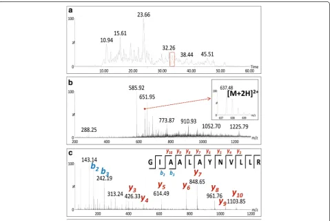
The protein identification in the dispersion of AgNPs
was performed starting from the protein tryptic lysis
followed by LC-MS/MS analysis [82
–
88]. An illustration
of the LC-MS/MS results obtained for proteins capping
AgNPs is shown in Fig. 4 and all identified proteins are
shown in Additional file 1: Table S1. The most intense
signals in the chromatograms of peptides were selected
for further fragmentation and, after obtaining their MS
spectra, three to five most intense m/z ions were
frag-mented in MS/MS spectra allowing us to associate an
amino acid sequence for a fragmentation pattern, as
exemplified for one of the identified peptides (Fig. 4).
Mass spectrometry analyses enabled the identification
of eight (8) proteins in the AgNPs dispersion and these
are presented in Additional file 1: Table S1. All of them,
secreted by
A. tubingensis
, display low isoelectric points,
ranging from 4.0 to 5.1, characteristic for acidic proteins.
Their molecular masses varied from 39 to 65.5 kDa.
A. tubingensis
was grown in broth whose pH was 6.5
to 6.8 and, therefore, the fungus extracellular proteins
should exhibit negative charge due to the deprotonation,
which could increase the zeta potential of the
synthe-tized AgNPs. Nevertheless, the positive zeta potential of
approximately 8 mV, which should be indicative of
low-charged surfaces, is probably a consequence of these
protein-capping deprotonation. Some published data on
chemical AgNPs and protein interactions also report
similar observations [50].
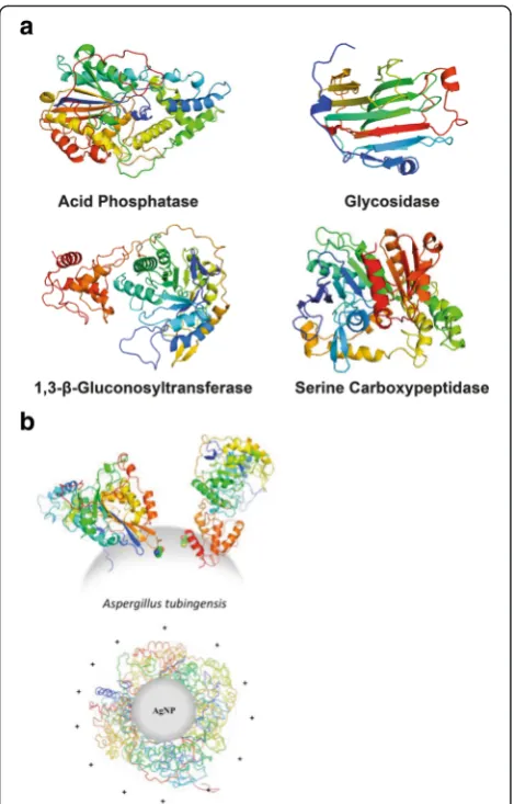
Among identified proteins, we have found glycoamilase
(1,4-
α
-D-glucanglucohydrolase, EC 3.2.1.3), acid
phosphat-ase (EC 3.1.3.2), serine carboxipeptidphosphat-ase (EC 3.4.21.26),
and glucanosyltransferase (EC 2.4) that are illustrated in
Fig. 5. All these proteins are involved in metabolic
path-ways of the fungi and belong to hydrolases [56, 89
–
93],
important for carbon, phosphorous, and nitrogen
up-take, respectively, and for the fungal growth.
Further-more, all identified hypothetic proteins also constitute
the secretome of
A. tubingensis
. Although of unknown
function, these proteins, which contain the signaling
se-quences at the N-terminal, are always secreted, and
their probable functions are associated with metabolic
supplies.
Conclusions
Silver nanoparticles were biosynthesized using the
se-creted proteins from the fungus
A. tubingensis
. This
fungal filtrate in contact with AgNO
3produced within
72 h AgNPs with 264.9 ± 3.2 nm in the hydrodynamic
diameter, 35 ± 10 nm in the nanoparticle diameter and
with a zeta potential of + 8.48 ± 0.45 mV. The
nanoparti-cle formation was followed by UV-Vis spectroscopy, and
the increase in the intensity of the SPR band was
ob-served during AgNPs synthesis. The presence of fungal
proteins in the AgNPs dispersion was verified by all
spectrometric and spectroscopic analyses used. The
FTIR along with the Raman data enabled us to identify
the amino I, II, and III bands of proteins adhered to
AgNP surface. Proteins formed covalent bonds with atoms
at the surface of AgNPs surface due to their cysteine
resi-dues (Ag
–
S bonds) most likely. Secondary and tertiary
structure features of proteins were preserved even when
they were chemically bound to Ag atoms at the surface of
the NPs. Eight proteins from
A. tubingensis
secretome
were identified by MS/MS. All data collected and analyzed
[image:6.595.305.540.86.453.2]strongly indicate that not all fungal proteins bind to
the formed AgNPs. However, some proteins enable the
synthesis of AgNPs and provide stability to the formed
nanosilver, not only through covalent bonds, but also
due to attraction of other proteins through hydrogen
bonds, electrostatic, or other supramolecular
interac-tions, forming a multilayer, as evidenced by zeta
po-tential measurements and size determinations of the
AgNPs.
Availability of Data and Materials
Mass spectrometry data treatments for the deconvolutions
of raw spectra were performed with Transform software
(Micromass, UK). MASCOT v.2.2 system (Matrix Science
Ltd. http://www.matrixscience.com) and the data bank
(UniProt http://www.uniprot.org/) searches were done in
order to identify fungal proteins.
Additional File
Additional file 1: Table S1.Aspergillus tubingensisidentified protein in the silver nanoparticles (AgNP) capping using LC-MS/MS. (DOC 58 kb)
Abbreviations
ADD, acquisition-dependent data; Ag, silver; AgNP, silver nanoparticles; DLS, dynamic light scattering; FF, fungal filtrate; FTIR, Fourier transform infrared spectroscopy; QTOF, quadrupole time-of-flight; TEM, transmission electron microscopy; UPLC, ultra performance liquid chromatography
Competing Interests
The authors declare that they have no competing interests.
Authors’Contributions
DB contributed by developing the study, analysis and interpretation of the results. PG and AO contributed to the analysis and interpretation of the results. SF carried out the manuscript preparation and data organization. PC and MS realized the Raman spectroscopy experiments and data interpretation. FG and AG performed the mass spectrometry analysis. DB and LT worked on the protein identification and characterization. LT has contributed in designing the study, supervising the experiments, understanding the results, and reviewing the manuscript. All authors read and approved the final manuscript.
Acknowledgements
We thank theFundação de Amparo à Pesquisa do Estado de São Paulo
(FAPESP, São Paulo, Brazil, Grants’s Numbers: 2010/14584-5, 2011/00222-8 and 2012/13119-3) andConselho Nacional de Desenvolvimento Científico e Tecnológico(CNPq, Brasília, Brazil) for financial supports and fellowships. And last, but not the least, we express our gratitude to Dr. Dahoumane who has carefully read and corrected our manuscript, thus making it more readable, and also for all given suggestions and comments.
Author details
1Laboratório de Química Biológica, Instituto de Química, Universidade
Estadual de Campinas, Campinas, SP, Brazil.2NanoBioss, SisNano, Universidade Estadual de Campinas, Campinas, SP, Brazil.3Departamento de Química Fundamental, Instituto de Química, Universidade de São Paulo, São Paulo, SP, Brazil.4Instituto de Ciências Exatas, Universidade Federal Fluminense, Volta Redonda, RJ, Brazil.5Laboratório de Bioquímica e Biofísica, Instituto Butantan, São Paulo, SP, Brazil.6Laboratório de Nanobiotecnologia, Faculdade de Ciências Farmacêuticas, Universidade de São Paulo, Riberão Preto, SP, Brazil.7Laboratório Dalton, Instituto de Química, Universidade Estadual de Campinas, Campinas, SP, Brazil.
Received: 15 March 2016 Accepted: 24 June 2016
References
1. Arun G, Eyini M, Gunasekaran P (2014) Green synthesis of silver nanoparticles using the mushroom fungusSchizophyllum communeand its biomedical applications. Biotechnol Bioprocess Eng 19:1083–1090
2. Sun T, Zhang YS, Pang B, Hyun DC, Yang M, Xia Y (2014) Engineered nanoparticles for drug delivery in cancer therapy. Angew Chem Int Ed 53: 12320–12364
3. Lu AH, Salabas EL, Schüth F (2007) Magnetic nanoparticles: synthesis, protection, functionalization and application. Angew Chem Int Ed 46:1222–4
4. Grisolia J, Viallet B, Amiens C, Baster S, Cordan AS, Leroy Y, Soldano C, Brugger J, Ressier L (2009) 99% random telegraph signal-like noise in gold nanoparticleμ-stripes. Nanotechnology 20:355303–9
5. Bullis K (2014) Nanoparticle networks promise cheaper batteries for storing renewable energy. MIT Technol Rev. http://www.technologyreview.com/ news/526811/nanoparticle-networks-promise-cheaper-batteries-for-storing-renewable-energy/. Accessed 30 Sep 2015
6. Bajaj A, Miranda OR, Kim IB, Phillips RL, Jerry DJ, Bunz UHF, Rotello VM (2009) Detection and differentiation of normal, cancerous, and metastatic cells using nanoparticle-polymer sensor arrays. Proc Natl Acad Sci U S A 106:10912–6
7. Baker S, Kumar KM, Santosh P, Rakshith D, Satish S (2015) Extracellular synthesis of silver nanoparticles by novelPseudomonas veroniiAS41G inhabitingAnnona squamosa L. and their bactericidal activity. Spectrochim Acta A Mol Biomol Spectrosc 136:1434–1440
8. Kuppusamy P, Ichwan SJA, Parine NR, Yusoff MM, Maniam GP, Govidan N (2015) Intracellular biosynthesis of Au and Ag nanoparticles using ethanolic extract ofBrassica oleracea L. and studies on their physicochemical and biological properties. J Environ Sci 29:151–7
9. Logeswari P, Silambarasan S, Abraham J (2015) Synthesis of silver nanoparticles using plants extract and analysis of their antimicrobial property. J Saudi Chem Soc 19:311–7
10. Sinha SN, Paul D (2015) Phytosynthesis of silver nanoparticles using
andrographis paniculataleaf extract and evaluation of their antibacterial activities. Spectrosc Lett 48:600–604
11. Velusamy P, Das J, Pachaiappan R, Vaseeharan B, Pandian K (2015) Greener approach for synthesis of antibacterial silver nanoparticles using aqueous solution of neem gum (Azadirachta indica L.). Ind Crops Prod 66:103–9 12. Tolaymat T, El Badawy AM, Genaidy A, Scheckel KG, Luxton TP, Suidan M
(2010) An evidence-based environmental perspective of manufactured silver nanoparticle in synthesis and applications: a systematic review and critical appraisal of peer-reviewed papers. Sci Tot Environ 408:999–1006 13. Li WR, Xie XB, Shi QS, Zeng HY, Ou-Yang YS, Chen YB (2010) Antibacterial activity and mechanism of silver nanoparticles onEscherichia coli. Appl Microbiol Biotechnol 85:1115–1122
14. Gajbhiye M, Kesharwani J, Ingle A, Gade A, Rai M (2009) Fungus-mediated synthesis of silver nanoparticles and their activity against pathogenic fungi in combination with fluconazole. Nanomedicine 5:382–6
15. Lara HH, Garza-Trevino EN, Ixtepan-Turrent L, Singh DK (2011) Silver nanoparticles are broad-spectrum bactericidal and virucidal compounds. J Nanobiotech 9:8
16. Morones JR, Elechiguerra JL, Camacho A, Holt K, Kouri JB, Ramirez JT, Yacaman MJ (2005) The bactericidal effect of silver nanoparticles. Nanotechnology 16:2346–2353
17. Galdiero S, Falanga A, Cantisani M, Ingle A, Galdiero M, Rai M (2014) Silver nanoparticles a novel antibacterial and antiviral agents. In: Torchilin V (ed) Handbook of nanobiomedical research: fundamentals, applications and recent developments. World Scientific Publishing Company, Singapure, pp 565–594
18. Le Ouay B, Stellacci F (2015) Antibacterial activity of silver nanoparticles: a surface science insight. Nano Today 10:339–354
19. Lara HH, Ayala-Nuñez NV, Ixtepan-Turrent L, Rodriguez-Padilla C (2010) Mode of antiviral action of silver nanoparticles against HIV-1. J Nanobiotechnol 8:1 20. Pal S, Tak YK, Song JM (2007) Does the antibacterial activity of silver
nanoparticles depend on the shape of the nanoparticle? A study of the Gram-negative bacteriumEscherichia coli. Appl Environ Microbiol 73:1712–1720 21. Ravindran A, Chandran P, Khan SS (2013) Biofunctionalized silver nanoparticles:
22. Ismail IM, Ewais HA (2015) Mechanistic and kinetic study of the formation of silver nanoparticles by reduction of silver(I) in the presence of surfactants and macromolecules. Transit Metal Chem 40:371–8
23. Lok CN, Ho CM, Chen R, He QY, Yu WY, Sun HZ, Tam PKH, Chiu JF, Che CM (2006) Proteomic analysis of the mode of antibacterial action of silver nanoparticles. J Proteome Res 5:916–924
24. Prabhu S, Poulose EK (2012) Silver nanoparticles: mechanism of antimicrobial action, synthesis, medical applications, and toxicity effects. Int Nano Lett 2:32 25. Iravani S, Korbekandi H, Mirmohammadi SV, Zolfaghari B (2014) Synthesis of
silver nanoparticles: chemical, physical and biological methods. Res Pharm Sci 9:385–406
26. Deepak V, Kalishwaralal K, Pandian SRK, Gurunathan S (2011) An Insight into the bacterial biogenesis of silver nanoparticles, industrial production and scale-up. In: Rai M, Duran N (eds) Metal nanoparticles in microbiology. Sringer-Verlag, Berlin, pp 17–35
27. Gupta IR, Anderson AJ, Rai M (2015) Toxicity of fungal-generated silver nanoparticles to soil-inhabitingPseudomonas putidaKT2440, a rhizospheric bacterium responsible for plant protection and bioremediation. J Hazard Mater 286:48–54
28. Rodrigues AG, Ping LY, Marcato PD, Alves OL, Silva MCP, Ruiz RC, Melo IS, Tasic L, De Souza AO (2013) Biogenic antimicrobial silver nanoparticles produced by fungi. Appl Microbiol Biotechnol 97:775–782
29. Krijsheld P, Altelaar AFM, Post H, Ringrose JH, Muller WH, Heck AJR, Wosten HAB (2012) Spatially resolving the secretome within the mycelium of the cell factoryAspergillus niger. J Proteome Res 11:2807–2818
30. Raliya R, Tarafdar JC (2012) Novel Approach for silver nanoparticle synthesis usingAspergillus terreusCZR-1: mechanism perspective. J Bionanosci 6:1–5 31. Apte M, Sambre D, Gaikawad S, Joshi S, Bankar A, Kumar AR, Zinjarde S
(2013) Psychrotrophic yeastYarrowia lipolyticaNCYC 789 mediates the synthesis of antimicrobial silver nanoparticles via cell-associated melanin. AMB Express 3:32
32. Hyllested JA, Palanco ME, Hagen N, Mogensen KB, Kneipp K (2015) Green preparation and spectroscopic characterization of plasmonic silver nanoparticles using fruits as reducing agentes. Beilstein J Nanotechnol 6:293–9
33. Sadeghi B, Gholamhoseinpoor F (2015) A study on the stability and green synthesis of silver nanoparticles usingZiziphora tenuior(Zt) extract at room temperature. Spectrochim Acta A Mol Biomol Spectrosc 134:310–5 34. Brayner R, Barberousse H, Hemadi M, Djedjat C, Yéprémian C, Coradin T,
Livage J, Fiévet F, Couté A (2007) Cyanobacteria as bioreactors for the synthesis of Au, Ag, Pd, and Pt nanoparticles via an enzyme-mediated route. J Nanosci Nanotech 1:2696–2708
35. Dahoumane SA, Wijesekera K, Filipe CDM, Brennan JD (2014) Stoichiometrically controlled production of bimetallic gold-silver alloy colloids using micro-alga cultures. J Colloid Interface Sci 416:67–72
36. Xie J, Lee JY, Wang DIC, Ting YP (2007) Silver nanoplates: from biological to biomimetic synthesis. ACS Nano 1:429–439
37. Abdeen S, Geo S, Sukanya, Praseetha PK, Dhanya RP (2014) Biosynthesis of silver nanoparticles from Actinomycetes for therapeutic applications. Int J Nano Dimens 5:155–162
38. Rai MK, Deshmukh SD, Ingle AP, Gade AK (2012) Silver nanoparticles: the powerful nanoweapon against multidrug-resistant bacteria. J Appl Microbiol 112:841
39. Otari SV, Patil RM, Ghosh SJ, Thorat ND, Pawar SH (2015) Intracellular synthesis of silver nanoparticle by actinobacteria and its antimicrobial activity. Spectrochim Acta A Mol Biomol Spectrosc 136:1175–1180 40. Gupta VK, Mach RL, Sreenivasaprasad S (2015) Fungal Biomolecules: sources,
applications and recent developments. 1st ed. Wiley-Blackwell, India, pp 117–136
41. Bhainsa KC, D’Souza SF (2006) Extracellular biosynthesis of silver nanoparticles using the fungusAspergillus fumigatus. Colloids Surf B Biointerfaces 47:160 42. Adkins Y, Lennard B (2004) Proteins and peptides. In: Neeser JR, German JB
(eds) Bioprocesses and biotechnology for functional foods and nutraceuticals. Marcel Dekker INC, New York, pp 149–174
43. Mohanpuria P, Rana NK, Yadav SK (2008) Biosynthesis of nanoparticles: technological concepts and future applications. J Nanopart Res 10:507–517 44. Mukherjee P, Ahmad A, Mandal D, Senapati S, Sainkar S, Khan MI,
Parischa R, Ajaykumar PV, Alam M, Kumar R, Sastry M (2001) Fungus-mediated synthesis of silver nanoparticles and their immobilization in the mycelial matrix: a novel biological approach to nanoparticle synthesis. Nano Lett 1:515–519
45. Iravani S (2011) Green synthesis of metal nanoparticles using plants. Green Chem 13:2638–2650
46. http://genome.jgi.doe.gov/Asptu1/Asptu1.home.html. Accessed 05 May 2016 47. Agnihotri S, Mukherji S, Mukherji S (2013) Immobilized silver nanoparticles
enhance contact killing and show highest efficacy: elucidation of the mechanism of bactericidal action of silver. Nanoscale 5:7328–7340 48. Kim JS, Kuk E, Yu KN, Kim JH, Park SJ, Lee HJ, Kim SH, Park YK, Park YH,
Hwang CY, Kim YK, Lee YS, Jeong DH, Cho MH (2007) Antimicrobial effects of silver nanoparticles. Nanomedicine 3:95–101
49. Klaus T, Joerger R, Olsson E, Granvqist CG (1999) Silver-based crystalline nanoparticles, microbially fabricated. Proc Natl Acad Sci U S A 96:13611–4 50. Käkien A, Ding F, Chen P, Mortimer M, Kahru A, Ke PC (2013) Interaction of
firefly luciferase and silver nanoparticles and its impact on enzyme activity. Nanotechnology 24:345101
51. Banerjee V, Das KP (2013) Interaction of silver nanoparticles with proteins: a characteristic protein concentration dependent profile of SPR signal. Colloids Surf B Biointerfaces 111:71–9
52. Bondarenko O, Ivask A, Kakinen A, Kurvet I, Kahru A (2013) Particle-cell contact enhances antibacterial activity of silver nanoparticles. PLoS One 8:64060 53. Quaresma P, Soares L, Contar L, Miranda A, Osorio I, Carvalho P, Franco R,
Pereira E (2009) Green photocatalytic synthesis of stable Au and Ag nanoparticles. Green Chem 11:1889–1893
54. Korbekandi H, Iravani S, Abbasi S (2012) Optimization of biological synthesis of silver nanoparticles usingLactobacillus caseisubsp. casei. J Chem Technol Biotechnol 87:932–7
55. Narayanan KB, Sakthivel N (2010) Biological synthesis of metal nanoparticles by microbes. Adv Colloid Interfac 156:1–13
56. Christakopoulos P, Kekos D, Macris BJ, Claeyssens M, Bhat MK (1995) Purification and mode of action of a low molecular mass endo-1,4-β -D-glucanase fromFusarium oxysporum. J Biotechnol 39:85–93
57. Christakopoulos P, Nerinckx W, Kekos D, Macris B, Claeyssens M (1996) Purification and characterization of two low molecular mass alkaline xylanases fromFusarium oxysporum. J Biotechnol 51:181–9
58. Durán N, Marcato PD, De Conti R, Alves OL, Costa FTM, Brocchi M (2010) Potential use of silver nanoparticles on pathogenic bacteria, their toxicity and possible mechanisms of action. J Braz Chem Soc 21:949–959 59. Wen YM, Geitner NK, Chen R, Ding F, Chen PY, Andorfer RE, Govindan PN,
Ke PC (2013) Binding of cytoskeletal proteins with silver nanoparticles. RSC Adv 3:22002–7
60. Chung YC, Chen IH, Chen CJ (2008) The surface modification of silver nanoparticles by phosphoryl disulfides for improved biocompatibility and intracellular uptake. Biomaterials 29:1807
61. Sharma VK, Yngard RA, Lin Y (2009) Silver nanoparticles: green synthesis and their antimicrobial activities. Adv Colloid Interface 145:83
62. Chaloupka K, Malam Y, Seifalian AM (2010) Nanosilver as a new generation of nanoproduct in biomedical applications. Trends Biotechnol 28:580 63. Aymonier C, Schlotterbeck U, Antonietti L, Zacharias P, Thomann R, Tiller JC,
Mecking S (2002) Hybrids of silver nanoparticles with amphiphilic hyperbranched macromolecules exhibiting antimicrobial properties. Chem Commun (Camb) 8: 3018-9
64. Yamanaka M, Hara K, Kudo J (2005) Bactericidal actions of a silver ion solution onEscherichia coli, studied by energy-filtering transmission electron microscopy and proteomic analysis. Appl Environ Microbiol 71:7589–7593 65. Rai M, Yadav A, Gade A (2009) Silver nanoparticles as a new generation of
antimicrobials. Biotechnol Adv 27:76–83
66. Bradford MM (1976) A rapid and sensitive method for the quantitation of microgram quantities of protein utilizing the principle of protein-dye binding. Anal Biochem 72:248–254
67. Durán N, Marcato PD, Durán M, Yadav A, Gade A, Rai M (2011) Mechanistic aspects in the biogenic synthesis of extracellular metal nanoparticles by peptides, bacteria, fungi, and plants. Appl Microbiol Biotechnol 90:1609–1624
68. Gaikwad SC, Birla SS, Ingle AP, Gade AK, Marcato PD, Rai M, Duran N (2013) Screening of differentFusariumspecies to select potential species for the synthesis of silver nanoparticles. J Braz Chem Soc 24:1974–1982
70. Oliveira C, Santos-Filho N, Menaldo D, Boldrini-França J, Giglio J, Calderon J, Stábeli R, Rodrigues F, Tasic L, Silva S, Soares A (2011) Structural and functional characterization of aγ-type phospholipase A2 inhibitor from
Bothrops jararacuçusnake plasma. Curr Top Med Chem 11:2509–2519 71. Fattori J, Prando A, Assis LHP, Aparício R, Tasic L (2011) Structural insights
on two hypothetical secretion chaperones fromXanthomonasaxonopodis pv. citri. Protein J 6:126–135
72. Delgado AV, González-Caballero F, Hunter RJ, Koopal LK, Lyklema J (2005) Measurement and interpretation of electrokinetic phenomena. Pure Appl Chem 77:1753–1805
73. Treguer M, Rocco F, Lelong G, Nestour AL, Cardinal T, Maali A, Lounis B (2005) Fluorescent silver oligomeric clusters and colloidal particles. Solid State Sci 7:812–8
74. Germar FV, Galan A, Llorca O, Carrascosa JL, Valpuesta JM, Mantele W, Muga A (1999) Conformational changes generated in GroEL during ATP hydrolysis as seen by time-resolved infrared spectroscopy. J Biol Chem 274:5508–5513 75. Corbin J, Methot N, Wang HH, Baenziger JE, Blanton MP (1998) Secondary
structure analysis of individual transmembrane segments of the nicotinic acetylcholine receptor by circular dichroism and fourier transform infrared spectroscopy. J Biol Chem 273:771
76. Haris PI, Chapman D (1995) The conformational analysis of peptides using fourier transform IR spectroscopy. Biopolymers 37:251–263
77. Pelton JP, McLean LR (2000) Spectroscopic methods for analysis of protein secondary structure. Anal Biochem 277:167–176
78. NiFu F, DeOlieveira DB, Trumble WR, Sarkar HK, Singh BR (1994) Secondary structure estimation of proteins using the Amide III region of fourier transform infrared spectroscopy: application to analyze calcium-binding-induced structural changes in calsequestrin. Appl Spectrosc 48:1432–1441 79. Tuma R (2005) Raman spectroscopy of proteins: from peptides to large
assemblies. J Raman Spectrosc 36:307–319
80. George J, Thomas J (1999) Raman spectroscopy of protein and nucleic acid assemblies. Annu Rev Biophys Biomol Struct 28:1–27
81. Thomas GS (1977) Laser Raman scattering as a probe of protein structure. Ann Rev Biochem 46:553–557
82. Trauger SA, Webb W, Suizdak G (2002) Peptide and protein analysis with mass spectrometry. Spectroscopy 16:15–28
83. Tonack S, Neoptolemos JP, Costello E (2010) Analysis of serum proteins by LC-MS/MS. Methods Mol Biol 658:281–291
84. Kubota K, Kosaka T, Ichikawa K (2009) Shotgun protein analysis by liquid chromatography-tandem mass spectrometry. Methods Mol Biol 519:483–494 85. Wu CC, MacCoss MJ (2002) Shotgun proteomics: tools for the analysis of
complex biological systems. Curr Opin Mol Ther 4:242–250 86. Druzhinina IS, Shelest E, Kubicek CP (2012) Novel traits ofTrichoderma
predicted through the analysis of its secretome. FEMS Microbiol Lett 337:1–9 87. Vodisch M, Scherlach K, Winkler R, Hertweck C, Braun HP, Roth M, Haas H, Werner ER, Brakhage AA, Kniemeyer O (2011) Analysis of theAspergillus fumigatusproteome reveals metabolic changes and the activation of the pseurotin A biosynthesis gene cluster in response to hypoxia. J Proteome Res 10:2508–2524
88. Adav SS, Chao LT, Sze SK (2012) Quantitative secretomic analysis of
Trichoderma reeseistrains reveals enzymatic composition for lignocellulosic biomass degradation. Mol Cell Proteomics 11:1
89. Rodriguez A, Perestelo F, Carnicero A, Regalado V, Perez R, De la Fuente G, Falcon MA (1996) Degradation of natural lignins and lignocellulosic substrates by soil-inhabiting fungi imperfecti. FEMS Microbiol Ecol 21:213–9 90. Sutherland JB, Pometto AL, Crawford DL (1983) Lignocellulose degradation
byFusariumspecies. Can J Bot 61:1194–8
91. Edwards KJ, Gihring TM, Banfield JF (1999) Seasonal variations in microbial populations and environmental conditions in an extreme acid mine drainage environment. Appl Environ Microbiol 65:3627–3632
92. Reddy MS, Kumar S, Babita K, Reddy MS (2002) Biosolubilization of poorly soluble rock phosphates byAspergillus tubingensisandAspergillus niger. Biores Tech 84:187–9
93. Krishna P, Reddy MS, Patnaik SK (2005)Aspergillus tubingensisreduces the pH of the bauxite residue (red mud) amended soils. Water Air Soil Poll 167:201–9
Submit your manuscript to a
journal and benefi t from:
7 Convenient online submission
7Rigorous peer review
7 Immediate publication on acceptance 7Open access: articles freely available online 7 High visibility within the fi eld
7Retaining the copyright to your article