Open Access
Research
Reduced peripheral arterial blood flow with preserved cardiac
output during submaximal bicycle exercise in elderly heart failure
Chirapa Puntawangkoon
3, Dalane W Kitzman
3, Stephen B Kritchevsky
4,
Craig A Hamilton
1, Barbara Nicklas
5, Xiaoyan Leng
2,7, Peter H Brubaker
8and
W Gregory Hundley*
3,6Address: 1Department of Biomedical Engineering, Wake Forest University School of Medicine, Medical Center Boulevard, Winston-Salem, North
Carolina, 27157, USA, 2Department of Biostastical Sciences, Wake Forest University School of Medicine, Medical Center Boulevard,
Winston-Salem, North Carolina, 27157, USA, 3Department of Internal Medicine - Cardiology Section, Wake Forest University School of Medicine, Medical
Center Boulevard, Winston-Salem, North Carolina, 27157, USA, 4Department of Internal Medicine - Geriatrics & Gerontology, Wake Forest
University School of Medicine, Medical Center Boulevard, Winston-Salem, North Carolina, 27157, USA, 5Department of Human Genomics, Wake
Forest University School of Medicine, Medical Center Boulevard, Winston-Salem, North Carolina, 27157, USA, 6Department of Radiology, Wake
Forest University School of Medicine, Medical Center Boulevard, Winston-Salem, North Carolina, 27157, USA, 7Department of Public Health
Sciences, Wake Forest University School of Medicine, Medical Center Boulevard, Winston-Salem, North Carolina, 27157, USA and 8Department
of Health and Exercise Science, Wake Forest University School of Medicine, Medical Center Boulevard, Winston-Salem, North Carolina, 27157, USA
Email: Chirapa Puntawangkoon - [email protected]; Dalane W Kitzman - [email protected];
Stephen B Kritchevsky - [email protected]; Craig A Hamilton - [email protected]; Barbara Nicklas - [email protected]; Xiaoyan Leng - [email protected]; Peter H Brubaker - [email protected]; W Gregory Hundley* - [email protected] * Corresponding author
Abstract
Background: Older heart failure (HF) patients exhibit exercise intolerance during activities of daily living. We hypothesized that reduced lower extremity blood flow (LBF) due to reduced forward cardiac output would contribute to submaximal exercise intolerance in older HF patients.
Methods and Results: Twelve HF patients both with preserved and reduced left ventricular ejection fraction (LVEF) (aged 68 ± 10 years) without large (aorta) or medium sized (iliac or femoral artery) vessel atherosclerosis, and 13 age and gender matched healthy volunteers underwent a sophisticated battery of assessments including a) peak exercise oxygen consumption (peak VO2), b) physical function, c) cardiovascular magnetic resonance (CMR) submaximal exercise measures of aortic and femoral arterial blood flow, and d) determination of thigh muscle area. Peak VO2 was reduced in HF subjects (14 ± 3 ml/kg/min) compared to healthy elderly subjects (20 ± 6 ml/kg/min) (p = 0.01). Four-meter walk speed was 1.35 ± 0.24 m/sec in healthy elderly verses 0.98 ± 0.15 m/sec in HF subjects (p < 0.001). After submaximal exercise, the change in superficial femoral LBF was reduced in HF participants (79 ± 92 ml/min) compared to healthy elderly (222 ± 108 ml/min; p = 0.002). This occurred even though submaximal stress-induced measures of the flow in the descending aorta (5.0 ± 1.2 vs. 5.1 ± 1.3 L/min; p = 0.87), and the stress-resting baseline difference in aortic flow (1.6 ± 0.8 vs. 1.7 ± 0.8 L/min; p = 0.75) were similar between the 2 groups. Importantly, the difference in submaximal exercise induced superficial femoral LBF between the 2 groups persisted after accounting for age, gender, body surface area, LVEF, and thigh muscle area (p ≤ 0.03).
Conclusion: During CMR submaximal bike exercise in the elderly with heart failure, mechanisms other than low cardiac output are responsible for reduced lower extremity blood flow.
Published: 18 November 2009
Journal of Cardiovascular Magnetic Resonance 2009, 11:48 doi:10.1186/1532-429X-11-48
Received: 12 August 2009 Accepted: 18 November 2009
This article is available from: http://www.jcmr-online.com/content/11/1/48
© 2009 Puntawangkoon et al; licensee BioMed Central Ltd.
Background
Heart failure (HF) patients often exhibit reduced exercise-induced lower extremity blood flow (LBF) that correlates with their severe exercise intolerance[1,2]. The failure of muscle blood flow to increase during maximal exercise in patients with HF has been shown to be mediated in part by reduced maximal exercise-induced cardiac output[2,3]. In the elderly, HF limits many activities of daily living, which are performed during submaximal as opposed to maximal exercise[4]. Understanding the relationship between cardiac output and LBF during submaximal exer-cise in the elderly would be useful for designing treatment strategies that could preserve the ability of older individu-als to perform activities of daily living.
Accordingly, we hypothesized that reduced cardiac output would contribute to reduced superficial femoral arterial LBF during submaximal exercise. To address this hypoth-esis, we performed cardiovascular magnetic resonance (CMR) in elderly participants in which aorta and superfi-cial femoral arterial blood flow was measured before and after submaximal exercise. In these same participants, we also performed assessments of middle thigh skeletal mus-cle area, physical function, and peak exercise capacity using expired gas analysis.
Methods
Study Population
The study was approved by the Institutional Review Board of the Wake Forest University School of Medicine, and all participants provided informed consent. The study popu-lation consisted of 25 individuals aged 57-82 years: 13 were healthy subjects, and 12 participants had HF. The diagnosis of HF was based on clinical criteria that included: (1) a HF clinical score from National Health and Nutrition Examination Survey-I of ≥ 3,[5] and (2) those utilized previously, including a history of acute pul-monary edema or the occurrence of at least 2 of the fol-lowing that improved with diuretic therapy without another identifiable cause: dyspnea on exertion, paroxys-mal nocturnal dyspnea, orthopnea, bilateral lower extremity edema, or exertional fatigue[5-7]. A range of left ventricular ejection fraction (LVEF) was recruited into the study. The healthy community-dwelling volunteers were aged ≥ 55 years, took no heart failure medications, had no chronic medical disease, had a normal physical examina-tion, had a systolic and diastolic blood pressure below 140 and 90 mmHg, respectively, normal pulmonary func-tion tests, and normal serum chemistries with normal rest and exercise stress echocardiogram.
Participants were ineligible for enrollment into the HF or healthy group if they had; (1) cognitive impairment; (2) alcoholic consumption > 2 drinks/day within the preced-ing 2 months; (3) current tobacco use; (4) an inability to
exercise; (5) any historical, physical exam, or imaging dence of peripheral or carotid vascular disease; (6) evi-dence of myocardial ischemia or infarction within 12 months of study participation; (7) a contraindication for CMR scanning (e.g. claustrophobia, pacemakers, defibril-lators or other implanted electronic device); (8) a stroke within 6 months; (9) active treatment cancer; (10) anemia (<11 gm hemoglobin), (11) renal insufficiency defined as a serum creatinine >2.5 gm/dl, (12) atherosclerosis within the thoracic aorta, (13) non-contrast CMR angiographic evidence of >30% luminal narrowings in the iliac or fem-oral arteries, or (14) moderate or severe valvular heart dis-ease.
Study Design
Participants reported in the morning in the post-prandial state after abstention from all caffeine containing prod-ucts for at least 12 hours. Heart failure participants also withheld medications after midnight. Each participant underwent knee extensor strength tests, a 4-meter walk speed assessment, and a chair rise test. After these tests, they participated in a maximal exercise test (upright posi-tion on an electronically braked bicycle, Medical Graph-ics, Minneapolis, MN) as previously described in which maximal oxygen consumption and anaerobic threshold was determined by expired gas analysis[5]. During this maximal test, the initial workload began at 12.5 watts for 2 minutes, was advanced to 25 watts for 2 minutes and then by 25-watts increments in 3-minute stages thereafter.
CMR of flow in thoracic aorta and superficial femoral artery
CMR was performed with the use of a 1.5-T, GE Twin Speed Scanner (General Electric Medical Systems, Wauke-sha, WI). Electrocardiographic monitoring leads and a nonferromagnetic brachial blood pressure cuff (In vivo Monitoring Systems, Orlando, FL) were applied for mon-itoring heart rate and systemic blood pressure throughout the exam. Phase-array surface coils were applied across the chest and around the leg to maximize signal to noise on the images.
According to previously published techniques,[10] inter-leaved, phase-contrast gradient-echo sequences were
posi-tioned perpendicular to the course of the artery of interest for the purpose of determining cardiac cycle dependent changes in flow. As shown in Figures 1 and 2, the loca-tions for measurement of flow within the arteries of inter-est were in the axial plane, 4 cm distal to aortic valve level for the ascending thoracic aorta, and 20 cm distal to fem-oral head for the superficial femfem-oral artery. Measurements in the descending thoracic aorta were obtained from the axial slice, which was used to obtain the ascending aorta flow measurements (Figure 1). Phase-contrast CMR parameters included an 8-mm-thick slice, a 256 × 256 matrix, a 20 cm (for the superficial femoral artery) to 32 cm (for the aorta) field of view (FOV), a 40-degree flip angle, an 13.5-ms repetition time (TR), a 4-ms echo time
Methodology used to measure flow in the ascending aorta in one of the study patients Figure 1
(TE), and VENC of 150 m/sec. The scan duration was 12 to 15 seconds.
MR of mid thigh cross-sectional area
Upon completion of the flow sequences, multislice axial images were acquired along the length of the femur. Imag-ing parameters for these acquisitions included double inversion recovery fast spin-echo images with a 7-mm-thick slice, an echo train length of 32, a TR of 2 × the R-wave to R-R-wave interval, a 650 msec inversion time, a 42 msec TE, a 20 cm FOV, and a 256 × 256 matrix (pixel sizes = 0.78 mm × 0.78 mm). Using the Tomo-Slice software as previously described, the areas of skeletal muscle, adi-pose, and other (bone, interstitial, vascular) tissue were determined (Figure 2)[11].
All CMR data were analyzed by individuals blinded to patient condition (HF versus healthy), individual compo-nents of the CMR exam (thigh muscle area versus flow), or
the measures of physical function or exercise capacity. Also, individuals reporting data on physical function exer-cise capacity were blinded to the results of CMR analyses.
Statistical analysis
Baseline population characteristics of healthy and HF groups were compared using Student's t-test (for continu-ous variables) or the Chi-square test (for categorical varia-bles). Distributions of the outcome measurements (physical performance, exercise capacity, mid thigh com-position and arterial blood flows) were examined, and appropriate transformations were made if necessary. Since superficial femoral LBF is affected by height, BSA, and mid-thigh cross-sectional muscle area, three adjusted ver-sions of superficial femoral LBF were obtained by dividing each factor. Student's t-tests were used to compare the 2 groups (HF and healthy) on these outcomes. In order to adjust for other confounding factors such as age, gender, and LVEF, we also fitted two separate analysis of covari-Methodologies used to acquire superficial femoral artery blood flow and mid-thigh muscle and fat composition
Figure 2
ance models to compare superficial femoral arterial blood flow measurements (rest, stress and the difference between rest and stress) between the 2 groups after adjust-ing for age, gender, or body surface area, height, LVEF, and thigh muscle area, respectively. All tests were two-sided with significance levels of 0.05. All analyses were per-formed with SAS 9.1 (Cary, NC). All authors had full access to the data and take full responsibility for the integ-rity of the data, and have read and agree to the manuscript as written.
All authors had full access to the data and take full respon-sibility for the integrity of the data, and have read and agree to the manuscript as written.
Results
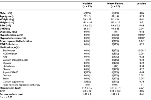
All participants completed the study protocol and their demographic data are displayed in Table 1. Participants with HF displayed a higher incidence of hypertension (67%) and remote myocardial infarction (33%), com-pared to the healthy older individuals. Elderly HF partici-pants trended toward reduced knee extensor strength
(maximum torque 94.5 ± 58.6 vs. 124.5 ± 45.1 Nm; p = 0.18) and delayed chair raise (11.7 ± 2.2 vs. 13.5 ± 1.9 m/ sec, p = 0.06), respectively, compared to healthy older individuals. Four-meter walk speed (0.98 ± 0.15 vs. 1.35 ± 0.24 m/sec, p < 0.001) was reduced in elderly HF partic-ipants relative to the healthy older individuals, respec-tively. Heart failure participants demonstrated reduced peak exercise capacity (peak VO2, p = 0.01) compared to healthy subjects (Table 2).
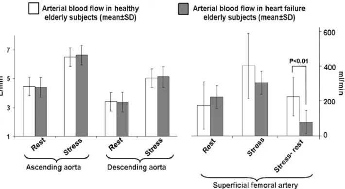
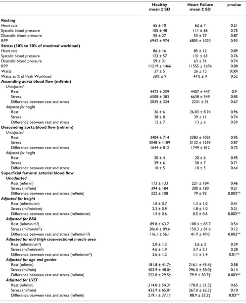
[image:5.612.59.553.370.700.2]After submaximal exercise, the change in superficial fem-oral artery LBF was lower in HF participants (79 ± 92 ml/ min) compared to healthy participants (222 ± 108 ml/ min; p = 0.002). This occurred even though resting, stress induced, and the stress-rest difference in the descending aortic flow were similar between the 2 groups (Figure 3). Peak VO2 was correlated with peak superficial femoral arterial flow after submaximal exercise (r = 0.67; p = 0.003). The watts achieved during CMR exercise were cor-related with peak superficial femoral artery blood flow (r = 0.51; p = 0.01), but not with the stress-rest difference in superficial femoral artery flow (r = -0.34; p = 0.11).
Table 1: Demographic data (mean ± standard deviation)
Healthy (n = 13)
Heart Failure (n = 12)
p-value
Male, n(%) 6(46%) 6(50%) 0.85
Age (years) 67 ± 5 70 ± 8 0.29
Weight (kg) 76 ± 11 81 ± 14 0.41
Height (cm) 171 ± 10 169 ± 10 0.5
BSA (m2) 1.9 ± 0.2 1.9 ± 0.2 0.64
LVEF(%) 66 ± 7 48 ± 25 0.04*
Diabetes, n(%) 0(0%) 1(8%) 0.48
Hypertension, n (%) 0(0%) 8(67%) 0.001*
Hypercholesterolemia 0(0%) 8(67%) 0.001*
Prior myocardial infarction 0(0%) 4(33%) 0.04*
COPD/Asthma 0(0%) 2(17%) 0.22
Medication, n(%)
Betablocker 0(0%) 8(67%) <0.001*
ACE inhibitor 0(0%) 6(50%) 0.01*
ARB 0(0%) 2(17%) 0.22
Calcium channel blocker 1(8%) 3(25%) 0.32
Digoxin 0(0%) 2(17%) 0.22
Hydralazine 0(0%) 1(8%) 0.48
Nitrate 0(0%) 2(17%) 0.22
Aspirin/NSAID 9(69%) 6(50%) 0.43
Diuretic 0(0%) 6(50%) 0.01*
Statin 0(0%) 5(42%) 0.01*
Calcium supplement 5(38%) 2(17%) 0.38
Prior hormone replacement therapy 1(8%) 0(0%) 1
Hemoglobin (g/dl) 14.9 ± 1.7 13.1 ± 1.4 0.02*
BNP 20 ± 12 118 ± 132 0.06
Serum sodium level 143 ± 2 142 ± 2 0.61
* = p < 0.05
To determine if the presence of prior myocardial infarc-tion, diabetes, hypertension, or hypercholesterolemia could have accounted for the differences in flow observed in the superficial femoral artery of our HF and healthy participants, we adjusted our results for the presence of these factors. After these adjustments, submaximal exer-cise induced difference (stress-rest) in superficial femoral artery LBF persisted between our healthy and HF groups (difference significant at p ≤ 0.05 after including all clini-cal factors). Also, the similarity between measures of ascending and descending aorta blood flow persisted between HF and healthy elderly participants (p > 0.2 after considering all clinical factors). Since body habitus and particularly thigh muscle size or mass can be different in participants with HF relative to normals, we performed
analyses that accounted for body habitus and thigh mus-cle area. Images of the middle thigh demonstrated trends toward reductions in thigh muscle area and elevations in subcutaneous fat and intermuscular fat in the elderly HF participants versus the healthy participants (0.1<p ≤ 0.15 for all areas assessed thigh components). After adjusting our blood flow measurements for age, gender, body sur-face area, and thigh muscle area, the differences in stress-rest measures of superficial femoral arterial LBF persisted (p = 0.002 to 0.003 for all).
[image:6.612.54.555.100.212.2]The 13 healthy elderly participants exhibited a LVEF of 66 ± 7%, which was on average higher than the participants with HF (average LVEF of 48 ± 25%). As has been previ-ously shown in elderly individuals with HF,[12] the LVEF
Table 2: Maximal (Peak) upright bicycle exercise and gas exchange analysis
Healthy Heart Failure p-value mean ± SD mean ± SD
Peak work load (watts) 97.8 ± 19.1 82.7 ± 22.3 0.002
Peak VO2 (ml/kg/min) 19.8 ± 5.7 13.9 ± 3.2 0.01
Max RER 1.03 ± 0.33 1.04 ± 0.07 0.95
Peak Heart rate 138 ± 28 111 ± 20 0.01
Peak Systolic blood pressure 195 ± 24 156 ± 30 0.01
Peak Diastolic blood pressure 81 ± 9 78 ± 8 0.51
Peak RPP 28295 ± 7412 17842 ± 5616 0.003
Abbreviations: PMHR = percent maximum heart rate (adjusted for age); RER = Respiratory exchange ratio; RPP = rate pressure project; VO2 = oxygen uptake
Bar graphs demonstrating rest and submaximal exercise induced measures of arterial blood flow (mean ± standard deviation) in the ascending aorta, descending aorta, and the superficial femoral artery
Figure 3
[image:6.612.129.484.442.636.2]in our HF participants ranged from severely reduced to preserved (15% to 86%). These findings are commensu-ruate with the syndrome of heart failure and a normal LVEF (HFNEF), heart failure and a reduced LVEF (HFREF), and a mixture of the two. With this range of LVEF in our study, we were able to perform analyses that evaluated the potential influence of LVEF on our results. Importantly, after adjusting our stress-rest changes in superficial femo-ral artery LBF for resting LVEF, the differences between our HF and healthy elderly groups persisted (p = 0.03) indicat-ing that the difference observed in superficial femoral arterial flow was present across the range of LVEF studied.
Discussion
Older patients with HF have particularly severe exercise intolerance, the primary symptom of chronic HF[12,13]. Studies in younger patients with HF have suggested that reduced LBF can contribute to exercise intolerance[14,15]. In the present study, we studied older patients with HF and compared them to an age-matched group of healthy elderly subjects. The characteristics of the HF participants in this study were representative of prior populations of elderly HF[12,16,17]. Using expired gas analysis during exhaustive upright exercise, we found that elderly HF par-ticipants exhibited severely decreased exercise capacity (Table 2), which is consistent with previous reports from our group and others[12,18,19]. All participants (HF and healthy) in this study were carefully screened to exclude factors known to alter arterial LBF, including evidence of peripheral arterial atherosclerosis.
Using phase contrast CMR to measure flow in the superfi-cial femoral artery, and the ascending and descending aorta at rest and immediately after supine submaximal exercise, we found that older patients with HF have signif-icantly reduced LBF despite similar responses in cardiac output (Figure 3). This phenomenon persisted after accounting for a wide variety of potentially confounding factors, including thigh muscle area, LVEF, gender, body size, and age. This finding occurred even though HF par-ticipants received medical therapy with angiotensin con-verting enzyme inhibitors or receptor blockers that could have improved blood flow (Table 3). These data suggest that older HF patients have abnormal distribution of blood flow to the peripheral vascular system even though cardiac output is preserved, and that reduced LBF may be an important contributor to their severe exercise intoler-ance.
The findings of the present study are supported by those of prior studies, which have demonstrated that LBF is reduced during exercise in younger and middle aged patients with exercise intolerance,[14,15] even though submaximal cardiac output increases. The present study increases our understanding of the relationship between
LBF and exercise capacity by extending these observations to older patients who comprise the majority of the popu-lation with HF, and who often have complex etiologies of HF with more severe exercise intolerance compared to younger patients[12]. Lower extremity blood flow has been shown to decrease with normal aging[15,20]. Importantly, in this study using highly precise measures of blood flow and well characterized participants, differ-ences in LBF between our HF and healthy elderly persist after accounting for the clinical conditions (hypertension, hypercholesterolemia, and diabetes) known to effect car-diac output and blood flow in the elderly.
The normal physiological response to exercise involves the control of LBF. With the onset of exercise, selective vasoconstriction temporarily decreases blood flow to splanchnic and other non-critical arterial beds[21,22]. In combination with selective vasodilatation of the femoral and distal leg arteries, this allows efficient distribution of cardiac output to the exercising leg muscles in order to facilitate physical work[23,24]. It is known that normal aging is associated with significant alterations in periph-eral arterial blood flow responses at rest and after a variety of stressors, including exercise [21-24]. Thus, inclusion of an age-matched healthy control group was an important design feature of the present study. Our results indicate that older HF patients have reduced leg blood flow with exercise beyond that which is associated with normal aging.
Table 3: Hemodynamic data during supine submaximal bicycle and blood flow measurement
Healthy mean ± SD
Heart Failure mean ± SD
p-value
Resting
Heart rate 65 ± 10 62 ± 7 0.51
Systolic blood pressure 105 ± 48 111 ± 56 0.75
Diastolic blood pressure 55 ± 27 53 ± 27 0.87
RPP 6942 ± 974 6805 ± 1023 0.92
Stress (30% to 50% of maximal workload)
Heart rate 86 ± 16 85 ± 12 0.89
Systolic blood pressure 123 ± 57 131 ± 63 0.76
Diastolic blood pressure 59 ± 31 63 ± 31 0.74
RPP 11219 ± 1466 11555 ± 1696 0.88
Watts 37 ± 5 26 ± 15 0.001
Watts as % of Peak Workload 38% ± 9 41% ± 9 0.52
Ascending aorta blood flow (ml/min)
Unadjusted
Rest 4473 ± 259 4407 ± 447 0.9
Stress 6508 ± 383 6638 ± 549 0.85
Difference between rest and stress 2035 ± 324 2231 ± 31 0.67
Adjusted for height
Rest 26 ± 6 26.03 ± 8.74 0.96
Stress 38 ± 8 39 ± 11 0.74
Difference between rest and stress 12 ± 7 13 ± 6 0.59
Descending aorta blood flow (ml/min)
Unadjusted
Rest 3404 ± 714 3383 ± 1021 0.95
Stress 5048 ± 1189 5132 ± 1293 0.87
Difference between rest and stress 1644 ± 812 1749 ± 812 0.75
Adjusted for height
Rest 20 ± 4 20 ± 6 0.95
Stress 29 ± 6 30 ± 7 0.71
Difference between rest and stress 10 ± 5 10 ± 5 0.64
Superficial femoral arterial blood flow
Unadjusted
Rest (ml/min) 173 ± 133 221 ± 184 0.46
Stress (ml/min) 394 ± 184 300 ± 180 0.21
Difference between rest and stress (ml/min) 222 ± 108 79 ± 92 0.002** Adjusted for height
Rest (ml/min/cm) 1.0 ± 0.7 1.3 ± 1.0 0.41
Stress (ml/min/cm) 2.3 ± 0.9 1.8 ± 1.0 0.21
Difference between rest and stress (ml/min/cm) 1.3 ± 0.6 0.5 ± 0.6 0.002** Adjusted for BSA
Rest (ml/min/m2) 89.8 ± 63.7 108.4 ± 83.7 0.54
Stress (ml/min/m2) 206.0 ± 89.6 150.3 ± 81.6 0.12
Difference between rest and stress (ml/min/m2) 116.1 ± 56.1 41.9 ± 49.0 0.002** Adjusted for mid thigh cross-sectional muscle area
Rest (ml/min/cm2) 2.0 ± 1.3 2.6 ± 2 0.39
Stress (ml/min/cm2) 4.6 ± 1.9 3.7 ± 2.1 0.28
Difference between rest and stress (ml/min/cm2) 2.6 ± 1.2 1.1 ± 1.4 0.01** Adjusted for age and gender
Rest (ml/min) 181.8 ± 41.7† 216.1 ± 43.4† 0.58
Stress (ml/min) 403.9 ± 48.0† 296.0 ± 50.0† 0.14
Difference between rest and stress (ml/min) 222.0 ± 29.5† 79.9 ± 30.7† 0.003** Adjusted for LVEF
Rest (ml/min) 214.8 ± 54.3† 178.0 ± 51.5† 0.65
Stress (ml/min) 433.9 ± 65.0† 267.0 ± 62.3† 0.10
Difference between rest and stress (ml/min) 219.1 ± 37.1† 88.9 ± 35.2† 0.03**
† = mean ± standard error, ** = p < 0.03
Although the control of LBF in the elderly is not com-pletely understood, several studies have implicated increased sympathetic nerve activity, vascular smooth muscle cell intimal medial thickening, endothelial dys-function, and chronic cytokine activation as mechanisms which alter redistribution of blood flow during exer-cise[23,31]. We have previously shown that older HF patients have reduced aortic distensibility which corre-lates with exercise intolerance,[18,32] and that older HF patients with a reduced LVEF have impaired flow-medi-ated arterial dilation (FMD), indicative of endothelial dys-function[16]. In this study we did not measure FMD in the subjects. The HF subjects demonstrate a similar increase in brachial artery blood pressure with exercise as control subjects. Despite this, the relative increase in LBF was blunted. Recognizing that mean arterial pressure in the brachial artery is indeed representative of central aortic mean arterial pressure (especially in older subjects where significant atheroslcerosis was an exclusion), these data suggest that a potential mechanism for the relatively blunted increase in LBF in the HF subjects is an impaired active hyperemia and/or a relative inability of the HF sub-jects to drop their systemic vascular resistance to exer-cise[33].
An important consideration regarding our results pertains to the fact that alterations in the control of LBF are modi-fiable. Beere et al.,[34] have shown that exercise condi-tioning partially reverses the normal age-related reduction in LBF during exercise, resulting in improved exercise capacity. In HF patients, exercise training and angiotensin converting enzyme inhibition improve exercise induced LBF and endothelial dysfunction, which leads to improve-ments in exercise intolerance[15,35-37].
Since the LVEF was lower in our HF group relative to our healthy older participants, due to inclusion of participants with HFREF in addition to those with HFNEF, we sus-pected that cardiac output would be reduced with sub-maximal exercise. This did not occur. Although the LVEF of the HF group was lower than that observed in the healthy group, the lower submaximal exercise induced difference (stress-rest) in superficial femoral arterial blood in the HF participants persisted after adjustment for LVEF (p = 0.03). Our data indicate that the LVEF per se and the impact of LVEF on cardiac output, are not responsible for the reduced submaximal exercise induced superficial fem-oral arterial flow observed in our HF participants. Our data does not allow us to conclude whether other indirect consequences of a poor LVEF, or the differential influence in factors, such as autonomic dysfunction or heightened inflammatory cytokines, which may be disproportion-ately present in individuals with HFNEF versus HFREF, may have been responsible for the reduction in
submaxi-mal exercise induced superficial femoral LBF observed in our HF subjects.
This study has potential limitations. First, we did not per-form our CMR blood flow measurements during maximal exercise; thus we cannot prove that reduced LBF was the sole cause of the severely reduced exercise capacity of the older HF patients. Second, we did not set an absolute workload for our participants; this resulted in the HF par-ticipants performing a lower absolute measure of work compared to controls. Absolute determinations could have been highly variable (25% to near 100%) of maxi-mum work for our HF participants dependant on their level of conditioning. We particularly wanted to avoid having HF participants come close to their anaerobic thresholds, a point during exercise when physiology changes, during our exercise protocol. Instead, we used a relative workload value based on all participants exhibit-ing maximum efforts (respiratory exchange ratios > 1.02), and then taking 30%-50% of this level to ascribe the work needed to perform submaximal exercise during our proto-col. This has the effect of producing comparable levels of submaximal work across all of the study participants.
Third, we cannot definitively exclude subclinical athero-sclerosis as a potential contributor to the reduced LBF in the superficial femoral artery. We vigorously screened all participants for atherosclerosis in the large arteries (carotid, femoral, iliac, and thoracic aorta) with ultra-sound and CMR. We did not perform brachial/ankle pres-sure assessments which can screen for peripheral arteriosclerosis. Importantly, the intergroup differences in leg blood flow persisted after adjusting for risk factors for atherosclerosis, including hypercholesterolemia, hyper-tension, diabetes, and prior myocardial infarction. Fourth, most of our participants were Caucasian; thus, we have little data on those of other race.
Finally, both a strength and a weakness of the current study was that the HF subjects held their HF medications the night before the study. This was a strength in that the potentially confounding effects of these medications were removed, thus allowing the study of 'native' cardiac and vascular function. A potential weakness of this strategy is that HF patients are prescribed these therapies to improve survival; thus they frequently take them. It is conceivable that the magnitude of the LBF differential may have been less, if for example, an ACEI or ARB had been continued in the HF participants at the time of exercise CMR.
In conclusion, in elderly patients with HF, functional impairment, and exercise intolerance, submaximal exer-cise induced femoral arterial blood flow is reduced even though cardiac output is preserved. This finding occurs after accounting for thigh muscle area, age, gender, and co-morbidities associated with poor vascular function. In contrast to our original hypothesis, these results indicate that mechanisms other than low cardiac output are responsible for reduced LBF during submaximal exercise in the elderly with HF. For these reasons, future studies should address whether a) inappropriate distribution of LBF in the absence of abnormalities of submaximal exer-cise induced changes in cardiac output are present in eld-erly disabled individuals with exercise intolerance, and b) therapies that improve LBF without necessarily modifying LVEF can reduce exercise intolerance and disability observed in elderly HF.
Competing interests
Drs. Hundley and Hamilton have an ownership stake in MRI Cardiac Services, Inc. that creates software in MRI image management and display.
Authors' contributions
CP acquired, analyzed, and interpreted the data, and drafted the manuscript. DWK participated in its design and coordination, handled funding and supervision, acquired, analyzed, and interpreted the data, and helped to draft the manuscript. SBK handled funding and super-vision. CAH aided in acquiring, analyzing, and interpret-ing the data. BN aided in acquirinterpret-ing, analyzinterpret-ing, and interpreting the data. XI performed the statistical analysis. PHB aided in acquiring, analyzing, and interpreting the data. WGH conceived of the study, participated in its design and coordination, handled funding and supervi-sion, acquired, analyzed, and interpreted the data, and helped to draft the manuscript. All authors made critical revision of the manuscript for important intellectual con-tent and approved of the final manuscript.
Acknowledgements
We acknowledge the expertise of Deanna Carr who assisted in preparation of the manuscript. Research supported in part by National Institutes of
Health grants M01RR07122, RO1AG18915, RO1HL076438, P30AG21332 and R41AG030248.
References
1. Mancini DM, Walter G, Reichek N, Lenkinski R, McCully KK, Mullen JL, Wilson JR: Contribution of skeletal muscle atrophy to exer-cise intolerance and altered muscle metabolism in heart fail-ure. Circulation 1992, 85(4):1364-73.
2. Wilson JR, Martin JL, Schwartz DC, Ferraro N: Exercise intoler-ance in patients with chronic heart failure: Role of impaired nutritive flow to skeletal muscle. Circulation 1984,
69(6):1079-1087.
3. Sullivan M, Cobb FR: Central hemodynamic response to exer-cise in patients with chronic heart failure. Chest 1992, 101(5 Suppl):340S-346S.
4. Buchner D, Beresford S, Larson EB, LaCroix AZ, Wagner EH: Effects of physical activity on health status in older adults. II. Inter-ventions studies. Annu Rev Public Health 1992, 13:469-488. 5. Marburger CT, Brubaker PH, Pollock WE, Morgan TM, Kitzman DW:
Reproducibility of cardiopulmonary exercise testing in eld-erly patients with congestive heart failure. Am J Cardiol 1998,
82(7):905-909.
6. Rich MW: Epidemiology, pathophysiology, and etiology of congestive heart failure in older adults. Am J Geriatr Cardiol 1997, 45:968-974.
7. Gandhi SK, Powers JE, Nomeir AM, Fowle K, Kitzman DW, Rankin KM, Little WC: The pathogenesis of acute pulmonary edema associated with hypertension. N Engl J Med 2001, 344(1):17-22. 8. Kitzman DW: Heart failure in the elderly: Systolic and
diasto-lic dysfunction. Am J Geriatr Cardiol 1995, 5(1):20-26.
9. Sullivan M, Higginbotham MB, Cobb FR: Exercise training in patients with chronic heart failure delays ventilatory anaer-obic threshold and improves submaximal exercise perform-ance. Circulation 1989, 79(2):324-329.
10. Alexander MR, Kitzman DW, Khaliq S, Darty SN, Hamilton CA, Her-rington DM, Link KM, Hundley WG: Determination of femoral artery endothelial function by phase contrast magnetic res-onance imaging. Am J Cardiol 2001, 88(9):1070-1074.
11. Mitsiopoulos N, Baumgartner RN, Heymsfield SB, Lyons W, Gallagher D, Ross R: Cadaver validation of skeletal muscle measure-ment by magnetic resonance imaging and computerized tomography. J Appl Physiol 1998, 85(1):115-122.
12. Kitzman DW, Little WC, Brubaker PH, Anderson RT, Hundley WG, Marburger CT, Brosnihan B, Morgan TM, Stewart KP: Pathophysio-logical characterization of isolated diastolic heart failure in comparison to systolic heart failure. JAMA 2002,
288(17):2144-2150.
13. Clark AL, Harrington D, Chua TP, Coats AJ: Exercise capacity in chronic heart failure is related to the aetiology of heart dis-ease. Heart 1997, 78(6):569-571.
14. Clark A, Volterrani M, Swan JW, Hue D, Hooper J, Coats AJ: Leg blood flow, metabolism and exercise capacity in chronic sta-ble heart failure. Int J Cardiol 1996, 55(2):127-135.
15. Sullivan M, Knight JD, Higginbotham MB, Cobb FR: Relation between central and peripheral hemodynamics during exer-cise in patients with chronic heart failure: muscle blood flow is reduced with maintenance of arterial perfusion pressure.
Circulation 1989, 80(4):769-781.
16. Hundley WG, Bayram E, Hamilton CA, Hamilton EA, Morgan TM, Darty SN, Stewart KP, Link KM, Herrington DM, Kitzman DW: Leg flow-mediated arterial dilation in elderly patients with heart failure and normal left ventricular ejection fraction. Am J Phys-iol Heart Circ PhysPhys-iol 2007, 292(3):H1427-H1434.
17. Fleg JL, O'Connor F, Gerstenblith G, Becker LC, Clulow J, Schulman SP, Lakatta EG: Impact of age on the cardiovascular response to dynamic upright exercise in healthy men and women. J Appl Physiol 1995, 78(3):890-900.
18. Hundley WG, Kitzman DW, Morgan TM, Hamilton CA, Darty SN, Stewart KP, Herrington DM, Little WC: Cardiac cycle dependent changes in aortic area and aortic distensibility are reduced in older patients with isolated diastolic heart failure and corre-late with exercise intolerance. J Am Coll Cardiol 2001,
38(3):796-802.
19. Kitzman DW, Higginbotham MB, Cobb FR, Sheikh KH, Sullivan MJ:
pre-Publish with BioMed Central and every scientist can read your work free of charge
"BioMed Central will be the most significant development for disseminating the results of biomedical researc h in our lifetime."
Sir Paul Nurse, Cancer Research UK
Your research papers will be:
available free of charge to the entire biomedical community
peer reviewed and published immediately upon acceptance
cited in PubMed and archived on PubMed Central
yours — you keep the copyright
Submit your manuscript here:
http://www.biomedcentral.com/info/publishing_adv.asp
BioMedcentral
served left ventricular systolic function: Failure of the Frank-Starling mechanism. J Am Coll Cardiol 1991, 17(5):1065-1072. 20. Dinenno FA, Tanaka H, Stauffer BL, Seals DR: Reductions in basal
limb blood flow and vascular conductance with human age-ing: Role for augmented α-adrenergic vasoconstriction. Jour-nal of Physiology 2001, 536(3):977-983.
21. Lakatta EG, Levy D: Arterial and cardiac aging: major share-holders in cardiovascular disease enterprises: Part I: aging arteries: a "set up" for vascular disease. Circulation 2003,
107(1):139-146.
22. Leithe ME, Hermiller JB, Magorien RD, Unverferth DV, Leier CV: The effect of age on central and regional hemodynamics. Geron-tology 1984, 30(4):240-246.
23. Dinenno FA, Jones PP, Seals DR, Tanaka H: Limb blood flow and vascular conductance are reduced with age in healthy humans: relation to elevations in sympathetic nerve activity and declines in oxygen demand. Circulation 1999,
100(2):164-170.
24. Julius S, Amery A, Whitlock LS, Conway J: Influence of age on the hemodynamic response to exercise. Circulation 1967,
36(2):222-230.
25. Flamm SD, Taki J, Moore R, Lewis SF, Keech F, Maltais F, Ahmad M, Callahan R, Dragotakes S, Alpert N, Strauss HW: Redistribution of regional and organ blood-volume and effect on cardiac-func-tion in relacardiac-func-tion to upright exercise intensity in healthy-human subjects. Circulation 1990, 81(5):1550-1559.
26. Wade OL, Combes B, Childs AW, Wheeler HO, Cournand A, Brad-ley SE: The effect of exercise on the splanchnic blood flow and splanchnic blood volume in normal man. Clin Sci (Colch) 1956,
15:457-463.
27. Osada T, Katsumura T, Hamaoka T, Inoue S, Esaki K, Sakamoto A, Murase N, Kajiyama N, Shimomitsu T, Iwane H: Reduced blood flow in abdominal viscera measured by Doppler ultrasound during one-legged knee extension. J Appl Physiol 1999,
86(2):709-719.
28. Marchesi S, Vaudo G, Lupattelli G, Lombardini R, Roscini AR, Bro-zzetti M, Siepi D, Mannarino E: Fat distribution and endothelial function in normal-overweight menopausal women. J Clin Pharm Ther 2007, 32(5):477-82.
29. Szmitko PE, Teoh H, Stewart DJ, Verma S: Adiponectin and cardi-ovascular disease: State of the art? Am J Physiol Heart Circ Physiol 2007, 292(3):H1655-H1633.
30. Piña IL, Apstein CS, Balady GJ, Belardinelli R, Chaitman BR, Duscha BD, Fletcher BJ, Fleg JL, Myers JN, Sullivan MJ: American Heart Association Committee on exercise, rehabilitation, and pre-vention. Exercise and heart failure: A statement from the American Heart Association committee on exercise, reha-bilitation, and prevention. Circulation 2003, 107(12):1210-1225. 31. Lakatta EG: Arterial and cardiac aging: major shareholders in
cardiovascular disease enterprises: Part III: cellular and molecular clues to heart and arterial aging. Circulation 2003,
107(3):490-497.
32. Rerkpattanapipat P, Hundley WG, Link KM, Brubaker PH, Hamilton CA, Darty SN, Morgan TM, Kitzman DW: Relation of aortic dis-tensibility determine by magnetic resonance imaging in patients >60 years of age to systolic heart failure and exer-cise capacity. Am J Cardiol 2002, 90(11):1221-1225.
33. Mason DT, Zelis R, Longhurst J, Lee G: Cardiocirculatory responses to muscular exercise in congestive heart failure.
Prog Cardiovasc Dis 1977, 19(6):475-489.
34. Beere PA, Russell SD, Morey MC, Kitzman DW, Higginbotham MB:
Aerobic exercise training can reverse age-related peripheral circulatory changes in healthy older men. Circulation 1999,
100(10):1085-1094.
35. Hornig B, Arakawa N, Haussmann D, Drexler H: Differential effects of quinaprilat and enalaprilat on endothelial function of conduit arteries in patients with chronic heart failure. Cir-culation 1998, 98(25):2842-2848.
36. Hornig B, Maier V, Drexler H: Physical training improves endothelial function in patients with chronic heart failure.
Circulation 1996, 93(2):210-214.
37. Hambrecht R, Fiehn E, Weigl C, Gielen S, Hamann C, Kaiser R, Yu J, Adams V, Niebauer J, Schuler G: Regular physical exercise cor-rects endothelial dysfunction and improves exercise capacity in patients with chronic heart failure. Circulation 1998,
98(24):2709-2715.
38. Klein WM, Bartels LW, Bax L, Graaf Y, Mali WP: Magnetic reso-nance imaging measurement of blood volume flow in periph-eral arteries in healthy subjects. J Vasc Surg 2003,
38(5):1060-1066.
39. Hundley WG, Li HF, Lange RA, Pfeifer DP, Meshack BM, Willard JE, Landau C, Willett DL, Hilis LD, Peshock RM: Assessment of left-to-right intracardiac shunting by velocity-encoded, phase-difference magnetic resonance imaging: A comparison with oximetric and indicator dilution techniques. Circulation 1995,
91(12):2955-2960.
40. Hundley WG, Li HF, Hillis LD, Meshack BM, Lange RA, Willard JE, Landau C, Peshock RM: Quantitation of cardiac output with velocity-encoded, phase-difference magnetic resonance imaging. Am J Cardiol 1995, 75(17):1250-1255.
41. Silber HA, Ouyang P, Bluemke DA, Gupta SN, Foo TK, Lima JA: Why is flow-mediated dilation dependent on arterial size? Assess-ment of the shear stimulus using phase-contrast magnetic resonance imaging. Am J Physiol Heart Circ Physiol 2005,
288(2):H822-H828.