0095-1137/07/$08.00
⫹
0
doi:10.1128/JCM.01986-06
Copyright © 2007, American Society for Microbiology. All Rights Reserved.
Multicenter Comparison of the VITEK 2 Yeast Susceptibility Test with
the CLSI Broth Microdilution Reference Method for Testing
Fluconazole against
Candida
spp.
䌤
M. A. Pfaller,
1* D. J. Diekema,
1G. W. Procop,
2and M. G. Rinaldi
3University of Iowa College of Medicine, Iowa City, Iowa
1; Cleveland Clinic Foundation, Cleveland, Ohio
2; and
University of Texas Health Science Center, San Antonio, Texas
3Received 25 September 2006/Returned for modification 4 December 2006/Accepted 26 December 2006
A fully automated commercial antifungal susceptibility test system (VITEK 2 yeast susceptibility test;
bioMerieux, Inc., Hazelwood, Mo.) was compared in three different laboratories with Clinical and
Labo-ratory Standards Institute (CLSI) reference broth microdilution (BMD) method by testing two quality
control strains and a total of 426 isolates of
Candida
spp. (103 to 135 clinical isolates in each laboratory
plus 80 challenge isolates in one laboratory) against fluconazole. Reference BMD MIC endpoints were
established after 24 and 48 h of incubation. VITEK 2 endpoints were determined spectrophotometrically
after 10 to 26 h of incubation (mean, 13 h). Excellent essential agreement (within two dilutions) between
the VITEK 2 and the 24- and 48-h BMD MICs was observed. The overall agreement values were 97.9 and
93.7%, respectively. Both intra- and interlaboratory agreement was 100%. The overall categorical
agree-ment between VITEK 2 and BMD was 97.2% at the 24-h BMD time point and 88.3% at the 48-h BMD time
point. Decreased categorical agreement at 48 h was attributed to trailing growth observed with
Candida
glabrata
. The VITEK 2 system reliably detected fluconazole resistance among
Candida
spp. and
demon-strated excellent quantitative and qualitative agreement with the reference BMD method.
Standardized broth microdilution (BMD) susceptibility
test-ing of fluconazole against
Candida
spp. has been available
since 1997 (19, 21, 30). The establishment of a panel of quality
control (QC) strains and validated, clinically useful
interpre-tive breakpoints (19, 20, 27, 30, 32) has allowed this method to
be used worldwide (3, 6–8, 12, 17, 21, 33).
The Clinical and Laboratory Standards Institute (CLSI;
for-merly National Committee for Clinical Laboratory Standards
[NCCLS]) BMD method for testing fluconazole has served as
a touchstone for the development of both broth- and
agar-based procedures designed to provide simple, flexible, and
commercially available alternative susceptibility testing
meth-ods for use in the clinical laboratory (8–12, 16, 17, 21, 23–25).
The performance of the various commercially available
anti-fungal testing systems has been variable (8, 10, 16, 17), and
prior to the present study only two, the Sensititre YeastOne
System (Trek, Cleveland, OH) and the Etest (AB BIODISK,
Solna Sweden), have been approved by the U.S. Food and
Drug Administration (FDA) for in vitro susceptibility testing
of fluconazole against
Candida
spp. (21, 26).
Although spectrophotometric reading of BMD MIC
end-points has been shown to be valid and feasible for use in the
clinical laboratory (11, 12, 16, 20, 21), this approach has not
been incorporated into a commercially available testing method.
Recently, bioMerieux (Hazelwood, MO) has developed a yeast
susceptibility test that determines growth
spectrophotometri-cally and allows fully automated antifungal susceptibility
test-ing of fluconazole against
Candida
using the VITEK 2
micro-biology system. The fully automated VITEK 2 system allows
for the standardization of all of the critical parameters known
for antifungal susceptibility testing: inoculum preparation,
fill-ing of the device, duration and temperature of incubation, and
endpoint determination. The yeast susceptibility test, coupled
with the rapid and accurate yeast identification capabilities
already available on the VITEK 2 (4), would allow clinical
laboratories to perform both yeast identification and antifungal
susceptibility testing using a fully automated and completely
standardized format. Preliminary studies by Zambardi et al.
(G. Zambardi et al., Abstr. 45th Intersci. Conf. Antimicrob.
Agents Chemother., abstr. M-1619, 2005) have shown both
essential and categorical agreements of
ⱖ
90% in a comparison
of VITEK 2 MICs with reference BMD MICs for fluconazole
and
Candida
spp. The VITEK 2 results were available in
ⱕ
15
h compared to 48 h for the reference BMD method.
The purpose of the present study was to validate the
per-formance of the VITEK 2 yeast susceptibility test with
flucon-azole against a broad range of
Candida
spp. in three
indepen-dent laboratories. The VITEK 2 results were compared to
those from a frozen reference BMD panel performed
accord-ing to CLSI guidelines. It is notable that after the completion
of the present study the VITEK 2 yeast susceptibility test for
fluconazole was approved for clinical use by the U.S. FDA
(bioMerieux press release, 27 September 2006).
MATERIALS AND METHODS
Study design.The study was designed to compare the MIC results for flucon-azole obtained by the VITEK 2 yeast susceptibility test to those obtained by the M27-A2 BMD method (20) in the three laboratories. Each laboratory tested at least 100 clinical isolates ofCandidaspp. (range, 103 to 135 isolates) by the VITEK 2 system and the CLSI frozen reference BMD panel (a total of 346
* Corresponding author. Mailing address: Medical Microbiology
Di-vision, C606 GH, Department of Pathology, University of Iowa
Col-lege of Medicine, Iowa City, IA 52242. Phone: (319) 356-8615. Fax:
(319) 356-4916. E-mail: [email protected].
䌤
Published ahead of print on 10 January 2007.
796
on May 16, 2020 by guest
http://jcm.asm.org/
clinical isolates). In addition, a challenge set of 80 well-characterized stock isolates was tested by both methods in one of the laboratories. Intra- and interlaboratory reproducibility was determined by testing a panel of 10Candida spp. isolates in triplicate on three separate days in each of the participating laboratories. The MIC results obtained with the VITEK 2 system after 10 to 26 h of incubation (depending on the organism growth rate) were compared to those obtained with the reference BMD panel read after both 24 and 48 h of incuba-tion.
Test organisms.The test organisms included two American Type Culture Col-lection (ATCC) strains that have been established as QC strains (Candida parapsi-losisATCC 22019 andC. kruseiATCC 6258) by the CLSI (5, 20). A challenge set of 80 isolates ofCandidaspp. selected to provide strains with on-scale MIC results and to represent both clinically important species and resistance mechanisms were tested in one of the participating laboratories. The challenge set included 32 isolates ofC. albicans, 6 isolates ofC. dubliniensis, 14 isolates ofC. glabrata, 3 isolates of C. guilliermondii, 5 isolates ofC. krusei, 5 isolates ofC. lusitaniae, 1 isolate ofC. norvegensis, 7 isolates ofC. parapsilosis, 2 isolates ofC. pelliculosa, and 5 isolates ofC. tropicalis.An additional 346 clinical isolates ofCandidaspp. were also tested. The clinical isolates included 166 isolates ofC. albicans, 2 isolates ofC. dubliniensis, 69 isolates ofC. glabrata, 46 isolates ofC. krusei, 4 isolates ofC. lusitaniae, 36 isolates ofC. parapsilosis, and 23 isolates ofC. tropicalis. These were all recent clinical isolates and were selected to represent the clinically prevalent species, including fluconazole-resistant strains. Reproducibility within and among laboratories was assessed by using a panel of 10Candidaisolates:C. glabratastrain 304201,C. glabratastrain 304927,C. haemuloniistrain 304848,C. kruseistrain 304204,C. kruseistrain 304845,C. kruseistrain 304850,C. lipolytica strain 204856,C. lusitaniaestrain 304205,C. norvegensisstrain 304852, andC. pelliculosastrain 304847. These isolates were selected to provide on-scale flu-conazole MICs ranging from 2 to 32g/ml. All isolates were identified by standard methods (14). Prior to testing, each isolate was passaged at least twice on Sabouraud dextrose agar (Remel, Lenexa, KS) to ensure purity and viability. Antifungal agents and microdilution panels.The VITEK 2 cards containing serial twofold dilutions of fluconazole (range, 1 to 64g/ml) were provided by the manufacturer. The frozen BMD reference panels containing serial twofold dilutions of fluconazole (range, 0.12 to 128g/ml) were provided by Trek Di-agnostic Systems (Cleveland, OH). The VITEK 2 cards were shipped in sealed packages and stored at 2 to 8°C until testing was performed. The BMD panels were shipped frozen in sealed packages and were stored at⫺70°C until the day of the test.
Inoculum preparation.Stock inoculum suspensions of theCandidaspp. were obtained from 24-h cultures on Sabouraud dextrose agar at 35°C. The inoculum suspensions for the VITEK 2 were prepared in sterile saline to a turbidity equal to a 2.0 McFarland standard by using the bioMerieux DensiChek instrument. The inoculum suspensions for the reference BMD were prepared by diluting a portion of the 2.0 McFarland suspension prepared for the VITEK 2 to match the turbidity of a 0.5 McFarland.
CLSI broth microdilution method.Reference BMD testing was performed exactly as outlined in CLSI document M27-A2 (20) with a final inoculum con-centration of 1.5⫻103⫾
1.0⫻103
cells/ml and RPMI 1640 medium buffered to pH 7.0 with 0.165 M morpholinepropanesulfonic acid buffer. The panels were incubated in air at 35°C and observed for the presence or absence of growth at 24 and 48 h. The fluconazole MIC was read as the lowest concentration that produced a prominent decrease in turbidity (ca. 50% reduction in growth) relative to the drug-free control (20).
VITEK 2 yeast susceptibility test.The standardized 2.0 McFarland inoculum suspension was placed into a VITEK 2 cassette along with a sterile polystyrene test tube and a yeast susceptibility test card for each organism. The loaded cassettes were then placed into the VITEK 2 instrument, and the respective yeast suspensions were diluted appropriately, after which the cards were filled, incu-bated, and read automatically. The time of incubation varied from 10 to 26.1 h, based on the rate of growth in the drug-free control well, and the results were expressed as MICs in micrograms per milliliter.
Quality control. Quality control was ensured by testing the CLSI-recom-mended quality control strainsC. parapsilosisATCC 2209 andC. krusei6258 (5, 20). These isolates were tested between 22 and 29 times in each of the three laboratories (total number of results⫽294), and all (100%) MICs were in the respective reference ranges.
Analysis of results.The MIC results obtained with the VITEK 2 yeast sus-ceptibility test were compared to those of the reference BMD panels read at 24 and 48 h. As with previous studies (9–12, 24, 25), high off-scale MIC results were converted to the next highest concentration, and low off-scale MIC results were left unchanged. Discrepancies among MIC endpoints of more than two dilutions (two wells) were used to calculate the essential agreement (EA). Interlaboratory
and intralaboratory agreement, assessed with the 10-isolate reproducibility panel, was defined when MIC results were within a three-dilution range. The CLSI interpretive breakpoints for fluconazole (susceptible [S],ⱕ8g/ml; sus-ceptible dose dependent [SDD], 16 to 32g/ml; resistant [R],ⱖ64g/ml) were used to obtain categorical agreement (CA) percentages between the MICs de-termined by VITEK 2 and the reference BMD (20, 27). Very major errors (VME) were identified when the reference MIC indicated R, and the VITEK 2 MIC indicated S. Major errors (ME) were identified when the isolate was classified as R by the VITEK 2 and S by the reference method. Minor errors were determined when the results of one of the test methods was either S or R and that of the other was SDD. The MIC results obtained forC. kruseiwere used as such (in micrograms per milliliter) for the purpose of assessing EA but were forced into the R category, as required by the CLSI (20), when determining CA.
RESULTS AND DISCUSSION
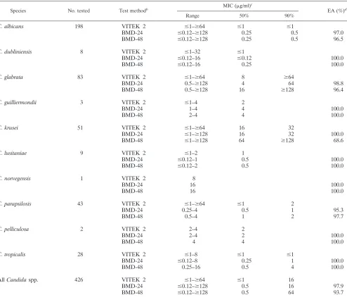
Table 1 summarizes the in vitro fluconazole susceptibilities
of 426 isolates of
Candida
spp. as determined by the VITEK 2
system and the reference BMD read at 24 and 48 h. Due to the
similarity in results obtained with the VITEK 2 compared to
the 24- and 48-h BMDs for both the challenge isolates (80
isolates, 100 and 97.5% EA, respectively) and the clinical
iso-lates (346 isoiso-lates, 97.4 and 92.8% EA, respectively), the
re-sults for the two organism sets were combined in Table 1. In
general, MIC results for fluconazole were typical of each
spe-cies of
Candida
(22), with the lowest MICs obtained with both
VITEK 2 and BMD observed for
C. albicans
and the highest
MICs observed for
C. glabrata
and
C. krusei
. Notably, BMD
MICs read at 24 h of incubation tended to be approximately
fourfold lower than those read at 48-h for both
C. glabrata
(MIC
50, 4
g/ml versus 16
g/ml, respectively) and
C. krusei
(MIC
50, 16
g/ml versus 64
g/ml, respectively), suggesting
significant trailing with these two species.
The overall EA between the VITEK 2 and the BMD MICs
ranged from 97.9% when the 24-h BMD result was used as a
reference to 93.7% when the 48-h BMD result was used as
reference (95.2 and 83.0%, respectively, for on-scale results).
Of the discrepancies noted between the VITEK 2 and 24-h
BMD MIC results, the MICs generated by the VITEK 2 were
higher than those obtained by BMD in all nine instances
(100%). In contrast, of the 27 discrepancies observed between
THE VITEK 2 and 48-h BMD MIC results, the MICs
gener-ated by the VITEK 2 were lower than those obtained by BMD
in 21 instances (77.8%). The latter discrepancies occurred
al-most exclusively with
C. glabrata
and
C. krusei
, emphasizing the
impact of trailing on BMD results obtained with these two
species.
The mean time to result for the VITEK 2 system was 13 h,
with a range from 10 to 26 h. Only one isolate, a clinical isolate
of
C. parapsilosis
, failed to grow in the VITEK 2 system, and all
isolates grew sufficiently well in the BMD panel to be read
after 24 h of incubation. Similar results were obtained at all
three study sites.
Regarding the individual species of
Candida
, the EA
be-tween the VITEK 2 results and either the 24-h or the 48-h
BMD MICs was
⬎
95% for all species with the exception of
C.
krusei
. Whereas the EA for this species was 100% when
VITEK 2 results were compared to the 24-h BMD MICs, it was
only 68.6% when the 48-h BMD MICs were used as the
ref-erence result. Again, these discrepancies were due to higher
MICs obtained with the BMD method than were obtained with
the VITEK 2 system for this species. Given the CLSI
on May 16, 2020 by guest
http://jcm.asm.org/
mendation that
C. krusei
should be considered resistant to
fluconazole irrespective of the MIC, these discrepancies
should not pose a problem clinically (20).
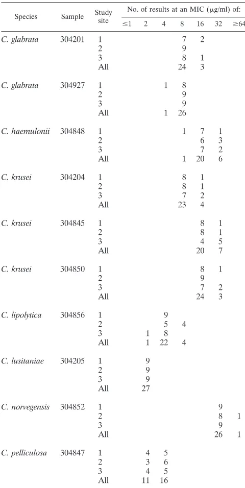
The VITEK 2 fluconazole MIC results were highly
repro-ducible, as determined by replicate testing of a panel of 10
Candida
spp. isolates in the three laboratories (Table 2). Both
intra- and interlaboratory reproducibility was 100% for all 10
organisms. This high level of reproducibility underscores the
excellent level of test standardization achieved with this
auto-mated microbiology system.
CA between the VITEK 2 and BMD methods was
as-sessed by combining the data obtained with the clinical and
challenge organism collections in all three laboratories
(Ta-ble 3). Excellent CA was observed for all comparisons with
the exception of
C. glabrata
and
C. norvegensis
. The overall
CA for the comparison of VITEK 2 results with 24-h BMD
results was 97.2% with no very major or major errors. Only
C. glabrata
(89.2%) and
C. norvegensis
(0.0%) among the 10
species tested showed less than 90% absolute CA with the
24-h BMD results. All of the errors observed with
C.
gla-brata
were minor errors and were the result of isolates
[image:3.585.47.541.89.511.2]determined to be susceptible by BMD and SDD by VITEK
2. This is not surprising given the tendency of fluconazole
MICs for this species to fall close to the susceptible
break-point: 82% of MICs determined by VITEK 2 and 78% of
MICs determined by 24-h BMD fell between 4 and 16
g/ml
(data not shown). Importantly, all isolates of
C. glabrata
testing as resistant by the BMD method at 24 h were also
TABLE 1. Fluconazole susceptibilities of 426 isolates of
Candida
spp. as determined by the VITEK 2 yeast
susceptibility test and by CLSI BMD methods
aSpecies No. tested Test methodb MIC (g/ml)
c
EA (%)d
Range 50% 90%
C. albicans
198
VITEK 2
ⱕ
1–
ⱖ
64
ⱕ
1
ⱕ
1
BMD-24
ⱕ
0.12–
ⱖ
128
0.25
0.5
97.0
BMD-48
ⱕ
0.12–
ⱖ
128
0.25
0.5
96.5
C. dubliniensis
8
VITEK 2
ⱕ
1–32
ⱕ
1
BMD-24
ⱕ
0.12–16
ⱕ
0.12
100.0
BMD-48
ⱕ
0.12–16
0.25
100.0
C. glabrata
83
VITEK 2
ⱕ
1–
ⱖ
64
8
ⱖ
64
BMD-24
0.5–
ⱖ
128
4
64
98.8
BMD-48
0.5–
ⱖ
128
16
ⱖ
128
96.4
C. guilliermondii
3
VITEK 2
ⱕ
1–4
2
BMD-24
1–4
4
100.0
BMD-48
2–4
4
100.0
C. krusei
51
VITEK 2
ⱕ
1–
ⱖ
64
16
32
BMD-24
ⱕ
1–
ⱖ
128
16
32
100.0
BMD-48
ⱕ
1–
ⱖ
128
64
ⱖ
128
68.6
C. lusitaniae
9
VITEK 2
ⱕ
1–2
1
BMD-24
ⱕ
0.12–1
0.5
100.0
BMD-48
ⱕ
0.12–2
0.5
100.0
C. norvegensis
1
VITEK 2
8
BMD-24
16
100.0
BMD-48
16
100.0
C. parapsilosis
43
VITEK 2
ⱕ
1–
ⱖ
64
ⱕ
1
2
BMD-24
0.25–4
0.5
1
95.3
BMD-48
0.5–4
1
2
97.7
C. pelliculosa
2
VITEK 2
2–4
2
BMD-24
2–4
2
100.0
BMD-48
4
4
100.0
C. tropicalis
28
VITEK 2
ⱕ
1–8
ⱕ
1
ⱕ
1
BMD-24
ⱕ
0.12–8
0.25
1
100.0
BMD-48
0.25–16
0.5
4
100.0
All
Candida
spp.
426
VITEK 2
ⱕ
1–
ⱖ
64
ⱕ
1
16
BMD-24
ⱕ
0.12–
ⱖ
128
0.5
16
97.9
BMD-48
ⱕ
0.12–
ⱖ
128
0.5
64
93.7
a
Isolates included both clinical (n⫽346) and challenge (n⫽80) sets.
b
BMD-24 and BMD-48, BMD performed at 24 h and 48 h of incubation, respectively.
c
50% and 90%, MICs encompassing 50% and 90% of isolates tested, respectively.
d
% EA is the EA (⫾2 log2dilutions) between VITEK 2 and BMD MICs.
on May 16, 2020 by guest
http://jcm.asm.org/
resistant with the VITEK 2 system. Only one isolate of
C.
norvegensis
was tested and was found to be susceptible by
VITEK 2 (MIC
⫽
8
g/ml) and SDD by 24-h BMD (MIC
⫽
16
g/ml).
Although the overall CA between VITEK 2 and the 48-h
BMD results was lower than that seen with the 24-h BMD
comparison (88.3% versus 97.2%), the vast majority of
er-rors were minor, and almost all were due to a shift in the
MICs for
C. glabrata
from susceptible at 24 h to SDD at 48 h
with the BMD method (Table 3). There were no ME and
only one VME (0.2% of all isolates and 1.5% of all resistant
isolates), the latter being a
C. glabrata
result. Again, with the
exception of
C. glabrata
and
C. norvegensis
, the CA between
the VITEK 2 and 48-h BMD results exceeded 95% for all
species of
Candida
. All but one of the errors observed with
C. glabrata
were minor and can be attributed to clustering of
fluconazole MICs around the S-SDD breakpoint (66% of
the 48-h BMD results fell in the range from 4 to 16
g/ml,
and 75% were in the range 8 to 32
g/ml) and the influence
of trailing growth seen with this species and fluconazole.
The overall pattern of results shown in Table 3 was also seen
with clinical isolates tested in each of the three laboratories
(Table 4). In each laboratory the categorical agreement was
ⱖ
95% when VITEK 2 results were compared to 24-h BMD
results and ranged from 88.0 to 89.6% with the comparison
between VITEK 2 and 48-h BMD. A shift from S to SDD was
observed in all three laboratories when 24-h BMD results were
compared to 48-h BMD results.
The findings of the present study document the excellent
degree of standardization and reproducibility that can be
achieved with the VITEK 2 yeast susceptibility test. This
sys-tem is the first automated approach to antifungal susceptibility
testing and as such provides the ultimate in test
standardiza-tion. In addition to providing highly reproducible results, the
VITEK 2 system was rapid, with a mean time to result of 13 h.
The availability of rapid quantitative antifungal susceptibility
data will be a major step in optimizing the therapy of invasive
candidal infections (1, 13, 15, 18).
The EA observed in the present study between VITEK 2
and BMD MICs demonstrates excellent quantitative
agree-ment between the methods for all species of
Candida
tested
and is comparable or superior to that reported for other
com-mercial systems (8–11, 16, 17, 24, 25). The highly automated,
hands-off nature of the system virtually eliminates the
subjec-tivity that affects all other test systems.
The use of a spectrophotometer to provide objective, and
earlier, readings of MICs is well established (8, 11, 12, 16).
It is also proposed as a means by which one can mitigate
falsely elevated fluconazole MICs due to trailing growth (2,
7, 8, 11, 12). Based on the comparative data presented
herein, it appears that the VITEK 2 yeast susceptibility test
accomplishes these objectives as well. Thus, although the
CA between the VITEK 2 and the 48-h BMD results was
lower than that observed for the 24-h BMD comparison, it
may be the latter that is more relevant clinically (2, 11, 16).
Earlier work has shown that the 24-h fluconazole MIC
end-point correlated better than the 48-h endend-point with sterol
quantification (2) and with treatment outcome both
clini-cally (29) and in a murine model of invasive candidiasis (31).
These findings suggest that fluconazole results for isolates of
Candida
spp. with significant trailing should be interpreted
on the basis of the lower MIC observed at the earlier (24-h)
time point (16, 20). Given the evidence of trailing, especially
with
C. glabrata
and
C. krusei
, at 48 h shown in Tables 1 and
3, the most appropriate comparator for the VITEK 2 may be
the 24-h BMD results. This comparison indicates that the
VITEK 2 provides highly accurate quantitative and
qualita-tive results for fluconazole and
Candida
spp.
The value of a rapid, automated, commercially available
antifungal susceptibility testing system such as the VITEK 2 is
not limited to the generation of fluconazole susceptibility data.
Recently, we have shown that fluconazole may serve as a
sur-TABLE 2. VITEK 2 Fluconazole MIC reproducibility within and
among three different laboratories
Species Sample Study site
No. of results at an MIC (g/ml) of: ⱕ1 2 4 8 16 32 ⱖ64
C. glabrata
304201
1
7
2
2
9
3
8
1
All
24
3
C. glabrata
304927
1
1
8
2
9
3
9
All
1
26
C. haemulonii
304848
1
1
7
1
2
6
3
3
7
2
All
1
20
6
C. krusei
304204
1
8
1
2
8
1
3
7
2
All
23
4
C. krusei
304845
1
8
1
2
8
1
3
4
5
All
20
7
C. krusei
304850
1
8
1
2
9
3
7
2
All
24
3
C. lipolytica
304856
1
9
2
5
4
3
1
8
All
1
22
4
C. lusitaniae
304205
1
9
2
9
3
9
All
27
C. norvegensis
304852
1
9
2
8
1
3
9
All
26
1
C. pelliculosa
304847
1
4
5
2
3
6
3
4
5
All
11
16
on May 16, 2020 by guest
http://jcm.asm.org/
[image:4.585.42.282.88.566.2]rogate marker for voriconazole susceptibility of
Candida
spp.
(28). Specifically, fluconazole MICs of
ⱕ
32
g/ml predict
sus-ceptibility and MICs of
ⱖ
64
g/ml predict resistance of
Can-dida
spp. to voriconazole with an absolute CA of 97%, 0.1%
VME, and 1.4% ME. Thus, clinical laboratories performing
antifungal susceptibility testing of fluconazole using the VITEK 2
can reliably use these results as surrogate markers of
suscep-tibility and resistance to voriconazole until commercial
FDA-approved voriconazole susceptibility tests become available.
In summary, the MICs of fluconazole can be determined in
an automated fashion in less than 15 h for most species of
Candida
with the VITEK 2 yeast susceptibility test. The
VITEK 2 system ensures that each test is performed in a highly
standardized manner and provides quantitative MIC results
that are reproducible and accurate. The use of
spectrophotom-TABLE 3. Categorical agreement between VITEK 2 yeast susceptibility test MICs and 24-h and 48-h CLSI BMD fluconazole
MICs for 426 isolates of
Candida
spp. in three laboratories
aSpecies (no. of strains
tested) Test method
% of MICs by category
% CA
% Errors
S SDD R VME ME Minor
C. albicans
(198)
VITEK 2
97.5
1.0
1.5
BMD-24
98.0
0.5
1.5
99.5
0.0
0.0
0.5
BMD-48
97.5
1.0
1.5
99.0
0.0
0.0
1.0
C. dubliniensis
(8)
VITEK 2
75.0
25.0
0.0
BMD-24
75.0
25.0
0.0
100.0
0.0
0.0
0.0
BMD-48
75.0
25.0
0.0
100.0
0.0
0.0
0.0
C. glabrata
(83)
VITEK 2
75.9
13.3
10.8
BMD-24
84.3
4.8
10.9
89.2
0.0
0.0
10.8
BMD-48
27.7
59.0
13.3
45.8
1.2
0.0
53.0
C. guilliermondii
(3)
VITEK 2
100.0
0.0
0.0
BMD-24
100.0
0.0
0.0
100.0
0.0
0.0
0.0
BMD-48
100.0
0.0
0.0
100.0
0.0
0.0
0.0
C. krusei
b(51)
VITEK 2
0.0
0.0
100.0
BMD-24
0.0
0.0
100.0
100.0
0.0
0.0
0.0
BMD-48
0.0
0.0
100.0
100.0
0.0
0.0
0.0
C. lusitaniae
(9)
VITEK 2
100.0
0.0
0.0
BMD-24
100.0
0.0
0.0
100.0
0.0
0.0
0.0
BMD-48
100.0
0.0
0.0
100.0
0.0
0.0
0.0
C. norvegensis
(1)
VITEK 2
100.0
0.0
0.0
BMD-24
0.0
100.0
0.0
0.0
0.0
0.0
100.0
BMD-48
0.0
100.0
0.0
0.0
0.0
0.0
100.0
C. parapsilosis
(43)
VITEK 2
97.7
2.3
0.0
BMD-24
100.0
0.0
0.0
97.7
0.0
0.0
2.3
BMD-48
100.0
0.0
0.0
97.7
0.0
0.0
2.3
C. pelliculosa
(2)
VITEK 2
100.0
0.0
0.0
BMD-24
100.0
0.0
0.0
100.0
0.0
0.0
0.0
BMD-48
100.0
0.0
0.0
100.0
0.0
0.0
0.0
C. tropicalis
(28)
VITEK 2
100.0
0.0
0.0
BMD-24
100.0
0.0
0.0
100.0
0.0
0.0
0.0
BMD-48
96.4
3.6
0.0
96.4
0.0
0.0
3.6
All
Candida
spp. (426)
VITEK 2
81.5
3.8
14.7
BMD-24
83.3
2.0
14.7
97.2
0.0
0.0
2.8
BMD-48
71.8
12.9
15.3
88.3
0.2
0.0
11.5
a
Isolates include both clinical (n⫽346) and challenge (n⫽80) sets.
b
[image:5.585.47.542.88.513.2]C. kruseicategorical results forced to R regardless of MICs.
TABLE 4. Agreement between VITEK 2 yeast susceptibility test
and reference BMD MIC results for fluconazole against clinical
isolates of
Candida
spp. in each of three laboratories
Study site(no. of strains tested)
BMD incubation
time (h)
% of BMD by
category % CA % Errors
S SDD R VME ME Minor
1 (135)
24
86.0
0.7
13.3
98.5
0.0
0.0
1.5
48
75.6
11.1
13.3
89.6
0.0
0.0
10.4
2 (108)
24
81.5
0.9
17.6
97.2
0.0
0.0
2.8
48
69.4
11.1
19.5
88.0
0.9
0.0
11.1
3 (103)
24
79.6
2.9
17.5
95.1
0.0
0.0
4.9
48
70.0
12.5
17.5
89.3
0.0
0.0
10.7
on May 16, 2020 by guest
http://jcm.asm.org/
[image:5.585.44.283.607.724.2]etry to determine the MIC endpoint eliminates subjectivity and
minimizes the effect of trailing growth that compromises the
performance of systems relying on visual MIC determination.
The VITEK 2 system reliably identifies fluconazole resistance
among
Candida
spp. and demonstrates excellent quantitative
and qualitative agreement with the reference BMD method.
The introduction of the VITEK 2 system in the clinical
labo-ratory will be an important step toward the optimization of
antifungal therapy of candidiasis.
ACKNOWLEDGMENTS
This study was supported in part by a grant from bioMerieux, Inc.
We thank Linda Elliott for secretarial assistance in the preparation
of the manuscript. We acknowledge the excellent assistance of the
technical personnel in the Iowa, Ohio, and Texas laboratories.
REFERENCES
1.Alexander, B. D., and M. A. Pfaller. 2006. Contemporary tools for the diagnosis and management of invasive mycoses. Clin. Infect. Dis.43(Suppl. 1):S15–S27.
2.Arthington-Skaggs, B. A., W. Lee-Yong, M. A. Ciblak, J. P. Frade, M. E. Brandt, R. A. Hajjeh, L. E. Harrison, A. N. Sofair, and D. W. Warnock.2002. Comparison of visual and spectrophotometric methods of broth microdilu-tion MIC endpoint determinamicrodilu-tion and evaluamicrodilu-tion of sterol quantitamicrodilu-tion method for in vitro susceptibility testing of fluconazole and itraconazole against trailing and non-trailingCandidaisolates. Antimicrob. Agents Che-mother.46:2477–2481.
3.Asmundsdottir, L. R., H. Erlendsdottir, and M. Gottfredsson.2002. Increas-ing incidence of candidemia: results from a 20-year nationwide study in Iceland. J. Clin. Microbiol.40:3489–3492.
4.Aubertine, C. L., M. Rivera, S. M. Rohan, and D. H. Larone.2006. Com-parative study of the new colorimetric VITEK 2 Yeast Identification Card versus the older fluorometric card and of CHROMagar Candida as a source medium with the new card. J. Clin. Microbiol.44:227–228.
5.Barry, A. L., M. A. Pfaller, S. D. Brown, A. Espinel-Ingroff, M. A. Ghan-noum, C. Knapp, R. P. Rennie, J. H. Rex, and M. G. Rinaldi.2000. Quality control limits for broth microdilution susceptibility tests of ten antifungal agents. J. Clin. Microbiol.38:3457–3459.
6.Chen, Y. C., S. C. Chang, K. T. Luh, and W. C. Hsieh.2003. Stable suscep-tibility ofCandidabloodstream isolates to fluconazole despite increasing use during the past 10 years. J. Antimicrob. Chemother.52:71–77.
7.Cuenca-Estrella, M., W. Lee-Yang, M. A. Ciblak, B. A. Arthington-Skaggs, E. Mellado, D. N. Warnock, and J. L. Rodriguez-Tudela.2002. Comparative evaluation of NCCLS M27-A and EUCAST broth microdilution procedures for antifungal susceptibility testing ofCandidaspecies. Antimicrob. Agents Chemother.46:3644–3647.
8.Cuenca-Estrella, M., A. Gomez-Lopez, E. Mellado, and J. L. Rodriguez-Tudela.2005. Correlation between the procedure for antifungal susceptibil-ity testing forCandidaspp. of the European Committee on Antibiotic Sus-ceptibility Testing (EUCAST) and four commercial techniques. Clin. Microbiol. Infect.11:486–492.
9.Espinel-Ingroff, A., M. Pfaller, S. A. Messer, C. C. Knapp, S. Killian, H. A. Norris, and M. A. Ghannoum.1999. Multicenter comparison of the Sensititre YeastOne Colorimetric Antifungal Panel with the National Committee for Clinical Laboratory Standards M27-A reference method for testing clinical isolates of common and emergingCandidaspp., Cryp-tococcusspp., and other yeasts and yeast-like organisms. J. Clin. Micro-biol.37:591–595.
10.Espinel-Ingroff, A., M. Pfaller, S. A. Messer, C. C. Knapp, N. Holliday, and S. B. Killian.2004. Multicenter comparison of the Sensititire YeastOne Colorimetric Antifungal Panel with the NCCLS M27–A2 reference method for testing new antifungal agents against clinical isolates ofCandidaspp. J. Clin. Microbiol.42:718–721.
11.Espinel-Ingroff, A., F. Barchiesi, M. Cuenca-Estrella, A. Fothergill, M. A. Pfaller, M. Rinaldi, J. L. Rodriguez-Tudela, and P. E. Verweij.2005. Com-parison of visual 24-hour and spectrophotometric 48-hour MICs to CLSI reference microdilution MICs of fluconazole, itraconazole, posaconazole, and voriconazole forCandidaspp.: a collaborative study. J. Clin. Microbiol. 43:4535–4540.
12.Espinel-Ingroff, A., F. Barchiesi, M. Cuenca-Estrella, M. A. Pfaller, M. Rinaldi, J. L. Rodriguez-Tudela, and P. E. Verweij.2005. International and multicenter comparison of EUCAST and CLSI M27–A2 broth mi-crodilution methods for testing susceptibilities ofCandidaspp. to flucon-azole, itraconflucon-azole, posaconflucon-azole, and voriconazole. J. Clin. Microbiol. 43:3884–3889.
13.Garey, K. W., M. Reye, M. P. Pai, D. E. Mingo, K. J. Suda, R. S. Turpin, and
D. T. Bearden. 2006. Time to initiation of fluconazole therapy impacts mortality in patients with candidemia: a multi-institutional study. Clin. In-fect. Dis.43:25–31.
14.Hazen, K. C., and S. A. Howell.2003.Candida,Cryptococcus, and other yeasts of medical importance, p. 1693–1711.InP. R. Murray, E. J. Baron, J. H. Jorgensen, M. A. Pfaller, and R. H. Yolken (ed.), Manual of clinical microbiology, 8th ed. ASM Press, Washington, DC.
15.Magill, S. S., C. Shields, C. L. Sears, M. Choti, and W. G. Merz.2006. Triazole-cross resistance among Candidaspp.: occurrence among blood-stream isolates, and implication for antifungal therapy. J. Clin. Microbiol. 44:529–535.
16.Matar, M. J., L. Ostrosky-Zeichner, V. L. Pretznick, J. R. Rodriguez, E. Chan, and J. H. Rex.2003. Correlation between E-test, disk diffusion, and microdilution methods for antifungal susceptibility testing of fluconazole and voriconazole. Antimicrob. Agents Chemother.47:1647–1651.
17.Morace, G., G. Amato, F. Bistoni, G. Fadda, P. Marone, M. Montagna, S. Oliveri, L. Polonelli, R. Rigoli, I. Mancuso, S. La Face, L. Masucci, L. Romano, C. Napoli, D. Tato, M. G. Buscema, C. M. C. Belli, M. M. Piccirillo, S. Conti, S. Covan, F. Fanti, C. Cavanna, F. D’Alo, and L. Pitzurra.2002. Multicenter comparative evaluation of six commercial systems and the Na-tional Committee for Clinical Laboratory Standards M27-A broth microdi-lution method for fluconazole susceptibility testing of Candida species. J. Clin. Microbiol.40:2953–2958.
18.Morrell, M., V. J. Fraser, and M. J. Kollef.2005. Delaying empiric treatment ofCandidabloodstream infection until positive blood culture results are obtained: a potential risk factor for mortality. Antimicrob. Agents Che-mother.49:3640–3645.
19.National Committee for Clinical Laboratory Standards.1997. Reference method for broth dilution antifungal susceptibility testing of yeasts. M27-A. National Committee for Clinical Laboratory Standards, Villanova, PA. 20.National Committee for Clinical Laboratory Standards.2002. Reference
method for broth dilution antifungal susceptibility testing of yeasts: approved standard, 2nd ed., M27–A2. National Committee for Clinical Laboratory Standards, Wayne, PA.
21.Pfaller, M. A.2005. Antifungal susceptibility testing methods. Current Drug Targets6:929–943.
22.Pfaller, M. A., and D. J. Diekema.2004. Twelve years of fluconazole in clinical practice: global trends in species distribution and fluconazole sus-ceptibility of bloodstream isolates of Candida. Clin. Microbiol. Infect. 10(Suppl. 1):11–23.
23.Pfaller, M. A., K. C. Hazen, S. A. Messer, L. Boyken, S. Tendolkar, R. J. Hollis, and D. J. Diekema.2004. Comparison of results of fluconazole disk diffusion testing forCandidaspecies with results from a central reference laboratory in the ARTEMIS Global Antifungal Surveillance Program. J. Clin. Microbiol.42:3607–3612.
24.Pfaller, M. A., L. Boyken, R. J. Hollis, S. A. Messer, S. Tendolkar, and D. J. Diekema.2004. Clinical evaluation of a dried commercially prepared mi-crodilution panel for antifungal susceptibility testing of five antifungal agents againstCandidaspp. andCryptococcus neoformans. Diagn. Microbiol. Infect. Dis.50:113–117.
25.Pfaller, M. A., A. Espinel-Ingroff, and R. N. Jones.2004. Clinical evaluation of the Sensititre YeastOne colorimetric antifungal plate for antifungal sus-ceptibility testing of the new triazoles voriconazole, posaconazole, and ravuconazole. J. Clin. Microbiol.42:4577–4580.
26.Pfaller, M. A, R. N. Jones, the Microbiology Resource Committee of the College of American Pathologists.2006. Performance accuracy of anti-bacterial and antifungal susceptibility test methods: report from the Col-lege of American Pathologists (CAP) Microbiology Surveys Program (2001–2003). Arch. Pathol. Lab. Med.130:767–778.
27.Pfaller, M. A., D. J. Diekema, and D. J. Sheehan. 2006. Intepretive breakpoints for fluconazole andCandidarevisited: a blueprint for the future of antifungal susceptibility testing. Clin. Microbiol. Rev.19:435– 447.
28.Pfaller, M. A., S. A. Messer, L. Boyken, C. Rice, S. Tendolkar, R. J. Hollis, and D. J. Diekema.2007. The use of fluconazole as a “surrogate marker” to predict susceptibility and resistance to voriconazole among 13,338 clinical isolates ofCandidaspp. tested by Clinical and Laboratory Standards Institute recommended broth microdilution methods. J. Clin. Microbiol.45:70–75.
29.Revankar, S. G., W. R. Kirkpatrick, R. K. McAtee, A. W. Fothergill, S. W. Redding, M. G. Rinaldi, and T. F. Patterson. 1998. Interpretation of trailing endpoints in antifungal susceptibility testing by the National Committee for Clinical Laboratory Standards method. J. Clin. Microbiol. 36:153–156.
30.Rex, J. H., M. A. Pfaller, J. N. Galgiani, M. S. Bartlett, A. Espinel-Ingroff, M. A. Ghannoum, M. Lancaster, F. C. Odds, M. G. Rinaldi, T. J. Walsh, and A. L. Barry.1997. Development of interpretive breakpoints for antifungal susceptibility testing: conceptual framework and analysis of in vitro-in vivo correlation data for fluconazole, itraconazole, andCandidainfections. Clin. Infect. Dis.24:235–247.
on May 16, 2020 by guest
http://jcm.asm.org/
31.Rex, J. H., P. W. Nelson, V. L. Paetznick, M. Lozano-Chiu, A. Espinel-Ingroff, and E. J. Anaissie.1998. Optimizing the correlation between results of testing in vitro and therapeutic outcome in vivo for fluconazole by testing critical isolates in a murine model of invasive candidiasis. Antimicrob. Agents Chemother.42:129–134.
32.Rex, J. H., M. A. Pfaller, T. J. Walsh, V. Chaturvedi, A. Espinel-Ingroff, M. A. Ghannoum, L. L. Gosey, F. C. Odds, M. G. Rinaldi, D. J. Sheehan, and
D. W. Warnock.2001. Antifungal susceptibility testing: practical aspects and current challenges. Clin. Microbiol. Rev.14:643–658.
33.Tortorano, A. M., A. L. Rigoni, E. Biraghi, A. Prigatino, M. A. Viviani, et al.2003. The European Confederation of Medical Mycology (ECMM) survey of candidemia in Italy: antifungal susceptibility patterns of 261 non-albicans Candidaisolates from blood. J. Antimicrob. Chemother. 52:679–682.