0095-1137/08/$08.00⫹0 doi:10.1128/JCM.01351-07
Copyright © 2008, American Society for Microbiology. All Rights Reserved.
Emergence of Clonal Complex 17
Enterococcus faecium
in The Netherlands
䌤
Janetta Top,
1* Rob Willems,
1Saskia van der Velden,
1Miranda Asbroek,
1and Marc Bonten
1,2Department of Medical Microbiology, University Medical Center Utrecht, Utrecht, The Netherlands,1and Julius Center for
Health Studies and Primary Care, University Medical Center Utrecht, Utrecht, The Netherlands2
Received 6 July 2007/Returned for modification 11 September 2007/Accepted 22 October 2007
The global emergence of vancomycin-resistant Enterococcus faeciumhas been characterized as the clonal spread of clonal complex 17 (CC17)E. faecium. CC17 was defined upon multilocus sequence typing and is characterized by resistance to quinolones and ampicillin and the presence of the enterococcal surface protein (Esp) in the majority of isolates. The recently noticed increased incidence of vancomycin-susceptible CC17E. faeciuminfections in our hospital initiated a nationwide study to determine ecological changes among entero-coccal infections. The data and strain collections were obtained from 26 (38%) and 9 (14%) of 66 microbiology laboratories in The Netherlands.E. faeciumandE. faecaliswere distinguished by multiplex PCR; allE. faecium
isolates were genotyped by multiple-locus variable-number tandem-repeat analysis (MLVA), and the presence ofesp was identified by PCR. Average numbers of ampicillin-resistant enterococcal isolates from normally sterile body sites per hospital increased from 5 ⴞ 1 in 1994 to 25 ⴞ 21 in 2005. Among all enterococcal bloodstream infections, the proportions of ampicillin-resistantE. faecium(AREF) increased from 4% in 1994 to 20% in 2005 (P< 0.001). AllE. faecalisisolates were susceptible to ampicillin, whereas 78% of theE. faecium
isolates were resistant (49% of these containedesp). Genotyping revealed that 86% of AREF isolates belonged to CC17, including four dominant MLVA types found in>3 hospitals, accounting for 64% of the AREF isolates. Infections caused by CC17E. faeciumhas increased nationwide, especially in university hospitals due to the clonal spread of four MLVA types, and seems associated with acquisition of theespgene.
The emergence of vancomycin-resistant Enterococcus fae-cium(VREF) in the United States in the 1990s was preceded by the emergence of ampicillin-resistantEnterococcus faecium
(AREF) in the 1980s (8, 11, 27, 28). Molecular epidemiological studies of human- and animal-derivedE. faecium since then, revealed the existence of a genetic lineage, labeled clonal com-plex 17 (CC17), associated with nosocomialE. faecium out-breaks and infections in five continents. CC17 is characterized by ampicillin and quinolone resistance and the presence of a putative pathogenicity island, including the esp gene in the majority of isolates (2–4, 9, 12, 17–20, 31, 36). In retrospect, it seems likely that the acquisition of ampicillin resistance was an earlier step in hospital adaptation ofE. faecium, facilitating the subsequent emergence of VREF (18, 36).
Since 2000, infection rates of VREF are rising in European hospitals (see EARSS Annual Report 2005 [www.rivm.nl /earss]), suggesting that the increase of VREF in Europe fol-lows the American epidemiology with a 10-year delay. Little is known, though, about the molecular epidemiology of AREF.
In our hospital (the University Medical Center Utrecht [UMCU]), the proportion of invasive enterococcal infections caused by AREF increased from 2% in 1994 to 32% in 2005, with partial replacement of ampicillin-susceptible (Amps)E.
faecalis by E. faecium (75% AREF) among enterococcal bloodstream infections (32). Based on these local findings, a nationwide study was initiated to determine the ecological
changes among enterococcal infections from sterile body sites in hospitals in The Netherlands.
MATERIALS AND METHODS
Microbiology data.All microbiology laboratories (n⫽66) serving 9 university and 87 nonuniversity hospitals in The Netherlands were invited to submit data on
annual numbers of ampicillin-resistant (Ampr
) enterococci isolated from nor-mally sterile body sites identified between 1994 and 2005. Nornor-mally sterile body sites included blood, abdominal and cerebrospinal fluid, intravascular catheter tips, and pus and wound specimens. These data did not differentiate enterococci to the species level.
Furthermore, the laboratories were invited to provide, for each year, the first 30 enterococcal bloodstream isolates, irrespective of antibiotic susceptibility (1
per patient). A species-specific multiplex PCR based on theddlgene was
per-formed to distinguishE. faeciumandE. faecalisas previously described (6, 32).
Susceptibilities to ampicillin were determined by inoculation of Mueller-Hinton agar containing ampicillin at 16 mg/liter according to Clinical and Laboratory Standards Institute (formerly the National Committee for Clinical Laboratory Standards) guidelines.
Genotyping ofE. faecium isolates.AllE. faeciumisolates, including 2006 isolates, were genotyped by using multiple-locus variable-number tandem-repeat (VNTR) analysis (MLVA), as described previously (31) with minor modifica-tions (www.mlva.umcutrecht.nl). Identification of CC17-specific MLVA types (MTs) was performed by comparing each MLVA profile to the previously de-scribed seven different repeat combinations for VNTR-7, -8, and -10 with a positive predictive value of 87% and a specificity of 90% to belong to CC17 (31). The genetic relatedness of MTs was confirmed by multilocus sequence typing (MLST) on a subset of representative isolates (9). The obtained MLST profiles were clustered with 313 MLST profiles, representing 855 isolates from the da-tabase using the eBURST algorithm (7, 18). The presence of the putative
patho-genicity island was determined by PCR using theespgene as a marker (20).
Statistical analysis.Statistical analysis of the data was performed with SPSS 12.0.1 for Windows (SPSS, Inc., Chicago, IL) using the chi-square test. The data from university hospitals were compared to those from nonuniversity hospitals.
RESULTS
Microbiology data invasive Ampr
enterococci.Of 66 micro-biology laboratories serving 7 of 9 (78%) university hospitals
* Corresponding author. Mailing address: Department of Medical Mi-crobiology, University Medical Center Utrecht, G04.614, P.O. Box 85500, 3508 GA Utrecht, The Netherlands. Phone: 31-88-7557627. Fax: 31-30-2541770. E-mail: [email protected].
䌤Published ahead of print on 31 October 2007.
214
on May 16, 2020 by guest
http://jcm.asm.org/
(⬎500 beds) and 22 of 87 (25%) nonuniversity hospitals (250 to 500 beds [n⫽6],⬎500 beds [n⫽16]), 26 (39%) provided data on Amprenterococci from normally sterile body sites. The
data from our own hospital, already described previously (32), were included as well. The hospitals were dispersed through-out The Netherlands. Only one nonuniversity and three uni-versity hospitals could provide data going back as far as 1994.
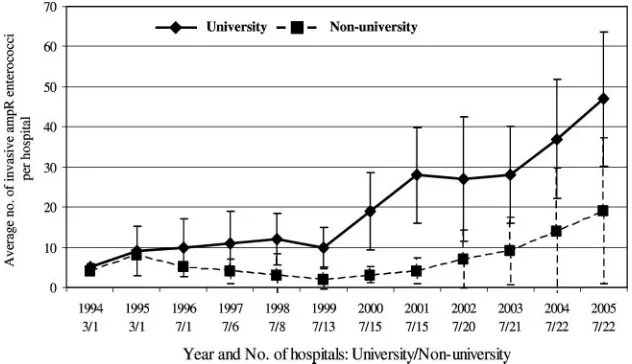
Average annual numbers of Ampr enterococci from
nor-mally sterile body sites per hospital increased from 5⫾ 1 in 1994 to 25⫾21 in 2005. The increase was most pronounced in university hospitals (from 5⫾1 in 1994 to 47⫾17 in 2005) (Fig. 1). The average annual numbers in nonuniversity hospi-tals increased from 4⫾0 in 1994 to 19⫾18 in 2005 (Fig. 1). Annual numbers per hospital varied between 1 and 14 for 250-to 500-bed hospitals and between 1 and 80 for larger hospitals (⬎500 beds).
E. faecium/E. faecalisratio among bloodstream isolates.In all, 1,573 enterococcal bloodstream isolates were obtained from nine hospitals (five nonuniversity and four university). Three of the four university hospitals provided isolates from 1994 onward. The oldest isolates obtained from a nonuniver-sity hospital were from 1999.
Species identification revealed 1,121E. faecalis, 303E. fae-cium, and 149 non-E. faecalisand non-E. faeciumisolates. The latter isolates were not further characterized. Discrepancies between the original identification, as provided by the submit-ting labs and identification based on theddlgene, were found in 116 (7%) isolates. All E. faecalis isolates were Amps,
whereas 237 of 303 (78%)E. faeciumisolates were Ampr.
The proportions of AREF among all enterococcal blood-stream isolates increased from 4% (1994) to 20% (2005) (P⫽
0.01), while the proportions of Amps E. faecalis decreased
from 89% (1994) to 77% (2005) (P ⫽ 0.5). Proportions of
Amps E. faecium remained ⬍9%, and no significant trend
could be observed over time. In university hospitals the pro-portions of AREF increased from 4% in 1994 to 27% in 2005 (P⬍0.001). For individual hospitals these proportions ranged from 0% in 1994 to 10% in 2005 (lowest) and from 27% in 1996 to 43% in 2005 (highest). In nonuniversity hospitals there
was a slight, but nonsignificant increase in the proportions of AREF from 6% in 1999 to 12% in 2005.
Genotyping E. faecium isolates. MLVA typing of 303 E. faeciumisolates revealed 61 different MTs among 263 isolates, including 41 MTs that had not been previously detected. In-complete MLVA profiles were obtained for 29 isolates due to repeatedly negative PCR results forⱖ1 of the VNTR loci. In 11 isolates none of the VNTR loci were PCR positive. All 40 isolates that could not be assigned a MT appeared to be Amps.
Twenty of the remaining 26 (77%) AmpsE. faeciumisolates
yielded a unique MT.
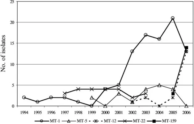
Sixty-seven percent (175 of 263) of typeable isolates be-longed to five MTs, including four MTs detected inⱖ3 hospi-tals: MT-1 (n⫽97 in 9 hospitals) MT-5 (n⫽19 in 5 hospitals),
MT-12 (n ⫽ 18 in 6 hospitals), and MT-159 (n ⫽ 17 in 5
hospitals), together accounting for 64% (151 of 237) of Ampr
isolates (Table 1). MT-22 (24 of 303 isolates) was detected in only one hospital, where it accounted for 29% (24 of 83) of all
E. faeciumisolates between 1999 and 2003 (Fig. 2).
Longitudinal analysis of the genotyping data revealed that MT-1 was already present in one hospital in 1994 and that its presence increased after 1999, with a documented presence in all nine hospitals (Fig. 2 and Table 1). MT-5 and MT-12 emerged from 1999 and 2002 onward (Fig. 2). The first MT-12 isolate was detected in one hospital in 2002, and it appeared in three other hospitals in 2006 (Table 1). Finally, MT-159 was found in two hospitals in 2005, with subsequent isolation in three additional hospitals in 2006.
The four most predominant MTs detected inⱖ3 hospitals
were closely related. MT-5 and MT-12 were single-locus vari-ants from MT-1, while MT-159 was a double-locus variant from MT-1 and a single-locus variant from MT-12 (Table 1). Identification of CC17-specific MTs based on different repeat combinations for VNTR-7, -8, and -10 (31) revealed that 86% (204 of 237) of Amprisolates belonged to CC17 (Table 1).
MLST was performed on 15 Ampr and 12 Amps isolates
[image:2.585.134.450.69.251.2](Table 2). The seven MT-159 isolates of different hospitals revealed a single sequence type (ST), ST-78. In contrast, seven MT-12 isolates from different years represented five different
FIG. 1. Average annual numbers of invasive Amprenterococci per hospital. Error bars denote standard deviations. University and
nonuni-versity hospitals were compared. For each year, the numbers of hospitals that provided data are indicated.
on May 16, 2020 by guest
http://jcm.asm.org/
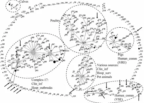
STs. All STs representing Amprisolates, except one (ST-324),
grouped within or were linked to CC17 (Fig. 3). The AmpsE.
faecium isolates, all MLVA nontypeable, revealed different STs, including seven new STs, ST-326 to -332, ST-334, and ST-100, -52, -272, and -296. Six STs clustered with other am-picillin- and vancomycin-susceptible human community iso-lates, including MLVA nontypeableE. faeciumisolates, four represented singletons, and one isolate grouped among poultry isolates, and one ST (ST-326) was linked to CC17 (Fig. 3).
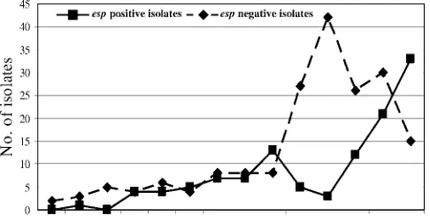
Determination ofespgene.Forty-nine percent (115 of 237) of AmprE. faeciumisolates contained theespgene, while none
of the Ampsisolates wasesppositive. In longitudinal analysis a
remarkable increase of esp-positive isolates occurred from 2004 onward (Fig. 4). The total numbers ofesp-negative iso-lates peaked in 2003 (n ⫽ 40) and decreased subsequently. Interestingly, all MT-12 isolates from 2002 and 2003 wereesp
negative, whereas all MT-12 isolates from 2005 onward con-tained theespgene. Similarly, the majority ofesp-positive iso-lates among MT-1 isoiso-lates (15 of 19 [79%]) were found be-tween 2004 and 2006. Before 2004, only 4 of 47 MT-1 isolates (9%) wereesppositive. Finally, 17 of 19 MT-5 isolates (89%) and all MT-159 isolates wereesppositive. These findings sug-gest that MT-1 and MT-12 isolates acquired theespgene and that the presence of this gene was associated with nosocomial spread. On the other hand,esp-positive MT-22 isolates were only found in one hospital and apparently disappeared in 2003,
and MT-5esp-positive isolates were detected in low numbers in five hospitals, without evidence of increased nosocomial spread during the years (Table 1).
DISCUSSION
The present study demonstrates a nationwide increase of CC17 AREF isolates obtained from normally sterile body sites in The Netherlands. The molecular epidemiology is character-ized by the emergence of several clones, with presumed intra-and interhospital spread. The presence of theespgene, previ-ously described as a marker of a putative pathogenicity island, seems strongly associated with the emergence of CC17 AREF.
The partial replacement of AmpsE. faecalisby CC17 AREF
has consequences for antimicrobial treatment of enterococcal infections and, more importantly, may set the stage for the emergence of vancomycin-resistantE. faecium.
Our study was based on the voluntary collaboration of mi-crobiological laboratories in The Netherlands and therefore has some potential limitations. In all, 39% of all laboratories provided information on annual numbers of Amprenterococci
[image:3.585.45.542.82.164.2]obtained from normally sterile body sites. Eight laboratories did not have computerized data information, and thirty hospi-tals never responded to our (once-repeated) request. Since we failed to obtain information from all laboratories, some selec-tion bias cannot be fully excluded. However, only one of the
[image:3.585.135.449.513.715.2]FIG. 2. Annual distribution of five predominant MTs. TABLE 1. Distribution of predominant MTs
MT
MLVA profile (no. of repeats) CC17-specific
MTsa
Total no. of isolates
No. of Ampr
isolates
No. of
esp-positive
isolates
No. of hospitals
VNTR-1 VNTR-2 VNTR-7 VNTR-8 VNTR-9 VNTR-10
1 5 7 3 3 2 3 ⫹ 97 97 19 9
5 5 7 3 2 2 3 ⫹ 19 19 17 5
12 5 7 3 3 1 3 ⫹ 18 18 16 6
22 5 7 4 2 2 1 – 24 24 24 1
159 5 7 3 3 1 2 ⫹ 17 17 17 5
a⫹
, MTs with the following repeat profiles for VNTR-7, -8, and -10: 3-3-3, 3-2-3, 3-3-2, 4-3-3, 3-4-3, 4-2-3, and 3-3-1.
on May 16, 2020 by guest
http://jcm.asm.org/
participating hospitals (large nonuniversity hospital) had iden-tified a nosocomial outbreak with AREF before our study request. On the other hand, three hospitals that did not par-ticipate reported the emergence of AREF infections (all MT-159) in 2006 (unpublished data).
Furthermore, one of the participating nonuniversity labora-tories, representing 4.4% of the total nonuniversity isolates and 1.9% of the total number of isolates, could not provide information on isolation sites and, therefore, some isolates might not reflect invasive infections. However, this would
ac-TABLE 2. MLST results for representative MTs
Resistance MT (no. of isolates typed) No. of
hospitals
esp
gene Yr MLST
Ampr MT-159 (7) 5 ⫹ 2005/2006 ST-78
MT-12 (3) 3 – 2002/2003 ST-18, ST-324, ST-325
MT-12 (1) 1 ⫹ 2005 ST-78
MT-12 (3) 3 ⫹ 2005/2006 ST-117
MT-22 (1) 1 ⫹ 2000 ST-16
Amps Nontypeable (12) 8 – NAa New: ST-326 to ST-332,
334; 100, ST-52, ST-272, ST-296
a
NA, not applicable.
FIG. 3. eBURST clustering of 18 MLST profiles, indicated by an arrow, representing 27 isolates from present study, with 313 MLST profiles representing 855E. faeciumisolates from the database (www.mlst.net). Each ST is represented as a node; the relative size of each node is indicative of its prevalence among the isolates, and lines connect single-locus variants (STs that differ in only one of the seven housekeeping genes). Dashed lines indicate connections between double-locus variants. The sources of specific clusters of STs are indicated, including CC17 comprising hospital outbreaks and clinical isolates. Clin_inf, isolates from clinical sites (mainly blood) from hospitalized patients; Hosp_outbreak, hospital outbreak isolates; Hosp_surv, feces isolates from hospitalized patients without an enterococcal infection and not associated with an enterococcal hospital outbreak; Human_comm, feces isolates from human volunteers not connected to hospitals.
on May 16, 2020 by guest
http://jcm.asm.org/
[image:4.585.47.547.303.657.2]count only for urine isolates, since surveillance for asymptom-atic carriage with Amprenterococci had not been performed in
any hospital. Few laboratories had stored enterococcal isolates, and nine could provide enterococcal bloodstream isolates. It is highly unlikely that hospitals preferably stored eitherE. faeca-lisorE. faeciumisolates and, therefore, the reported propor-tions of AREF probably reflect an unbiased estimate. For all of these reasons, we consider the present study to be a reliable reflection of the enterococcal epidemiology in The Nether-lands.
The increase and replacement of AREF was most pro-nounced in university hospitals and large nonuniversity hospi-tals (⬎500 beds), a finding that probably reflects differences in patient populations, compared to smaller hospitals. Hematol-ogy and transplant patients are generally considered at highest risk for enterococcal bacteremia (5, 32). In our hospital, the increase in AREF bloodstream infections was associated with increased fecal carriage of AREF among hospitalized patients (32). Point-prevalence studies revealed intestinal colonization with AREF in up to 35% of hospitalized patients, especially among high-risk patients on hematology and nephrology wards. Although colonization data are absent for other centers, the endemicity of intestinal colonization with AREF has prob-ably been established in multiple hospitals in The Netherlands. MLVA typing revealed four highly related types to be
re-sponsible for the nationwide emergence of AREF. MT-159E.
faeciumisolates first appeared in one hospital in 2005, with documented presence in five hospitals in 2006. However, out-breaks of AREF documented in three other hospitals and not included in the present study were also caused by MT-159 isolates (data not shown). MLST of representative MT-159 isolates (also from the three hospitals not included in the study) revealed ST-78. Nosocomial outbreaks of ST-78 have been described in Korea and Europe, including Germany and Italy (1, 14, 16, 24).
Interestingly, the majority (57%) of esp-positive isolates were found from 2004 onward, and this gene was contained in MT-1 and its genetically related variants MT-12 and -159. We consider theespgene to be a marker of a putative pathoge-nicity island (17). This sudden increase ofesp-positive isolates suggests that MT-1 and MT-12 acquired the putative pathoge-nicity island via conjugative transfer, as has been shown in vitro (23), which might contribute to an increased ability to spread
and cause infections. In a recent study, Esp expression on the surface ofE. faeciumvaried substantially between isolates and was correlated with initial adherence to polystyrene and bio-film formation (35). Therefore, a role for Esp in the early stages of colonization and subsequent infection has been hy-pothesized (35).
Previously, MLST of several MT-1 isolates indicated that MT-1 is comprised of multiple STs, including ST-17, the pre-sumed founder of CC17, thus representing a polyclonal pop-ulation (31, 36). The observation that particular MTs, such as the MT-12 isolates from the present study, are represented by different types determined by MLST and vice versa has been reported before (31), and probably results from differences in the frequency in occurrence of changes in repeat numbers compared to DNA polymorphisms, mutation, and recombina-tion in housekeeping genes.
MLVA typing of 40 AmpsE. faeciumisolates revealed
in-complete MLVA profiles. Southern blot hybridization of three representative isolates confirmed the absence of at least one of the VNTR regions (data not shown). MLST of 12 MLVA nontypeable isolates confirmed that the AmpsE. faecium
iso-lates are not linked to CC17 but clustered with other MLVA nontypeable AmpsE. faeciumisolates that were not involved in
hospital outbreaks.
In the United States, the emergence of AREF preceded the nationwide nosocomial epidemic of vancomycin-resistant en-terococci. A changingE. faecalis/E. faeciumratio in hospital infections was reported in three longitudinal microbiology-based studies (10, 22, 34). In Europe, several reports on the increase in invasive AREF have been published (4, 29, 33), but to our knowledge ours is the first nationwide study in Europe on the molecular epidemiology of AREF. The emergence of CC17 AREF, resulting in changingE. faecalis/E. faeciumratios among bloodstream isolates, and with 78% ofE. faecium
iso-lated determined to be Ampr will impact the treatment of
enterococcal infections. The preferred antibiotic for invasive enterococcal infections, amoxicillin, must now be replaced by vancomycin, linezolid, or daptomycin. Increased use of these agents may create selective antibiotic pressure, facilitating the emergence of VREF due to the horizontal transfer of vanco-mycin resistance genes (13, 30, 32), to mutations leading to resistance to linezolid (15, 26, 37), or to an as-yet-undescribed resistance to daptomycin (21, 25).
ACKNOWLEDGMENTS
We thank the following individuals and laboratories in The Nether-lands for providing data and/or isolates: J. H. Sloos, Laboratory for Med-ical Microbiology, MedMed-ical Center, Alkmaar; L. Spanjaard, MedMed-ical Mi-crobiology, Academic Medical Center, Amsterdam; E. R. Heddema, Medical Microbiology, VU Medical Center, Amsterdam; Medical Micro-biology and Infection Prevention, Gelre Hospitals, Apeldoorn; E. Mas-cini, Medical Microbiology, Rijnstate Arnhem; P. Willemse, Laboratory for Medical Microbiology and Infection Prevention, Amphia Hospital, Breda; R. Brimicombe, Haga Hospitals, The Hague; F. W. Sebens, Lab-oratory for Medical Microbiology and Infectious Diseases, Deventer; M. A. Schouten, Medical Microbiology, Gelderse Vallei Hospital, Ede; E. Roelofsen, Laboratory for Microbiology, Enschede; A. Demeulemeester, Laboratory for Medical Microbiology and Immunology, Goes; E. Mooi-Kokenberg, Medical Microbiology Groene Hart Hospital, Gouda; K. E. Veldkamp, Laboratory for Infectious Diseases, Groningen; J. P. Arends, Medical Microbiology, University Medical Centre, Groningen; C. G. van Mameren, Medical Microbiology, St. Jansdal Harderwijk; J. H. T. Wag-envoort, Atrium Medical Centre, Heerlen; M. A. Muijsken, Medical Mi-FIG. 4. Comparison of the annual distribution ofesp-positive and
-negative isolates.
on May 16, 2020 by guest
http://jcm.asm.org/
[image:5.585.43.286.70.196.2]crobiology Westfriesgasthuis, Hoorn; J. H. van Zeijl, Public Health Lab-oratory Friesland, Leeuwarden; L. Dijkshoorn, Department of Infectious Disease, Leiden University Medical Center, Leiden; B. M. de Jongh, Medical Microbiology and Immunology St. Antonius Hospital, Nieu-wegein; P. Sturm, Medical Microbiology, University Medical Center, Ni-jmegen; H. Dahmen, Medical Microbiology, Laurentius Hospital, Roer-mond; A. van Belkum, Medical Microbiology, Erasmus Medical Center, Rotterdam; A. Buiting, Medical Microbiology, St. Elisabeth Hospital Til-burg; Medical Microbiology, Veldhoven; and J. Berkhout, Medical Mi-crobiology, VieCuri Medical Center, Venlo.
REFERENCES
1.Bonora, M. G., M. Ligozzi, M. De Fatima, L. Bragagnolo, A. Goglio, G. C. Guazzotti, and R. Fontana.2004. Vancomycin-resistantEnterococcus
fae-ciumisolates causing hospital outbreaks in northern Italy belong to the
multilocus sequence typing C1 lineage. Microb. Drug Resist.10:114–123.
2.Bonten, M. J., R. Willems, and R. A. Weinstein.2001. Vancomycin-resistant enterococci: why are they here, and where do they come from? Lancet Infect.
Dis.1:314–325.
3.Coque, T. M., R. Willems, R. Canton, R. Del Campo, and F. Baquero.2002.
High occurrence ofespamong ampicillin-resistant and
vancomycin-suscep-tibleEnterococcus faeciumclones from hospitalized patients. J. Antimicrob.
Chemother.50:1035–1038.
4.Coque, T. M., R. J. Willems, J. Fortun, J. Top, S. Diz, E. Loza, R. Canton, and F. Baquero.2005. Population structure ofEnterococcus faeciumcausing bacteremia in a Spanish university hospital: setting the scene for a future increase in vancomycin resistance? Antimicrob. Agents Chemother.
49:2693–2700.
5.Dubberke, E. R., J. M. Hollands, P. Georgantopoulos, K. Augustin, J. F. DiPersio, L. M. Mundy, and H. J. Khoury. 2006. Vancomycin-resistant enterococcal bloodstream infections on a hematopoietic stem cell transplant
unit: are the sick getting sicker? Bone Marrow Transplant.38:813–819.
6.Dutka-Malen, S., S. Evers, and P. Courvalin.1995. Detection of glycopep-tide resistance genotypes and identification to the species level of clinically
relevant enterococci by PCR. J. Clin. Microbiol.33:24–27.
7.Feil, E. J., B. C. Li, D. M. Aanensen, W. P. Hanage, and B. G. Spratt.2004. eBURST: inferring patterns of evolutionary descent among clusters of re-lated bacterial genotypes from multilocus sequence typing data. J. Bacteriol.
186:1518–1530.
8.Grayson, M. L., G. M. Eliopoulos, C. B. Wennersten, K. L. Ruoff, P. C. De Girolami, M. J. Ferraro, and R. C. Moellering, Jr.1991. Increasing
resis-tance to beta-lactam antibiotics among clinical isolates ofEnterococcus
fae-cium: a 22-year review at one institution. Antimicrob. Agents Chemother.
35:2180–2184.
9.Homan, W. L., D. Tribe, S. Poznanski, M. Li, G. Hogg, E. Spalburg, J. D. Van Embden, and R. J. Willems.2002. Multilocus sequence typing scheme forEnterococcus faecium. J. Clin. Microbiol.40:1963–1971.
10.Iwen, P. C., D. M. Kelly, J. Linder, S. H. Hinrichs, E. A. Dominguez, M. E. Rupp, and K. D. Patil.1997. Change in prevalence and antibiotic resistance ofEnterococcusspecies isolated from blood cultures over an 8-year period.
Antimicrob. Agents Chemother.41:494–495.
11.Jones, R. N., H. S. Sader, M. E. Erwin, S. C. Anderson, et al.1995. Emerging multiply resistant enterococci among clinical isolates. I. Prevalence data from 97 medical center surveillance study in the United States. Diagn.
Microbiol. Infect. Dis.21:85–93.
12.Jureen, R., J. Top, S. C. Mohn, S. Harthug, N. Langeland, and R. J. Willems.
2003. Molecular characterization of ampicillin-resistantEnterococcus
fae-ciumisolates from hospitalized patients in Norway. J. Clin. Microbiol.41:
2330–2336.
13.Kawalec, M., M. Gniadkowski, M. Zaleska, T. Ozorowski, L. Konopka, and W. Hryniewicz.2001. Outbreak of vancomycin-resistantEnterococcus
fae-ciumof the phenotype VanB in a hospital in Warsaw, Poland: probable
transmission of the resistance determinants into an endemic
vancomycin-susceptible strain. J. Clin. Microbiol.39:1781–1787.
14.Klare, I., C. Konstabel, S. Mueller-Bertling, G. Werner, B. Strommenger, C. Kettlitz, S. Borgmann, B. Schulte, D. Jonas, A. Serr, A. M. Fahr, U. Eigner, and W. Witte.2005. Spread of ampicillin/vancomycin-resistantEnterococcus faeciumof the epidemic-virulent clonal complex-17 carrying the genesesp
andhylin German hospitals. Eur. J. Clin. Microbiol. Infect. Dis.24:815–825.
15.Kloss, P., L. Xiong, D. L. Shinabarger, and A. S. Mankin.1999. Resistance mutations in 23 S rRNA identify the site of action of the protein synthesis inhibitor linezolid in the ribosomal peptidyl transferase center. J. Mol. Biol.
294:93–101.
16.Ko, K. S., J. Y. Baek, J. Y. Lee, W. S. Oh, K. R. Peck, N. Lee, W. G. Lee, K. Lee, and J. H. Song.2005. Molecular characterization of vancomycin-resis-tantEnterococcus faeciumisolates from Korea. J. Clin. Microbiol.43:2303– 2306.
17.Leavis, H., J. Top, N. Shankar, K. Borgen, M. Bonten, J. van Embden, and
R. J. Willems. 2004. A novel putative enterococcal pathogenicity island
linked to theespvirulence gene ofEnterococcus faeciumand associated with
epidemicity. J. Bacteriol.186:672–682.
18.Leavis, H. L., M. J. Bonten, and R. J. Willems. 2006. Identification of high-risk enterococcal clonal complexes: global dispersion and antibiotic
resistance. Curr. Opin. Microbiol.9:454–460.
19.Leavis, H. L., R. J. Willems, J. Top, and M. J. Bonten.2006. High-level
ciprofloxacin resistance from point mutations ingyrAandparCconfined to
global hospital-adapted clonal lineage CC17 ofEnterococcus faecium. J. Clin.
Microbiol.44:1059–1064.
20.Leavis, H. L., R. J. Willems, J. Top, E. Spalburg, E. M. Mascini, A. C. Fluit, A. Hoepelman, A. J. de Neeling, and M. J. Bonten.2003. Epidemic and
nonepidemic multidrug-resistantEnterococcus faecium. Emerg. Infect. Dis.
9:1108–1115.
21.Lewis, J. S., A. Owens, J. Cadena, K. Sabol, J. E. Patterson, and J. H. Jorgensen.2005. Emergence of daptomycin resistance inEnterococcus
fae-ciumduring daptomycin therapy. Antimicrob. Agents Chemother.49:1664–
1665.
22.Murdoch, D. R., S. Mirrett, L. J. Harrell, J. S. Monahan, and L. B. Reller.
2002. Sequential emergence of antibiotic resistance in enterococcal
blood-stream isolates over 25 years. Antimicrob. Agents Chemother.46:3676–3678.
23.Oancea, C., I. Klare, W. Witte, and G. Werner.2004. Conjugative transfer of
the virulence gene,esp, among isolates ofEnterococcus faeciumand
Entero-coccus faecalis. J. Antimicrob. Chemother.54:232–235.
24.Peta, M., E. Carretto, D. Barbarini, A. Zamperoni, L. Carnevale, L. Perversi, M. Pagani, M. G. Bonora, R. Fontana, P. Marone, and M. Langer.2006.
Outbreak of vancomycin-resistantEnterococcusspp. in an Italian general
intensive care unit. Clin. Microbiol. Infect.12:163–169.
25.Poutsiaka, D. D., S. Skiffington, K. B. Miller, S. Hadley, and D. R. Snydman.
2007. Daptomycin in the treatment of vancomycin-resistantEnterococcus
faeciumbacteremia in neutropenic patients. J. Infect.54:567–571. 26.Prystowsky, J., F. Siddiqui, J. Chosay, D. L. Shinabarger, J. Millichap, L. R.
Peterson, and G. A. Noskin.2001. Resistance to linezolid: characterization of mutations in rRNA and comparison of their occurrences in
vancomycin-resistant enterococci. Antimicrob. Agents Chemother.45:2154–2156.
27.Rice, L. B.2001. Emergence of vancomycin-resistant enterococci. Emerg.
Infect. Dis.7:183–187.
28.Shepard, B. D., and M. S. Gilmore.2002. Antibiotic-resistant enterococci: the mechanisms and dynamics of drug introduction and resistance. Microbes.
Infect.4:215–224.
29.Simonsen, G. S., L. Smabrekke, D. L. Monnet, T. L. Sorensen, J. K. Moller, K. G. Kristinsson, A. Lagerqvist-Widh, E. Torell, A. Digranes, S. Harthug, and A. Sundsfjord.2003. Prevalence of resistance to ampicillin, gentamicin
and vancomycin inEnterococcus faecalisandEnterococcus faeciumisolates
from clinical specimens and use of antimicrobials in five Nordic hospitals. J.
Antimicrob. Chemother.51:323–331.
30.Suppola, J. P., E. Kolho, S. Salmenlinna, E. Tarkka, J. Vuopio-Varkila, and M. Vaara.1999.vanAandvanBincorporate into an endemic
ampicillin-resistant vancomycin-sensitiveEnterococcus faeciumstrain: effect on
inter-pretation of clonality. J. Clin. Microbiol.37:3934–3939.
31.Top, J., L. M. Schouls, M. J. Bonten, and R. J. Willems.2004. Multiple-locus variable-number tandem repeat analysis, a novel typing scheme to study the
genetic relatedness and epidemiology of Enterococcus faecium isolates.
J. Clin. Microbiol.42:4503–4511.
32.Top, J., R. Willems, H. Blok, M. de Regt, K. Jalink, A. Troelstra, B. Goorhuis, and M. Bonten.2007. Ecological replacement ofEnterococcus faecalisby multiresistant clonal complex 17Enterococcus faecium. Clin.
Mi-crobiol. Infect.13:316–319.
33.Torell, E., O. Cars, B. Olsson-Liljequist, B. M. Hoffman, J. Lindback, and L. G. Burman.1999. Near absence of vancomycin-resistant enterococci but high carriage rates of quinolone-resistant ampicillin-resistant enterococci among hospitalized patients and nonhospitalized individuals in Sweden.
J. Clin. Microbiol.37:3509–3513.
34.Treitman, A. N., P. R. Yarnold, J. Warren, and G. A. Noskin.2005. Emerging
incidence ofEnterococcus faeciumamong hospital isolates (1993 to 2002).
J. Clin. Microbiol.43:462–463.
35.van Wamel, W. J., A. P. Hendrickx, M. J. Bonten, J. Top, G. Posthuma, and R. J. Willems.2007. Growth condition-dependent Esp expression by Entero-coccus faeciumaffects initial adherence and biofilm formation. Infect.
Im-mun.75:924–931.
36.Willems, R. J., J. Top, M. van Santen, D. A. Robinson, T. M. Coque, F. Baquero, H. Grundmann, and M. J. Bonten.2005. Global spread of
vanco-mycin-resistantEnterococcus faeciumfrom distinct nosocomial genetic
com-plex. Emerg. Infect. Dis.11:821–828.
37.Woodford, N., L. Tysall, C. Auckland, M. W. Stockdale, A. J. Lawson, R. A. Walker, and D. M. Livermore.2002. Detection of oxazolidinone-resistant
Enterococcus faecalisandEnterococcus faeciumstrains by real-time PCR and PCR-restriction fragment length polymorphism analysis. J. Clin. Microbiol.
40:4298–4300.