Samples by Use of the Previ Isola System Compared to Manual
Inoculation in a Routine Laboratory: Finding a Cost-Effective and
Accurate Approach
Alexander Mischnik,aMarkus Mieth,bCornelius J. Busch,cStefan Hofer,cand Stefan Zimmermanna
Department of Infectious Diseases, Medical Microbiology and Hygiene, Heidelberg University Hospital, Heidelberg, Germanya; Department of General and Transplant Surgery, Heidelberg University Hospital, Heidelberg, Germanyb; and Department of Anesthesiology, Heidelberg University Hospital, Heidelberg, Germanyc
Automation of plate streaking is ongoing in clinical microbiological laboratories, but evaluation for routine use is mostly open. In the present study, the recovery of microorganisms from the Previ Isola system plated polyurethane (PU) swab samples is com-pared to manually plated control viscose swab samples from wounds according to the CLSI procedure M40-A (quality control of microbiological transport systems). One hundred twelve paired samples (224 swabs) were analyzed. In 80/112 samples (71%), concordant culture results were obtained with the two methods. In 32/112 samples (29%), CFU recovery of microorganisms from the two methods was discordant. In 24 (75%) of the 32 paired samples with a discordant result, Previ Isola plated PU swabs were superior. In 8 (25%) of the 32 paired samples with a discordant result, control viscose swabs were superior. The quality of colony growth on culture media for further investigations was superior with Previ Isola inoculated plates compared to manual plating techniques. Gram stain results were concordant between the two methods in 62/112 samples (55%). In 50/112 samples (45%), the results of Gram staining were discordant between the two methods. In 34 (68%) of the 50 paired samples with discor-dant results, Gram staining of PU swabs was superior to that of control viscose swabs. In 16 (32%) of the 50 paired samples, Gram staining of control viscose swabs was superior to that of PU swabs. We report the first clinical evaluation of Previ Isola automated specimen inoculation for wound swab samples. This study suggests that use of an automated specimen inoculation system has good results with regard to CFU recovery, quality of Gram staining, and accuracy of diagnosis.
A
ppropriate specimen collection and transport are important for accurate laboratory diagnosis of bacterial infections. To provide correct and rapid identification of pathogens, automation in clinical laboratories is ongoing to improve and accelerate de-tection of infectious agents. Swabs are often used in clinical labo-ratories (4,11), although aspirates of fluids and exudates are su-perior to samples collected on swabs (2). The gold standard is still culture of microorganisms to perform susceptibility testing. Gram staining is a critical test for the rapid presumptive diagnosis of infectious agents and serves to assess the quality of clinical speci-mens. The timely report of a Gram stain can give useful informa-tion and allows the laboratory to have opinforma-tions in triaging speci-mens.When culture media are plated manually by the technical staff, differences in quality of plating and quantity of microorganisms to be found cannot be completely avoided. Highly automated streak-ing machines have been introduced in clinical microbiological laboratories worldwide to contribute to more accurate, rapid, and cost-effective management of patient samples. The intention of automated streaking is easy and fast reading of plates. The overall advantage of automated streaking systems is the reproducible in-oculation process and a greater number of isolated colonies than that with inoculation by hand (7, 10). Up to now, only a few automated specimen streakers have been evaluated clinically (1). The Previ Isola system (bioMérieux, Marcy l’Etoile, France) was created for automated and standardized inoculation and streaking of plates. With the help of a circular applicator, a stan-dard quantity of inoculum is used every time and is streaked under controlled pressure on agar plates. In five silos, 270 agar plates can
be stored to deliver a sufficient loading capacity for quick process-ing of samples. Streakprocess-ing of 180 plates takes 1 h, guaranteeprocess-ing a high standard of plate processing. The Previ Isola system can be used not only for liquid specimens but also for swab systems with transport media such as liquid Amies medium to improve the diagnosis of aerobes, anaerobes, fastidious bacteria, and fungi (14). Use of polyurethane (PU) swabs with a foam bud is comfort-able for the patient and guarantees a maximum release of micro-organisms into the liquid phase of the medium. The evaluation of automated plate streakers for swab samples in routine use for clinical specimens is ongoing. Due to CLSI procedure M40-A (quality control of microbiological transport systems), evaluation of the newly manufactured swab systems has become standard-ized (12).
The aim of this study was to assess the performance of auto-mated specimen inoculation for wound swab samples in patients hospitalized in the surgery department in a routine clinical labo-ratory by Previ Isola in comparison to inoculation plating by the
Received19 August 2011 Returned for modification7 October 2011
Accepted3 June 2012
Published ahead of print12 June 2012
Address correspondence to Alexander Mischnik, alexander.mischnik@med .uni-heidelberg.de.
Supplemental material for this article may be found athttp://jcm.asm.org/.
Copyright © 2012, American Society for Microbiology. All Rights Reserved. doi:10.1128/JCM.05501-11
on May 16, 2020 by guest
http://jcm.asm.org/
technical staff. The quality of colony growth on agar plates, the recovery of microorganisms, and the quality of Gram staining but also cost-effectiveness was evaluated and directly compared. The aim of the study was not to evaluate different types of swabs for automated specimen inoculation. The manufacturer of Previ Isola cooperates with distinct producers of swab media; therefore, fur-ther evaluation of different swab media was not done in this study.
MATERIALS AND METHODS
Collection of specimens.A total of 224 swabs (112 paired samples) were collected from patients hospitalized in the surgical department of Heidel-berg University Hospital, HeidelHeidel-berg, Germany. Swab samples using PU soft foam bud Sigma Transwabs MW176S (PU swabs) submerged into liquid Amies medium (Medical Wire & Equipment Co. Ltd., Corsham, Wilts, United Kingdom) and polystyrene-plus-viscose Eurotubo swab samples without charcoal submerged into Amies medium (Deltalab, Rubí, Spain), which served as control swabs, were simultaneously ob-tained from the same site of a wound. Sampling was carried out by rotat-ing the swab for several seconds within the wound, removrotat-ing it, and plac-ing it directly in the swab transport medium. Localization of the wounds was predominantly abdominal, but other locations were also involved. Subsequently, the swab was carefully withdrawn to prevent contamina-tion with microflora and placed immediately into the transport tube con-taining the transport medium. All samples were collected by medical per-sonnel and transported to the microbiology laboratory at the hospital within 2 h.
Plating of PU swabs with Previ Isola system.Volumes of 18l from the PU swab transport medium were inoculated and plated by the Previ Isola system (bioMérieux, Marcy l’Etoile, France) onto Columbia agar with 5% sheep blood (Becton, Dickinson, Franklin Lakes, NJ), chocolate agar, MacConkey agar, Schaedler agar, and neomycin-vancomycin agar (bioMérieux). After plate inoculation, thioglycolate broth medium was inoculated manually with 2 drops of the transport medium. In the case of suspicion of fungal recovery, chromogenicCandidaagar (Becton, Dick-inson) was plated manually afterwards as a control medium.
Plating of control viscose swabs.The control viscose swabs were plated manually by experienced technical personnel. The same panel of plates as that for PU swabs was used. Thioglycolate broth medium was inoculated afterwards. As a control in the case of suspicion of fungal infection, chromogenicCandidaagar was plated last.
Gram staining.Gram staining was performed on both PU swabs and control viscose swabs after inoculation of broth medium. Gram staining of PU swabs was performed by depositing 1 to 2 drops of liquid Amies medium on a glass slide. The control viscose swabs were streaked onto the surface of a glass slide. The specimens were air dried on the slide, fixed with heat, and stained according to microbiological standards. Gram stains were evaluated by one experienced person. Gram stain smears were evaluated semiquantitatively in terms of bacterial morphotypes (Gram-positive cocci, Gram-negative rods, and yeast cells) and polymorphonu-clear cells (PMN) as follows: 0 (0/field), 1⫹(⬍5/field), 2⫹(5 to 20/field), and 3⫹(⬎20/field).
Incubation procedures.Columbia, chocolate, and MacConkey agars were incubated at 37°C in 5% CO2for 24 to 48 h. Schaedler and neomy-cin-vancomycin agars were incubated at 37°C in an anaerobic chamber (GasPak; Becton, Dickinson, Franklin Lakes, NJ) for 48 h. Chromogenic Candidaagar was incubated at 37°C in 5% CO2for 24 to 48 h.
Colony identification.Plates were reviewed between 24 and 48 h. Colonies were identified according to CLSI procedures by Vitek 2 (bio-Mérieux) and matrix-assisted laser desorption ionization–time of flight (MALDI-TOF) (Bruker Daltonics, Billerica, MA). Examination of plates was done by different and independent members of the technical and medical staff. Colony growth was counted as CFU per ml.
Quality control strains.ATCC strains (Escherichia coli25922, Kleb-siella pneumoniae 700603, Staphylococcus aureus 25923, Enterococcus faecalis29212,Pseudomonas aeruginosa27853,Bacteroides fragilis25285,
Candida glabrataMYA 2950, andCandida albicans 90028) were sus-pended in 0.9% sodium chloride and incubated for 15 min at 37°C. PU and viscose swabs were inserted into the suspension and plated on the above-mentioned panel of culture media.
Statistical analysis.Results of Gram staining were divided into four groups (PMN, Gram-positive cocci, Gram-negative rods, and yeast cells). Results of culture were divided into six groups (Enterobacteriaceae, en-terococci, staphylococci/streptococci,Candidaspecies, anaerobes, and other type of bacteria). Kappa () scores were calculated to determine interrate agreement between the two methods. Values of less than 0.20 were considered poor strength of agreement, values of 0.21 to 0.40 were considered fair strength of agreement, values of 0.41 to 0.60 were consid-ered moderate strength of agreement, and values of⬎0.61 were consid-ered good strength of agreement. Statistical analyses were performed us-ing Prism 3.03 (GraphPad Software).
Performance analysis.For performance analysis, the two methods were compared directly and qualitatively. Results of culture were consid-ered concordant if the change in CFU between grown microorganisms was not different by more than one scale and if no additional microorgan-isms were grown by one method. Results were considered discordant if the change in CFU between grown microorganisms was different by more than one scale or if additional microorganisms were grown by one method.
Gram staining results were compared directly and qualitatively. Re-sults were considered concordant if the same bacterial morphotypes and/or PMN were detected and if the semiquantitative count did not differ by more than one scale. Results were considered discordant if addi-tional bacterial morphotypes and/or PMN were detected or if the semi-quantitative count differed by more than one scale. Discordant results were separated into superior and inferior for culture and Gram staining.
RESULTS
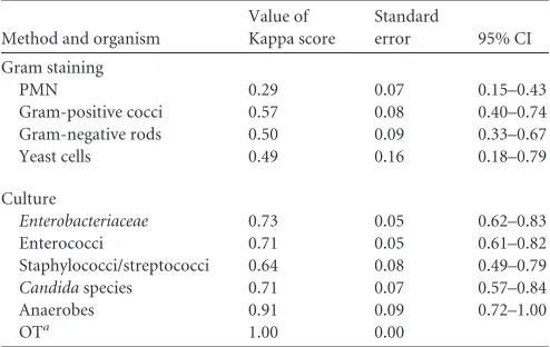
Interrate agreement between Previ Isola automated specimen inoculation and manual inoculation.Results of Gram staining had a fair strength of agreement for PMN ( ⫽0.29) and moder-ate strength of agreement for Gram-positive cocci ( ⫽0.57), Gram-negative rods ( ⫽0.50), and yeast cells ( ⫽0.49). Results of culture had a good strength of agreement between the two methods ([Enterobacteriaceae]⫽0.73,[enterococci] ⫽0.71,
[staphylococci/streptococci]⫽0.64,[Candidaspecies]⫽0.71,
[anaerobes]⫽0.91, and[other types]⫽1.00). Standard errors and upper and lower limits of the 95% confidence intervals (CI) are shown inTable 1.
[image:2.585.298.545.87.243.2]Performance in recovery of microorganisms.A total of 224
TABLE 1Kappa scores, standard errors, and 95% CIs for Gram staining and results of culture
Method and organism
Value of Kappa score
Standard
error 95% CI
Gram staining
PMN 0.29 0.07 0.15–0.43
Gram-positive cocci 0.57 0.08 0.40–0.74
Gram-negative rods 0.50 0.09 0.33–0.67
Yeast cells 0.49 0.16 0.18–0.79
Culture
Enterobacteriaceae 0.73 0.05 0.62–0.83
Enterococci 0.71 0.05 0.61–0.82
Staphylococci/streptococci 0.64 0.08 0.49–0.79
Candidaspecies 0.71 0.07 0.57–0.84
Anaerobes 0.91 0.09 0.72–1.00
OTa 1.00 0.00
aOT, other type.
on May 16, 2020 by guest
http://jcm.asm.org/
swabs were evaluated in the study, consisting of 112 paired sam-ples of Previ Isola plated PU and control viscose swabs. Seventeen paired samples (34 swabs) were sterile in culture. In 95 paired samples (190 swabs), microorganisms could be recovered from at least one of the swab systems. Recovery of the isolates is summa-rized in Table S6 in the supplemental material.
In 80/112 (71%) samples, results of culture were concordant between the two methods. In 32/112 (29%) samples, results of culture were discordant between the two methods. In 24 (75%) of the 32 paired samples with discordant results, Previ Isola plated PU swabs were superior to control viscose swabs. In 8 (25%) of the
32 paired samples, control viscose swabs were superior to Previ Isola plated PU swabs (Table 2).
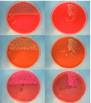
Quality of colony growth.Colony growth on Previ Isola and manually plated agar plates is shown inFig. 1. Previ Isola plated agar plates allowed better processing of colonies in most cases as colonies were more often individually distinct and less confluent. Different microorganisms could be distinguished more easily.
ATCC quality control strains.Both methods yielded good re-covery of the ATCC quality control strains.
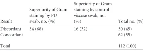
Evaluation of Gram stain quality/quantity.In 62/112 (55%) samples, results of Gram staining were concordant between the two methods. In 50/112 (45%) samples, results of Gram staining were discordant between the two methods. In 34 (68%) of the 50 paired samples with discordant results, Previ Isola plated PU swabs were superior to control viscose swabs. In 16 (32%) of the 50 paired samples, control viscose swabs were superior to Previ Isola plated PU swabs (Table 3).
DISCUSSION
[image:3.585.41.286.87.184.2]To the best of our knowledge, this is the first study to evaluate the performance of the Previ Isola system for wound swab samples in a routine clinical microbiological laboratory. Several instruments for automated processing have been recently introduced into the market but not evaluated critically until now (8). Swab systems
TABLE 2Overall results from culture by Previ Isola plated PU swabs versus manually plated control viscose swabs
Result
Superiority of Previ Isola plated PU swabs, no. (%)
Superiority of manually plated control viscose swabs,
no. (%) Total no. (%)
Discordant 24 (75) 8 (25) 32 (29)
Concordant 80 (71)
Total 112 (100)
FIG 1Colony growth on Columbia agar, chocolate agar, and MacConkey agar (top to bottom, respectively) streaked manually (left) and corresponding agar plates streaked by Previ Isola (right).
on May 16, 2020 by guest
http://jcm.asm.org/
[image:3.585.137.451.347.701.2]with semigel stabilization were shown to be effective for specimen collection and transport (9) and for maintaining viability of aer-obic and anaeraer-obic microorganisms (6,9). This study was in-tended to compare the recovery of microorganisms in clinical samples from wound samples plated by the Previ Isola system on PU swabs with the recovery in samples from manually plated con-trol viscose swabs. The type of swab used was not the subject of investigation.
Twenty-nine percent of results by culture were discordant be-tween the two methods, which is an outstanding percentage of difference when suggesting standardized methods of specimen collection. The superiority of Previ Isola plated agar plates might speak in favor of precise inoculation and a better distinction of colonies. Abdominal wounds, which are often mixed infections, were investigated; therefore, more individually distinct colonies on agar plates favor easy processing. Higher numbers of CFU might also indicate that absorption and release of microorganisms are better for identification of pathogens found in the wounds. Automated plated swabs might contribute to avoiding the use of expensive selective media like chromogenicCandidaagar. Several Previ Isola plated PU swab samples showed growth ofCandida
species which could be found in the set of plates used without using selective culture medium. Fungi could be detected as ap-posed colonies on other microorganisms which could be easily subcultivated because of the circular plating technique. Detection of fungal colonization or infection might therefore be improved by primarily avoiding selective culture media. For comparison, control viscose samples were plated on chromogenic Candida
agar, which led to comparable recovery of fungi. In a few samples with discordant results, control viscose swabs, including negative liquid broth cultures, were sterile, but few broth cultures of PU swabs were visibly grown (data not shown).
Kappa scores for Gram staining between the two methods seem to be convincing except for PMN. In analysis of the fre-quency table for PMN, a tendency toward a better detection of PMN could be seen (Table 4). This might be due to a light back-ground when examining Previ Isola plated PU swabs, possibly because the PU swab was not streaked onto the slide while prepar-ing the Gram stain, so that mechanical stress could be avoided. Details of PMN appeared to be better preserved in the Gram stains prepared from Previ Isola inoculation. Detection of PMN was superior with Previ Isola plated PU swabs (see Table S7 in the supplemental material).
The Previ Isola system is used together with PU swabs. To the best of our knowledge, other swab systems have not been evalu-ated for its use yet. It cannot be ruled out that various swab types show different levels of effectiveness in different clinical settings. Several swab types have been evaluated up to now (13). This study
was performed in a surgical department with a focus on abdomi-nal surgery. Predominant microorganisms found in the wound samples wereEnterococcusspecies,Enterobacteriaceae, and Can-didaspecies. Pathogenicity of the recovered microorganisms was not considered a criterion for evaluation.
There are numerous advantages of automated plate streakers. Processing is easy and can therefore be done even by inexperi-enced users. As shown inFig. 1, identification of suspicious colo-nies is easier, because most microorganisms can be processed di-rectly from primary plates without growing subcultures. Many colonies are already fractionated after plating, so that direct pro-cessing by Vitek or Phoenix or MALDI-TOF identification can be done without subcultures, which also improves the speed of pro-cessing. A system of automated specimen inoculation can be used easily and delivers standardized inoculation.
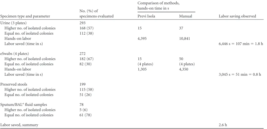
Preliminary studies have shown that streaking of liquid swabs with Previ Isola can provide potential time savings compared to the manual method (15 s versus 50 s per plate, respectively [3]).
These potential savings could most certainly be enhanced by using the LeanSigma method to optimize the integration of Previ Isola into the laboratory process and maintain control over each step in the process (Table 5). Moreover, it has been observed that the standardization of plate streaking with Previ Isola reduces the need for plate reincubation (0.8 to 1.1% with Previ Isola com-pared with 5 to 15% with traditional methods), since a higher proportion of isolated colonies than that in the manual methods is obtained (data from internal and external validation [bioMérieux]). However, further studies will be needed to provide more conclu-sive data.
There are some limitations to this study, including the lack of a gold standard to compare the methods for determination of sen-sitivity and specificity for different swab systems.
Limitations of the study might also be the modest number of samples investigated and the predominance of wound samples from abdominal surgery. Most wounds were superficial; there-fore, aerobic bacilli and fungi were dominant, whereas anaerobes were not frequently found, either with Previ Isola plated PU swabs or with control viscose swabs. The ability of liquid Amies medium to maintain the viability of aerobes, anaerobes, and fastidious bac-teria for up to 48 h at ambient and refrigerated temperatures (as required for compliance with CLSI M40-A) had been demon-strated; thus, we hypothesize that recovery of anaerobes might theoretically be equivalent to that of other swab systems. Loss of anaerobes during specimen transport is well known (5) but occurs for other swab systems likewise. We cannot rule out the possibility that one swab type may be more effective than another in clinical settings where other patient and context factors may have an im-pact on the swabbing procedure.
In conclusion, in this study a better recovery of
microorgan-TABLE 3Comparison of Gram staining by PU swab versus control viscose swab
Result
Superiority of Gram staining by PU swab, no. (%)
Superiority of Gram staining by control viscose swab, no.
(%) Total no. (%)
Discordant 34 (68) 16 (32) 50 (45)
Concordant 62 (55)
[image:4.585.40.284.88.175.2]Total 112 (100)
TABLE 4Frequency table for PMN in Gram stain for manually plated control viscose swabs versus Previ Isola plated PU swabs
Manual inoculation, scale of PMN
Previ Isola inoculation, scale of PMN
0 (0/field) 1⫹(⬍5/field) 2⫹(5–20/field) 3⫹(⬎20/field)
0 (0/field) 46 12 10 6
1⫹(⬍5/field) 7 15 6 2
2⫹(5–20/field) 3 2 1
3⫹(⬎20/field) 1 1
on May 16, 2020 by guest
http://jcm.asm.org/
[image:4.585.299.546.90.165.2]isms was observed for Previ Isola than for manual inoculation. The quality of colony growth and isolation is superior to that of manually streaked plates, which allows faster identification and susceptibility testing of microorganisms. Avoidance of selective culture media may even be possible. The diagnosis of infection might be improved and more cost-effective. The higher rate of recovery could increase the possibility of detecting potential pathogenic microorganisms.
ACKNOWLEDGMENTS
We thank the medical personnel and the technicians for their dedicated work. We thank bioMérieux for delivering data on cost-effectiveness and potential savings.
REFERENCES
1.Bourbeau PP, Swartz BL.2009. First evaluation of the WASP, a new automated microbiology plating instrument. J. Clin. Microbiol.47:1101– 1106.
2.Brook I. 1987. Comparison of two transport systems for recovery of aerobic and anaerobic bacteria from abscesses. J. Clin. Microbiol.25: 2020 –2022.
3.Bruno LC, Janda WM, Villarreal RI, Nguyen AK.2011. Evaluation of bioMerieux PREVI Isola automated plate streaker, poster 2152. Abstr. 111th Gen. Meet. Am. Soc. Microbiol.http://gm.asm.org/.
4.Citron DM, Warren YA, Hudspeth MK, Goldstein EJ.2000. Survival of aerobic and anaerobic bacteria in purulent clinical specimens maintained in the Copan Venturi Transystem and Becton Dickinson Port-a-Cul transport systems. J. Clin. Microbiol.38:892– 894.
5.Collee JG.1980. Factors contributing to loss of anaerobic bacteria in
transit from the patient to the laboratory. Infection8(Suppl. 2):S145– S147.
6.Drake C, Barenfanger J, Lawhorn J, Verhulst S.2005. Comparison of Easy-Flow Copan Liquid Stuart’s and Starplex Swab transport systems for recovery of fastidious aerobic bacteria. J. Clin. Microbiol.43:1301–1303. 7.Glasson JH, Guthrie LH, Nielsen DJ, Bethell FA.2008. Evaluation of an automated instrument for inoculating and spreading samples onto agar plates. J. Clin. Microbiol.46:1281–1284.
8.Greub G, Prod’hom G.2011. Automation in clinical bacteriology: what system to choose? Clin. Microbiol. Infect.17:655– 660.
9.Hindiyeh M, Acevedo V, Carroll KC.2001. Comparison of three trans-port systems (Starplex StarSwab II, the new Copan Vi-Pak Amies Agar Gel collection and transport swabs, and BBL Port-A-Cul) for maintenance of anaerobic and fastidious aerobic organisms. J. Clin. Microbiol.39:377– 380.
10. King GW, et al.2006. Automated agar plate streaker: a linear plater on Society for Biomolecular Sciences standard plates. J. Biomol. Screen.11: 704 –711.
11. Morosini MI, et al.2006. Evaluation of 4 swab transport systems for the recovery of ATCC and clinical strains with characterized resistance mech-anisms. Diagn. Microbiol. Infect. Dis.56:19 –24.
12. National Committee for Clinical Laboratory Standards.2003. Quality control of microbiological transport systems. Approved standard M40-A. National Committee for Clinical Laboratory Standards, Wayne, PA. 13. Saegeman V, et al.2011. Clinical evaluation of the Copan ESwab for
methicillin-resistant Staphylococcus aureus detection and culture of wounds. Eur. J. Clin. Microbiol. Infect. Dis.30:943–949.
[image:5.585.40.551.79.327.2]14. Van Horn KG, Audette CD, Sebeck D, Tucker KA.2008. Comparison of the Copan ESwab system with two Amies agar swab transport systems for maintenance of microorganism viability. J. Clin. Microbiol.46:1655– 1658.
TABLE 5Laboratory savings observed when comparing Previ Isola to manual inoculation
Specimen type and parameter
No. (%) of specimens evaluated
Comparison of methods, hands-on time in s
Labor saving observed
Previ Isola Manual
Urine (3 plates) 293
Higher no. of isolated colonies 168 (57) 15 37
Equal no. of isolated colonies 112 (38)
Hands-on labor 4,395 10,841
Labor saved (time in s) 6,446 s⫽107 min⫽1.8 h
eSwabs (4 plates) 272
Higher no. of isolated colonies 182 (67) 15 50
Equal no. of isolated colonies 82 (30) (4 plates) (4 plates)
Hands-on labor 1,305 4,350
Labor saved (time in s) 3,045 s⫽51 min⫽0.8 h
Preserved stools 199
Higher no. of isolated colonies 115 (58) Equal no. of isolated colonies 51 (26)
Sputum/BALafluid samples 78
Higher no. of isolated colonies 5 (6) Equal no. of isolated colonies 61 (78)
Labor saved, summary 2.6 h
aBAL, bronchoalveolar lavage.