R E S E A R C H A R T I C L E
Open Access
Does medial support decrease major
complications of unstable proximal humerus
fractures treated with locking plate?
Woo-Bin Jung
1, Eun-Sun Moon
1, Sung-Kyu Kim
1, David Kovacevic
2and Myung-Sun Kim
1*Abstract
Background:The purpose of this study was to evaluate the role of medial support and clinical factors responsible on outcomes and major complications associated with treatment of unstable proximal humerus fractures using a locking plate and suture augmentation.
Methods:Sixty-three cases in 62 patients (42 female, 20 male) were evaluated between September 2004 and October 2008. Cases were divided into either a medial support group (36 cases) or non-medial support group (27 cases). Clinical and radiographic evaluations included Neer’s evaluation criteria, the neck-shaft angle using the Paavolainen method, and complications. We analyzed the correlation between bone- and fracture- related complications and three independent clinical variables, such as the presence of medial support, fracture type, and osteoporosis by way of multivariate logistic regression.
Results:There were statistically significant differences in the overall incidence of complications based on the presence of medial support (p = 0.014) and preoperative fracture type (p = 0.018), but no differences based on the presence of osteoporosis (p = 0.157). According to multivariate logistic regression analysis, the restoration of medial support was the most reliable factor to prevent bone- and fracture- related complications. In addition, when we compared the incidence of bone- and fracture-related complications in the presence or absence of medial support among 30 patients with osteoporosis, the group with restoration of medial support had only one complication of humeral head osteonecrosis despite the presence of osteoporosis (5.9% vs. 46.2%, p = 0.025). According to Neer’s criteria, excellent or satisfactory clinical results accounted for seventy-three percent of the total cases (46 of 63 cases). Seventy-eight percent (49 of 55 cases) showed good radiographic results by the Paavolainen method. There were 14 complications in 13 of 63 cases (20.6%).
Conclusions:In the treatment of unstable proximal humerus fractures with locking plate technology and suture augmentation, we suggest that obtaining medial support is an important factor in preventing major bone- and fracture-related postoperative complications such as reduction loss or nonunion.
Keywords:Proximal humerus fracture, Locking plate, Complication, Medial support, Osteoporosis
* Correspondence:[email protected] 1
Department of Orthopaedic Surgery, Chonnam National University College of Medicine, 671, Jebong-Ro, Dong-Gu 501-757, Gwangju, South Korea Full list of author information is available at the end of the article
Background
In the operative treatment of unstable proximal humerus fractures, regardless of the type of fixation used, periopera-tive complications can occur, often leading to a worse clin-ical outcome than that obtained following nonoperative management. Especially in the patient with osteoporosis, there is no gold standard for surgical treatment, and the following complications have arisen following various treatment modalities: humeral head osteonecrosis, reduc-tion loss, nonunion, malunion, plate breakage, screw prob-lems, and infection [1-5].
The proximal humerus locking plate system has many advantages, including its anatomic design, low profile, the presence of suture holes, divergent angulation of locking screws, and high angular and rotational stability. It is believed that these locking plates provide improved fix-ation of proximal humerus fractures than the conventional plates, especially for bones that are osteoporotic.
Despite these advantages, it has been reported that locking plate technology for the surgical treatment of prox-imal humerus fractures has a high complication rate ran-ging from 16% to 36% [6-13]. Several major complications, such as reduction loss with or without screw perforation, nonunion, humeral head osteonecrosis, and metal break-age, may adversely affect clinical outcome.
Several studies [13-17] have stressed the importance of medial support in locked plating of proximal humerus fractures, and achieving mechanical support of the inferomedial region of the proximal humerus seems to be important for maintaining fracture reduction. We are aware of one report that identifies the clinical factors (i.e., medical co-morbidities, medial support, and head-neck-shaft angle) responsible for postoperative compli-cations following the operative treatment of unstable proximal humerus fractures using a locking plate and suture augmentation.
We hypothesized that medial support decreases the complications of proximal humerus fractures treated with locking plate technology and suture augmentation, especially in older patients with osteoporosis. The ob-jective of this study was to evaluate the role of medial support and clinical factors responsible on the outcomes and major complications associated with treatment of unstable proximal humerus fractures using a locking plate and suture augmentation.
Methods
Study design
A retrospective study was approved by the Institutional Review Board of Chonnam National University Hospital (CNUH) to analyze the clinical and radiographic out-comes of 62 patients (63 cases) who had been treated surgically for a proximal humerus fracture with locking plate fixation and tension band suture augmentation
between September 2004 and October 2008. Preopera-tively, the fracture pattern was determined according to the Neer classification [18] using a standardized preopera-tive surgical workup, which included conventional radiog-raphy, as well as 3-dimensional computed tomography (3D-CT) in all patients. Two fellowship-trained shoulder and elbow surgeons (ESM & MSK) used the same ana-tomic approach and surgical technique for all cases.
Inclusion criteria
Adult patients who sustained a closed, unstable proximal humerus fracture without neurovascular complication at the time of injury and subsequently underwent operative management were identified for review. Those patients who were treated with an anatomic proximal humeral locking plate and suture augmentation were included in this study. The implants used in this series included 40 proximal humerus locking compression plates (PH-LCP, Synthes, Switzerland) and 23 PHILOS plates (Synthes, Switerland).
Exclusion criteria
Adult patients were excluded from this study if it was de-termined that they sustained polytraumatic injuries or isolated proximal humerus fractures considered open, seg-mental or with diaphyseal extension. Patients with con-comitant fractures of the ipsilateral elbow or distal radius were excluded from this study as well as those patients with nonunions, pathologic fractures, or refractures in-volving the proximal humerus.
Operative technique and rehabilitation
anatomic reduction could be achieved, an oblique locking screw was not placed intentionally in the inferomedial quadrant of the proximal humerus. Conversely, in cases where inferomedial comminution was present, a reduction was of medial cortex was difficult, or the shaft could not be medialized and impacted into the head fragment, then we tried to place an oblique locking screw in the inferomedial quadrant of the proximal humerus to provide medial support.
Then suture augmentation in a tension band configur-ation was performed using the non-absorbable sutures that were passed through the rotator cuff tendons to secure the rotator cuff tendons to the holes in the locking plate. The surgical bed was irrigated with sterile normal saline and the soft tissue and skin were closed in a layered fashion.
After surgery, all shoulders were immobilized in a sling for the first 3 to 6 weeks, and passive range of motion exercise was allowed from 1 to 6 weeks postoperatively depending on fracture type, the degree of medial commin-ution, and intraoperative fixation stability. Elbow, wrist, and hand range of motion exercises were encouraged im-mediately. Stretching and resistive strengthening exercises were allowed when evidence of fracture healing was noted on follow-up radiographic imaging.
Clinical and radiographic evaluation
Routine follow-up was initially performed at 3 weeks, 6 weeks, 3 months, 6 months, and 12 months postopera-tively, and then annually. Clinical outcomes were deter-mined using Neer’s evaluation criteria [18] at the final follow-up visit. Neer’s criteria include the following: pain (35 points), function (30 points), range of motion (25 points), and anatomy (10 points), with a total of 100 points. Outcomes on the Neer scale are based on total number of points: excellent is > 89; satisfactory is 80–89; unsatisfactory is 70–79; and failure is < 70.
Preoperative imaging of the injured shoulder was obtained to assist with surgical planning and consisted of three orthogonal radiographic views (anteroposterior, scapular Y, and axillary views), 3-D CT, and fluoroscopic evaluation of the shoulder in all patients. Postopera-tively, we obtained serial follow-up radiographs (antero-posterior, scapular Y, and axillary views) at each visit. Preoperative and postoperative radiographs were retro-spectively analyzed in a serial fashion by two independ-ent observers (ESM and MSK) to evaluate the fracture type according to Neer’s classification, bony union, hu-meral neck-shaft angle, and complications.
We divided the cases into a medial support group (MS+ group) and a non-medial support group (MS- group) based on criteria suggested by Gardner et al. A fracture was considered to have medial support based on the fol-lowing: (1) the medial pillar of the proximal humerus was
anatomically reduced and not comminuted; (2) the shaft was medialized and impacted into the head fragment; (3) an oblique locking screw was placed directly into the inferomedial quadrant of the proximal humeral head frag-ment to within 5 mm of the subchondral bone [14].
Humeral neck-shaft angles were measured according to the Paavolainen method [19]. Briefly, a vertical axis was drawn from the superior border to the inferior bor-der of the articular surface and a line perpendicular to this line was made that ran through the center of the humeral head. The angle between this perpendicular line and the line bisecting the humeral shaft was defined as the humeral neck-shaft angle. Postoperatively, restoration of humeral neck-shaft angle to 130° ± 10° was defined as good, fair was defined as being between 100° to 120° and poor was defined as being < 100°. Further, the presence of associated osteoporosis was measured by dual energy X-ray absorptiometry (DEXA) and defined when the pa-tient had a T-score less than−2.5 involving the bone dens-ity of the hip and spine as established by the World Health Organization criteria for osteoporosis [20,21].
We evaluated the postoperative complications and cate-gorized them into four groups [11]; (1) surgical technique-related complications; (2) soft-tissue- and wound-technique-related complications; (3) implant- related complications; (4) bone-and fracture- related complications. Surgical technique-related complications attributed to the initial surgical procedure include primary screw perforation and plate irri-tation. Soft-tissue- and wound-related complications include superficial wound infection, deep wound infection, and neurological lesion. Implant-related complications in-clude screw loosening and plate breakage [11]. Finally, bone- and fracture-related complications include reduction loss without screw perforation, reduction loss with screw perforation. Among these complications, we considered reduction loss with or without screw perforation, osteo-necrosis of the humeral head, and nonunion, and metal breakage as major complications.
Overall clinical and radiographic outcomes as well as complications were evaluated and compared between the 2 groups. In addition, we analyzed the distributional pattern of complications based on the presence of medial support, the fracture type, and the presence of osteoporosis.
Statistical analysis
presence of medial support, preoperative fracture type, and the presence of osteoporosis) was performed. Finally, we performed the post-hoc power analysis (Statistics cal-culators, version 3.0). A p-value of <0.05 was considered statistically significant.
Results
Sociodemographic and clinical characteristics
There were 42 women and 20 men who underwent op-erative treatment of a proximal humerus fracture with locking plate technology and suture augmentation. One patient required open reduction and internal fixation for bilateral proximal humerus fractures. The average age was 62.2 years (range 18–92 years). The mean duration of follow-up was 13.4 months (range 12–38 months). The mechanism of shoulder injury was found to be a fall from a standing position in 33 patients, motor vehicle colli-sion in 23 patients, and a fall from a height or stairs in seven patients. There were 42 two-part fractures, 18 three-part fractures, and three four-three-part fractures based on the Neer classification. The demographic data showed no significant differences in terms of age, sex, osteoporosis, or fracture pattern.
Review of the initial postoperative radiographs demon-strated that medial support was established in 36 cases according to Gardner’s criteria [14], while medial support was not reestablished in 27 cases. According to fracture
type, there were 25 2-part fractures (69%), ten 3-part frac-tures (28%), and one 4-part fracture (3%) in the medial support group, while there were 17 2-part fractures (63%), eight 3-part fractures (30%), and two 4-part fractures (7%) in the non-medial support group (Table 1).
Complications
There were 14 total complications in 13 of 63 cases noted during the follow-up period. There was one fe-male patient who had two complications: (1) primary screw perforation during the index procedure, and (2) humeral head osteonecrosis at final follow-up (Table 2).
There were three surgical technique-related complica-tions: primary screw perforation of the humeral head that was unrecognized during surgery in two cases and plate irritation due to high plating in one case. There were no soft-tissue/wound-related complications such as superficial wound infection and deep infection or neuro-logical injury. There were nine bone- and fracture- related complications: five reduction losses without screw perfor-ation; one reduction loss with screw perforperfor-ation; two non-unions, and one humeral head osteonecrosis. Finally, there were two implant-related complications: one screw loos-ening and one plate breakage. Plate breakage occurred in a 58 year old male two months following the index procedure after he sustained a fall and landed on his outstretched, operative limb. This patient subsequently underwent
osteo-Table 1 Sociodemographic data and the clinical outcomes according to the presence of medial support
MS+ (n=36) MS- (n=27) Total (63) Av. Neer’s score P-value
Sex M: 9, F:27* M:11, F:16* 0.184
Age(yr) 62.5 61.7 62.2 0.739
Osteoporosis(%) 20 (56) 16 (59) 36 0.769
Fracture Type
2 part 25 (69) 17 (63) 42
3 part 10 (28) 8 (30) 18 0.673
4 part 1 (3) 2 (7) 3
Neer criteria
Excellent (>89) 14 (38.9) 7 (25.9) 21 (33.3)
Satisfactory(80–90) 15 (41.7) 10 (37.0) 25 (39.7) Unsatisfactory(70–79) 7 (19.4) 4 (14.9) 11 (17.5)
Failure(<70) 0(0) 6 (22.2) 6 (9.5)
Av. Neer’s score 85.7 ± 7.8 78.0 ± 14.2 82.4 ± 11.5 0.008
Paavolainen Score
Good (>130±10°) 32 (88.9) 17 (62.9) 49 (77.8) 84.9 ± 8.3
Fair (100-120°) 4 (11.1) 7 (26.0) 11 (17.5) 80.6 ± 9.9 0.004
Poor(<100°) 0 (0) 3 (11.1) 3 (4.8) 48.0 ± 6.1
Av. NSA 130.4 ± 9.5 123.5 ± 21.3 127.4 ± 15.9 0.574
Values are given as number (percentage).
*one female have bilateral proximal humerus fractures.
[image:4.595.58.539.440.705.2]synthesis using a proximal humerus locking plate with iliac crest bone graft, which resulted in bony union. In the case of screw loosening, the screw was removed at tive day three and bony union was achieved by postopera-tive week twelve.
There were statistically significant differences in the overall incidence of complications based on the presence of medial support (p = 0.014) and the preoperative frac-ture type (p = 0.018), but no differences were noted in the presence of osteoporosis (p = 0.157).
Among 14 total complications, four (two screw perfor-ation, one osteonecrosis, and one plate irritation) were seen in the medial support group, but only one was considered a major complication (humeral head osteonecrosis in an osteoporotic patient). The remaining ten complications were in the non-medial support group, nine of which were considered major complications (Table 2).
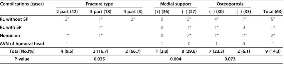
When we compared the nine major bone- and fracture-related complications, seven cases had osteoporosis, and six of these seven cases were in the non-medial support group. There were statistically significant differences in the overall incidence of complications based on the presence of medial support (p = 0.004) and preoperative fracture type (p = 0.035), but no differences based on the presence of osteoporosis (p = 0.073), sex (p = 0.301), or age (R2= 0.176, p = 0.095) (Table 3 and 4).
[image:5.595.61.542.110.277.2]Multivariate logistic regression analysis demonstrated that the presence of medial support was the only factor predictive of major bone- and fracture- related compli-cations (B = −2.761, p = 0.016). Preoperative fracture type (B = 1.011, p = 0.118) and the presence of osteopor-osis (B = −1.618, p = 0.086) did not have a statistically significant correlation with bone- and fracture- related complications.
Table 2 The distributional pattern of complications according to the fracture types, the presence of medial support, and osteoporosis
Complications (cases) Fracture type Medial support Osteoporosis
2 part (42) 3 part (18) 4 part (3) (+) (36) (−) (27) (+) (30) (−) (33) Total (63)
Screw perforation 2† 2 0 2 0 2
Plate irritation 1 1 0 0 1 1
RL without SP* 2 1 2 0 5 4 1 5
RL with SP* 1 0 1 1 0 1
Nonunion* 1 1 0 2 1 1 2
AVN of humoral head* 1† 1 0 1 0 1
Screw loosening 1 0 1 0 1 1
Plate breakage* 1 0 1 0 1 1
Total No. (%) 8 (19.0) 3 (16.7) 3 (100) 4 (11.1) 10 (37.0) 9 (30.0) 5 (15.2) 14 (22.2)
P-value 0.018 0.014 0.157
Values are given as number (percentage). *Clinically serious major complication.
†One female patient had these two complications simultaneously.
RL = reduction loss, SP = screw perforation, AVN = avascular necrosis.
Table 3 The distributional pattern focusing on the fracture- and bone- related complication
Complications (cases) Fracture type Medial support Osteoporosis
2 part (42) 3 part (18) 4 part (3) (+) (36) (−) (27) (+) (30) (−) (33) Total (63)
RL without SP 2* 1* 2* 0 5* 4* 1* 5*
RL with SP 1* 0 1* 1* 0 1*
Nonunion 1* 1* 0 2* 1* 1* 2*
AVN of humoral head 1 1 0 1 0 1
Total No.(%) 4 (9.5) 3 (16.7) 2 (66.7) 1 (2.8) 8 (29.6) 7 (23.3) 2 (6.1) 9 (14.3)
P-value 0.035 0.004 0.073
Values are given as number (percentage). *non-medial support group.
[image:5.595.57.541.597.707.2]Finally, when we compared the incidence of bone- and fracture- related complications based on the presence of medial support among 30 patients with osteoporosis, the group with medial support restoration had fewer complica-tions than the group with medial support failure (Table 4).
Clinical outcomes
According to Neer’s evaluation criteria, 21 of 63 cases (33.3%) showed excellent results, 25 cases (39.7%) were satisfactory, 11 cases (17.5%) were unsatisfactory, and six cases (9.5%) were failures. The excellent or satisfactory clinical results accounted for 73.0% of the all cases.
Those six cases that went on to clinical failure in-cluded nonunion (N=2), humeral head osteonecrosis, reduction loss without screw perforation (Figure 1), re-duction loss with screw perforation, and plate breakage. Twenty-nine cases (80.6%) showed excellent or satisfac-tory outcomes in the medial support group (36 cases) (Figure 2), while 17 cases (62.9%) demonstrated excellent or satisfactory outcomes in the non-medial support group (27 cases). The average Neer score of the medial support group was higher than that of the non-medial support group and this difference was statistically signifi-cant (MS+ group, 85.7 ± 7.8 (average ± SD); MS- group, 78.0 ± 14.2, (p = 0.008)) (Table 1).
Radiographic outcomes
All fractures were united at final follow-up, except in four cases (two nonunions, one osteonecrosis, and one plate breakage). Forty-nine of the 63 cases (77.8%) showed good results by Paavolainen method, 11 cases (17.5%) had fair results, and three cases (4.8%) had poor results. Comparing neck-shaft angle according to the presence of medial support at last follow-up, 32 cases (88.9%) were scored excellent and four cases(11.1%) were scored fair. In the non-medial support group, 17 cases (62.9%) were scored excellent, seven cases (26.0%) were scored fair, and three cases (11.1%) were scored poor. There were no statistically significant differences in
radiological outcome of neck-shaft angle between the two groups (MS+ group, 130.4 ± 9.5; MS- group, 123.5 ± 21.3, (p = 0.574)). However, according to the analysis of sub-group by Paavolainen method, The average Neer score of the poor group was lower than those of the Good or Fair group and this difference was statistically significant (Good, 84.9 ± 8.3 (average ± SD); Fair, 80.6 ± 9.9); Poor, 48.0 ± 6.1, (p = 0.004)) (Table 1).
Discussion
Although the optimal surgical treatment of proximal humerus fractures has not been determined, there have been many operative techniques described, including percutaneous fixation, conventional plate fixation, intra-medullary fixation with rods or pins, tension band wir-ing, and blade plate fixation, whose clinical outcomes have varied [12]. The current trend for treating these fractures utilizes locking plate technology as its lower profile may reduce impingement; its multiple divergent locking screw positions allow for improved fixation; and its biomechanical properties provide improved stability and load to failure. A recent biomechanical analysis in which blade-plate fixation was compared with locking plate fixation for the treatment of proximal humeral fractures demonstrated potential advantages with use of the locking plate technology [22].
In the current study, excellent or satisfactory clinical results were realized in 73.0% of cases, with good radio-graphical results in 77.8% of cases. These clinical and radiographic findings suggest that locking plate fixation and suture augmentation for proximal humerus fractures provide value as an operative treatment modality. However, room for improvement exists as 14 complications occurred in thirteen of 63 cases, representing a complication rate of 22%. These complications included screw perforation, plate irritation, loss of reduction, osteonecrosis, nonunion, screw loosening, and implant breakage (Table 2).
[image:6.595.56.541.113.223.2]There have been several reports about the complica-tions encountered with locking plate technology. Egol
Table 4 The relationship of osteoporosis and medial support confining to the fracture- and bone- related complications
Medial support group (N=36) Non-medial support group (N=27) Total
Osteoporosis (+)
AVN of humoral head* (1) RL without SP* (4)
7 (23.3) RL with SP* (1)
Nonunion* (1)
Osteoporosis (−) RL without SP* (1) 2 (6.1)
Nonunion* (1)
Total No. (%) 1 (11.1) 8 (37.0) 9 (100)
P-value 0.004
Values are given as number (percentage). *Clinically serious major complication.
et al. [8] reported complications in 12 of 51 patients (24%) following proximal humerus locking plate fixation at 16 months follow-up. The complications occurred in eight patients (16%), including intraarticular screw pene-tration, osteonecrosis, acute fracture, nonunion, and het-erotopic ossification. Similarly, Owsley et al. [10] reported a 36% complication rate in 53 patients, with intraarticular screw penetration occurring in 23% and a statistically sig-nificant higher radiographic complication rate noted in pa-tients older than 60 years of age. In a study by Lee et al. [9], 20% of 45 patients had postoperative complications that included loss of fixation, adhesive capsulitis, and deep infection, while Sudkamp et al. [11] reported various com-plications in 34% of 155 patients including: screw
penetration, plate impingement, infection, loss of reduc-tion with or without screw perforareduc-tion, humeral head osteonecrosis, nonunion, screw loosening, plate pullout, and implant breakage. Brunner et al. [7] reported an over-all complication rate of 35% and Badman et al. [12] presented 13 complications (16%) in 81 patients and reported varus collapse in 5 patients (6%), intraarticular screw penetration in 3 (3.7%) and osteonecrosis in 5 (6.2%). Königshausen et al. [13] reported 12 (23.1%) com-plications in 73 patients. The overall complication rate in the current study was not higher compared to previous re-ports (22% versus 16- 36%)
[image:7.595.60.541.91.511.2]Sudkamp et al. [11] classified the complications related to the locking plate into four categories. They reported Figure 1(A) Initial radiographs of 82 years old male with osteoporosis showed 2 part proximal humerus fracture.(B) Immediate
25 (40%) initial incorrect surgical technique-related com-plications amongst 62 total comcom-plications, which included primary intraoperative screw perforations of the humeral head in 21 cases and subacromial impingement due to significant cranial positioning of the plate in 4 cases. They suggested that adherence to proper surgical technique is necessary to avoid iatrogenic errors. Others, including Badman et al. [12] and Brunner et al. [7], have reported on primary and secondary intraarticular screw penetration into the glenohumeral joint and recommended that more accurate screw length measurement and shorter screw selection would prevent primary screw perforation.
In the current study, there were three (4.8%) initial in-correct surgical technique-related complications encoun-tered in 63 cases at the end of the operative procedure: primary intraoperative screw perforation in two cases and subacromial impingement in one case. We believe that confirming screw position in more than one plane using an image intensifier will decrease the incidence of these surgical technique-related complications.
Of the four types of complications, we believe that bone-and fracture- related complication are the most important to prevent because they can negatively impact clinical and radiographic outcomes. More importantly though, this type of complication is under the surgeon’s control, and thus can be avoided with meticulous surgical technique. In the current study, all six cases with failed clinical outcomes had bone- and fracture- related complications. Our find-ings mirror previous work in this field, including Sudkamp et al. [11] who reported twenty one (13.5%) bone- and fracture- related complications in 155 patients.
[image:8.595.58.537.89.431.2]support the medial buttress. However, none of the five pa-tients received tension band sutures between the rotator cuff and the plate to neutralize traction forces. Based on their observations, they suggested that traction forces from the rotator cuff should be neutralized using tension band sutures combined with screws supporting the medial calcar, especially when medial support is insufficient. Simi-larly, Badman et al. [12] documented that restoration of the medial calcar and supplemental suture fixation may decrease the incidence of hardware-related complications.
In our study, augmentation with a tension band con-struct using non-absorbable sutures through the rotator cuff to the holes in the plate was applied in all cases. However, despite augmentation with a tension band con-struct, there were six cases where secondary reduction loss with or without screw perforation occurred in pa-tients without medial support (Tables 3 and 4).
The incidence of humeral head osteonecrosis follow-ing lockfollow-ing plate fixation at short-term follow-up has been reported in 3.8% to 25% of cases [6,7,11,12]. In our current study, we had one case of humeral head osteo-necrosis (1.6%) in a patient with a two-part proximal hu-merus fracture-dislocation. It is thought that open reduction and internal fixation may increase the risk of osteonecrosis unless the medial capsular structures and metaphyseal bony attachments are maintained so as to preserve the humeral head blood supply [23]. We believe that we had a low incidence of osteonecrosis because we were able to utilize the locking plate system to indirectly reduce the fracture fragments and avoid additional soft tissue dissection and damage near the fracture site.
We experienced plate breakage in one case, when a patient fell and landed on his outstretched, operative limb at two months following his index procedure. The patient subsequently underwent osteosynthesis using a proximal humerus locking plate with iliac crest bone graft, leading to successful bony union. We believe this complication was due to implant fatigue failure and a deficient posteromedial calcar in the setting of a low en-ergy traumatic event. We believe that medial support may be important to resist implant fatigue.
Recently, much attention has been paid to the import-ance of the medial column for maintaining stable fixation of proximal humerus fractures [9,13-17,24-27]. Anatomic reduction and restoration of the medial calcar allow the medial column to both buttress and reduce the stresses of laterally-based plate fixation. Gardner et al. [14] first em-phasized this concept by noting that when mechanical support of the inferomedial region of the proximal hu-merus was obtained, fracture subsidence was significantly reduced postoperatively. They suggested that mechanical support of the medial column may be achieved either with placement of humeral head screws inferomedially or end-osteal fibular allograft strut augmentation when anatomic
cortical contact is not possible [14,26,27]. They reported that lack of medial support led to a 30% screw perforation rate compared to a 6% screw perforation rate for fractures with an intact medial column.
According to our experience, direct placement of an ob-lique long locking screw into the inferomedial quadrant of the proximal humeral head is considered as the more im-portant and substantive way to obtain the medial support when there is medial communition. In contrast, anatomical reduction of the medial calcar with good cortical contact, especially in patients without medial cortex comminution, may be additive in their ability to prevent postoperative complications.
Lee at al [9] reported that absence of comorbidity and the restoration of the medial metaphysis were the most reliable predictors of successful clinical outcomes, while Solberg et al. [24] recognized that the presence of a metaphyseal segment in the region of the medial calcar greater than 2 mm was associated with better clinical out-comes and independent of Neer fracture type. Others have corroborated the importance of restoring and maintaining the medial calcar to enhance mechanical stability and to avoid reduction loss [13,15,16,25]. In situations where the host bone is osteoporotic and anatomic reduction and res-toration of the posteromedial column cannot be achieved, it is recommended that augmentation with endosteal fibu-lar allograft struts or primary arthroplasty be considered [16]. Zhang et al. [15] reported clinical and radiological outcomes of seventy-two consecutive patients (mean 30.8-month follow-up, MS+ group: 29 patients; MS- group: 39 patients). They showed a statistically significant difference regarding the failure rate (23.1% in the MS- group vs. 3.4% in the MS+ group). They documented that the early loss of fixation was related to higher age and less initial neck-shaft angle of the patients. However, bone mineral density was not significantly associated with loss of fixation. They also observed a significantly lower final neck-shaft angle in the MS- group and greater secondary angle loss in the subgroup of Neer three-part (P = 0.033 and 0.015, respect-ively) and four-part fractures (P = 0.043 and 0.027). There-fore, they concluded that medial support for proximal humerus fractures seems to have no benefits in Neer two-part fractures, but the additional medial support screws inserted into the inferomedial region of the humeral head may help to enhance mechanical stability in complex frac-tures and allow for better maintenance of reduction.
treatment of proximal humerus fractures using a locking plate and suture augmentation. There appears to be a trend towards significance for osteoporosis on multivari-ate regression analysis with p-values approaching 0.05. Further, a subgroup analysis of 30 cases with osteopor-osis demonstrated that medial support restoration led to significantly less major bone- and fracture-related complications compared to medial support loss. Thus, even osteoporotic patients can benefit from achieving and maintaining medial support so they decrease their chances of having a major complication such as reduc-tion loss or nonunion.
The limitations of the current study are its retrospect-ive nature and small sample size, potentially introducing bias and β-error. We did not perform a priori power calculation analysis for sample size estimation as this work is a preliminary report of our experience. A post-hoc power analysis demonstrated that the observed power for the addition of the set of independent vari-ables was 0.64458902. (Statistics calculators, version 3.0). Future work will focus on performing a prospective, ran-domized study that is adequately powered to have suffi-cient sample size to detect differences between the two groups. There was no control group in the present study; therefore, we cannot determine if another treatment me-thod would have led to different results. In addition, we used two different types of locking plates (PH-LCP and PHILOS plate). These plates differ in profile and plate design, including the number of locking holes and the thickness of the plate, both of which can affect the overall outcome. Although we tried to place the oblique long locking screw into the inferomedial quadrant of the prox-imal humeral head intentionally after noticing the role of the medial support reported by Gardner et al. [14], some size mismatching between the humerus and the locking plates precluded the placement of the oblique long locking screw at the intended location. This led to both the MS+ and MS- groups comprising cases treated with both differ-ent types of locking plates, which can affect the overall outcome also. With regards to the presence or absence of osteoporosis, regional osteoporosis in the hip or spine was used in this study as a surrogate for the presence of local osteoporosis at the proximal humerus. But, this may be not the ideal method to assess local osteoporosis of the proximal humerus. Additionally, we divided the patients to two groups based on the presence or absence of osteo-porosis (i.e., t-score <−2.5). We did not see any linear cor-relations between the t-scores and the other possible factors. Finally, although we describe a generalized post-operative rehabilitation protocol in the Methods, most cases had an individualized protocol for length of sling immobilization and timing of passive range of motion exercises. This depended on the fracture type, degree of medial comminution, and intraoperative fixation stability.
These factors may have affected the clinical and radio-graphic outcomes.
Conclusions
In the treatment of unstable proximal humerus frac-tures, locking plate technology and suture augmentation are considered a useful treatment modality based on ad-equate clinical and radiographic outcomes. We found that the presence of medial support is a critical, modifi-able factor that the surgeon can control in preventing major complications. Therefore, we suggest that restoring medial support is important in preventing major bone-and fracture-related complications such as reduction loss or nonunion in the operative management of unstable proximal humerus fractures.
Competing interests
The authors declare that they have no competing interests.
Authors’contributions
MSK designed this study, review the literatures and drafted the manuscript. JWB have made substantial contributions to acquisition of data, analysis and interpretation of data. ESM, SKK and DK have been involved in drafting the manuscript and revising it critically for important intellectual contents. All authors read and approved the final manuscript.
Acknowledgements
No funding was received for this research. None of the authors have any conflict of interest or disclosures to report in relation to this work.
Author details
1Department of Orthopaedic Surgery, Chonnam National University College
of Medicine, 671, Jebong-Ro, Dong-Gu 501-757, Gwangju, South Korea.
2Department of Orthopaedic Surgery, Cleveland Clinic Foundation,
Cleveland, OH, USA.
Received: 4 January 2012 Accepted: 13 March 2013 Published: 22 March 2013
References
1. Clifford PC:Fractures of the neck of the humerus: a review of the late results.Injury1980,12(2):91–95.
2. Cofield RH:Comminuted fractures of the proximal humerus.Clin Orthop Relat Res1988,230:49–57.
3. Elkowitz SJ, Juckerman JD:Decision making for the treatment of proximal humerus fractures.Techniques in Shoulder and Elbow Surg2002,4:234–250. 4. Mills HJ, Horne G:Fractures of the proximal humerus in adults.J Trauma
1985,25(8):801–805.
5. Park M, Seong B, Lee S, Lee T, Shin S, Kim H:The result of treatment in fracture of the proximal humerus.J Korean Fracture Soc2002, 12(2):299–306.
6. Bjorkenheim JM, Pajarinen J, Savolainen V:Internal fixation of proximal humeral fractures with a locking compression plate: a retrospective evaluation of 72 patients followed for a minimum of 1 year.Acta Orthop Scand2004,75(6):741–745.
7. Brunner F, Sommer C, Bahrs C, Heuwinkel R, Hafner C, Rillmann P, Kohut G, Ekelund A, Muller M, Audige L, Babst R:Open reduction and internal fixation of proximal humerus fractures using a proximal humeral locked plate: a prospective multicenter analysis.J Orthop Trauma2009, 23(3):163–172.
8. Egol KA, Ong CC, Walsh M, Jazrawi LM, Tejwani NC, Zuckerman JD:Early complications in proximal humerus fractures (OTA Types 11) treated with locked plates.J Orthop Trauma2008,22(3):159–164.
10. Owsley KC, Gorczyca JT:Fracture displacement and screw cutout after open reduction and locked plate fixation of proximal humeral fractures corrected.J Bone Joint Surg Am2008,90(2):233–240.
11. Sudkamp N, Bayer J, Hepp P, Voigt C, Oestern H, Kaab M, Luo C, Plecko M, Wendt K, Kostler W, Konrad G:Open reduction and internal fixation of proximal humeral fractures with use of the locking proximal humerus plate. Results of a prospective, multicenter, observational study.J Bone Joint Surg Am2009,91(6):1320–1328.
12. Badman B, Frankle M, Keating C, Henderson L, Brooks J, Mighell M:Results of proximal humeral locked plating with supplemental suture fixation of rotator cuff.J Shoulder Elbow Surg2011,20(4):616–624.
13. Konigshausen M, Kubler L, Godry H, Citak M, Schildhauer TA, Seybold D: Clinical outcome and complications using a polyaxial locking plate in the treatment of displaced proximal humerus fractures. A reliable system?Injury2012,43(2):223–221.
14. Gardner MJ, Weil Y, Barker JU, Kelly BT, Helfet DL, Lorich DG:The importance of medial support in locked plating of proximal humerus fractures.J Orthop Trauma2007,21(3):185–191.
15. Zhang L, Zheng J, Wang W, Lin G, Huang Y, Zheng J, Edem Prince GA, Yang G:The clinical benefit of medial support screws in locking plating of proximal humerus fractures: a prospective randomized study.Int Orthop
2011,35(11):1655–1661.
16. Krappinger D, Bizzotto N, Riedmann S, Kammerlander C, Hengg C, Kralinger FS:Predicting failure after surgical fixation of proximal humerus fractures.Injury2011,42(11):1283–1288.
17. Thanasas C, Kontakis G, Angoules A, Limb D, Giannoudis P:Treatment of proximal humerus fractures with locking plates: a systematic review.
J Shoulder Elbow Surg2009,18(6):837–844.
18. Neer CS 2nd:Displaced proximal humeral fractures. I. Classification and evaluation.J Bone Joint Surg Am1970,52(6):1077–1089.
19. Paavolainen P, Bjorkenheim JM, Slatis P, Paukku P:Operative treatment of severe proximal humeral fractures.Acta Orthop Scand1983,54(3):374–379. 20. Brown JP, Josse RG, Scientific Advisory Council of the Osteoporosis Society
of Canada:2002 clinical practice guidelines for the diagnosis and management of osteoporosis in Canada.CMAJ2002,167(10 Suppl):S1–34. 21. DePalma A:Surgery of the shoulder.3rd edition. Philadelphia: Lippincott; 1983. 22. Siffri PC, Peindl RD, Coley ER, Norton J, Connor PM, Kellam JF:
Biomechanical analysis of blade plate versus locking plate fixation for a proximal humerus fracture: comparison using cadaveric and synthetic humeri.J Orthop Trauma2006,20(8):547–554.
23. Tan SL, Balogh ZJ:Indications and limitations of locked plating.Injury
2009,40(7):683–691.
24. Solberg BD, Moon CN, Franco DP, Paiement GD:Locked plating of 3- and 4-part proximal humerus fractures in older patients: the effect of initial fracture pattern on outcome.J Orthop Trauma2009,23(2):113–119. 25. Osterhoff G, Baumgartner D, Favre P, Wanner GA, Gerber H, Simmen HP,
Werner CM:Medial support by fibula bone graft in angular stable plate fixation of proximal humeral fractures: an in vitro study with synthetic bone.J Shoulder Elbow Surg2011,20(5):740–746.
26. Gardner MJ, Boraiah S, Helfet DL, Lorich DG:Indirect medial reduction and strut support of proximal humerus fractures using an endosteal implant.
J Orthop Trauma2008,22(3):195–200.
27. Gardner MJ, Lorich DG, Werner CM, Helfet DL:Second-generation concepts for locked plating of proximal humerus fractures.Am J Orthop (Belle Mead NJ)2007,36(9):460–465.
doi:10.1186/1471-2474-14-102
Cite this article as:Junget al.:Does medial support decrease major
complications of unstable proximal humerus fractures treated with locking plate?.BMC Musculoskeletal Disorders201314:102.
Submit your next manuscript to BioMed Central and take full advantage of:
• Convenient online submission
• Thorough peer review
• No space constraints or color figure charges
• Immediate publication on acceptance
• Inclusion in PubMed, CAS, Scopus and Google Scholar
• Research which is freely available for redistribution