Electronic Theses and Dissertations Theses, Dissertations, and Major Papers
2017
Physiological, Psychological, and Behavioural Sequelae of
Physiological, Psychological, and Behavioural Sequelae of
Concussion in a Classical Conditioning Paradigm
Concussion in a Classical Conditioning Paradigm
Sabrina Freund University of Windsor
Follow this and additional works at: https://scholar.uwindsor.ca/etd
Recommended Citation Recommended Citation
Freund, Sabrina, "Physiological, Psychological, and Behavioural Sequelae of Concussion in a Classical Conditioning Paradigm" (2017). Electronic Theses and Dissertations. 7355.
https://scholar.uwindsor.ca/etd/7355
This online database contains the full-text of PhD dissertations and Masters’ theses of University of Windsor students from 1954 forward. These documents are made available for personal study and research purposes only, in accordance with the Canadian Copyright Act and the Creative Commons license—CC BY-NC-ND (Attribution, Non-Commercial, No Derivative Works). Under this license, works must always be attributed to the copyright holder (original author), cannot be used for any commercial purposes, and may not be altered. Any other use would require the permission of the copyright holder. Students may inquire about withdrawing their dissertation and/or thesis from this database. For additional inquiries, please contact the repository administrator via email
Physiological, Psychological, and Behavioural Sequelae of Concussion in a
Classical Conditioning Paradigm
By
Sabrina Freund
A Dissertation
Submitted to the Faculty of Graduate Studies through the Department of Psychology in Partial Fulfillment of the Requirements for
the Degree of Doctor of Philosophy at the University of Windsor
Windsor, Ontario, Canada
2017
Physiological, Psychological, and Behavioural Sequelae of Concussion in a Classical Conditioning Paradigm
by
Sabrina Freund
APPROVED BY:
__________________________________________________ L. Mainwaring, External Examiner
University of Toronto
__________________________________________________ K. Milne
Department of Kinesiology
__________________________________________________ L. Buchanan
Department of Psychology
__________________________________________________ L. Erdodi
Department of Psychology
__________________________________________________ C. Abeare, Advisor
Department of Psychology
DECLARATION OF ORIGINALITY
I hereby certify that I am the sole author of this thesis and that no part of this
thesis has been published or submitted for publication.
I certify that, to the best of my knowledge, my thesis does not infringe upon
anyone’s copyright nor violate any proprietary rights and that any ideas, techniques,
quotations, or any other material from the work of other people included in my thesis,
published or otherwise, are fully acknowledged in accordance with the standard
referencing practices. Furthermore, to the extent that I have included copyrighted
material that surpasses the bounds of fair dealing within the meaning of the Canada
Copyright Act, I certify that I have obtained a written permission from the copyright
owner(s) to include such material(s) in my thesis and have included copies of such
copyright clearances to my appendix.
I declare that this is a true copy of my thesis, including any final revisions, as
approved by my thesis committee and the Graduate Studies office, and that this thesis has
ABSTRACT
Mild traumatic brain injury (mTBI) has been identified as a major public health
concern that places individuals at risk for psychological distress, including anxiety
(Mooney & Speed, 2001). Research employing rodent models of mTBI have suggested
that changes in aversive conditioning underlie this increased risk, and separate models
examining psychological and behavioural factors have identified dysfunctional illness
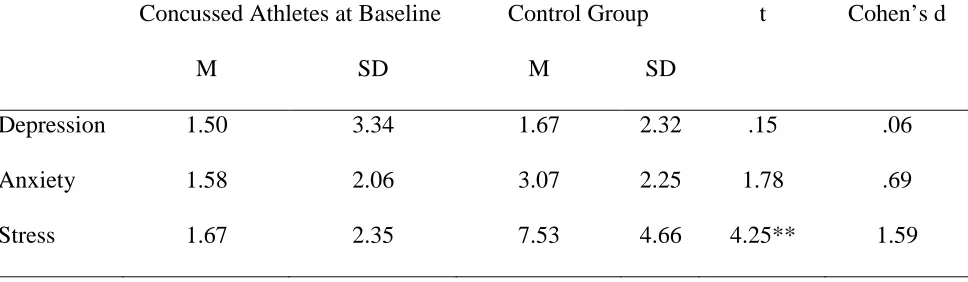
representations and coping as potential mechanisms. The present study included 30
participants (15 concussed athletes, 15 non-concussed non-athletes) that were matched on
age, education, and both past and current anxiety and depression. All participants
completed measures of coping and emotional symptoms (depression, anxiety, and stress),
provided two salivary cortisol samples (at the beginning and end of the experiment), and
completed two classical conditioning tasks (pleasant and aversive) while heart rate and
skin conductance responses were recorded. Background information, including history of
head injuries, was collected for all participants. Concussed athletes completed an
additional measure of illness representations. The results indicate that athletes
demonstrated faster reaction times to the conditioned stimulus during the acquisition
phase of the aversive task, and higher expectancy ratings to the conditioned stimulus
during the generalization phase of both the pleasant and aversive task. Further
exploratory analyses also revealed a pattern in which athletes had higher expectancy
ratings to the conditioned stimulus in the first trial of both the generalization and
extinction phases of both tasks. There were no differences in any of the other measures of
coping strategies were found to partially mediate the relationship between illness beliefs
of personal control and post-concussive symptoms. In addition, correlations between
cyclical timeline beliefs and poor outcome were identified. Implications and directions
DEDICATION
ACKNOWLEDGEMENTS
First and foremost, I would like to thank my family and my partner. To my
parents, your faith in my abilities has never wavered, and your support throughout this
journey – physically, emotionally, financially - has meant everything to me. To Kat, you
probably understand the anxiety I felt during this process better than anyone else. Our
sister-to-sister conversations always left me feeling understood and hopeful! To Chris,
thanks for always making me laugh and helping me to realize the important things in life.
I couldn’t ask for a better little brother! To my partner Mike, there are not enough words.
Your understanding and support have far exceeded my expectations throughout this
process. You’ve been there to celebrate every success, to calm every nerve, and to wipe
away every tear. I love you.
This dissertation would not have been possible without the support and guidance
of my research advisor, Dr. Christopher Abeare. Chris, I joined your research lab 9 years
ago as an undergraduate. You’ve helped me grow as a researcher, a clinician, and a
person ever since. Your belief in my ability to complete a doctorate in neuropsychology
more often than not exceeded my own, and your advice and kind words never failed to
reduce my anxiety or to motivate me. I couldn’t have done this without you!
There have been many people who have been involved in this research and
without them I would not have been able to complete my dissertation. I would like to
thank Dr. Lori Buchanan, Dr. Laszlo Erdodi, and Dr. Kevin Milne for serving on my
dissertation committee and offering guidance and insight at various points. Thank you as
well to Dr. Lynda Mainwaring for providing your expertise as my external reader. Thank
you to my research assistant Ben Guins for taking over data collection when I could no
longer be physically present in Windsor. I would also like to thank the wonderful
administrative staff within the Psychology department.
Finally, I would like to thank all of my friends and colleagues. To my University
of Windsor Psychology friends, we’ve made it! These years have been hard, but we’ve
woven in periods of laughter and countless good memories, and formed friendships that
outside of school, thank you for understanding when I was too busy or stressed to see
you, but always being there when I needed you. Your support has meant more than you
TABLE OF CONTENTS
DECLARATION OF ORIGINALITY iii
ABSTRACT iv
DEDICATION vi
ACKNOWLEDGEMENTS vii
LIST OF TABLES xiii
LIST OF FIGURES xv
CHAPTER 1: INTRODUCTION 1
CHAPTER 2: LITERATURE REVIEW 3
Concussion 3
Definition, Prevalence, and Cause 3
Biomechanics of Concussion 8
Neuropathology 10
Neurochemical Changes 11
Structural Changes 13
Functional Changes 17
Concussion Outcome 19
Factors Affecting Outcome 22
Post-Concussion Syndrome 24
Anxiety 26
Section I. Impact of Biological Variables: Aversive Conditioning and the HPA 30
The Role of the Hypothalamic-Pituitary-Adrenal (HPA) Axis 30
Aversive Conditioning and the Fear Circuit 32
The Relationship between Fear and Anxiety 32
Classical Conditioning 33
The Role of Aversive Conditioning in Anxiety 36
Fear Circuit 38
Amygdala 39
Hippocampus 43
Insula 45
Role of the Traumatic Event 45
Rodent Models of Aversive Conditioning following mTBI 46
Section II. Impact of Psychological Variables: Illness Representations and Coping 48
Common Sense Model 48
Components of Illness Representations 50
Coping 54
Common Sense Model: A Mediational Model 61
Current Study 63
Hypothesis1 64
Hypothesis 2 65
Hypothesis 3 65
Hypothesis 4 65
Hypothesis 5 65
CHAPTER 3: METHOD 66
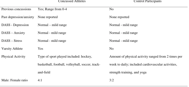
Participants 66
Method 67
Questionnaires 67
Intake Interview 67
Brain Injury Screening Questionnaire – Adapted 67
Post-Concussion Symptom Scale 68
Depression Anxiety Stress Scale 70
Brief COPE 70
Illness Perception Questionnaire – Revised 71
Physiological Measures 71
Aversive Conditioning Task 72
Pleasant Conditioning Task 74
Procedure 75
Clinical Participants 75
Control Participants 76
Data Manipulation 77
Participants 79
Control Group 79
Concussed Athlete Group 80
Descriptives 80
Statistical Analyses 82
Data Cleaning and Testing Assumptions 82
Affective Outcomes following Concussion 83
Pre-Acquisition Trial 83
Affective Ratings 84
Hypothesis1 86
Hypothesis 2 93
Hypothesis 3 99
Hypothesis 4 100
Hypothesis 5 104
CHAPTER 5: DISCUSSION 108
Classical Conditioning/Associative Learning 109
Acquisition and Extinction 109
Generalization 114
Cortisol-Related Stress Response 114
Common Sense Model 116
Integrating Psychological and Physiological Factors in Understanding Outcome 119
Study Limitations 120
Strengths of Study 121
Conclusions 123
REFERENCES 125
APPENDICES 153
Appendix A: Intake Interview 153
Appendix B: Brain Injury Screening Questionnaire – Adapted 160
Appendix C: Depression, Anxiety, and Stress Scale 163
Appendix E: Brief COPE 168
Appendix F: Illness Perception Questionnaire – Revised 170
LIST OF TABLES
Table 1. Questionnaires 69
Table 2. Comparison of Concussed Athlete and Control Participant
Groups 81
Table 3. Baseline Measures of Depression, Anxiety, and Stress 82
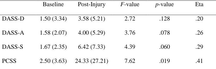
Table 4. Changes in DASS and PCSS Scores from Baseline to
Post-Injury in the Concussed Athlete Group 83
Table 5. Comparisons of Expectancy Ratings, Autonomic Response,
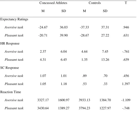
and Reaction Time during the Pre-Acquisition Trial 84
Table 6. Affective Ratings of the Conditioned Stimulus during the
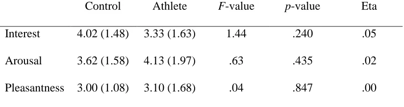
Aversive Conditioning Task 85
Table 7. Affective Ratings of the Conditioned Stimulus during the
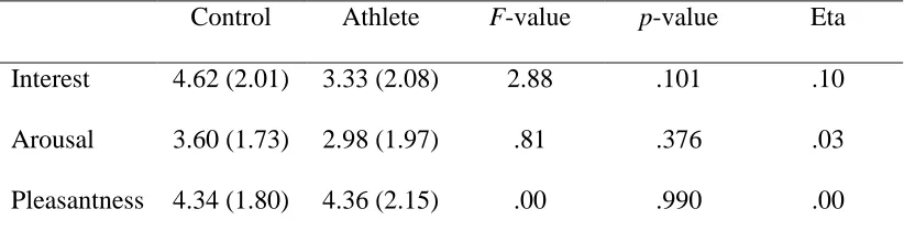
Pleasant Conditioning Task 86
Table 8. Correlations between Days since Injury and Mean Expectancy Ratings during the Acquisition, Generalization, and Extinction Phases of the Aversive and Pleasant Conditioning Tasks 93
Table 9. Mean Heart Rate (BPM) and Heart Rate Response during First Three Seconds of Conditioned Stimulus Presentation in
the Aversive Task 94
Table 10. Mean Skin Conductance (µs) and Skin Conductance Response during First Three Seconds of Conditioned Stimulus
Presentation in the Aversive Task 95
Table 11. Mean Heart Rate (BPM) and Heart Rate Response during First Three Seconds of Conditioned Stimulus Presentation in the
Pleasant Task 96
Table 12. Mean Skin Conductance (µs) and Skin Conductance Response during First Three Seconds of Conditioned Stimulus
Presentation in the Pleasant Task 97
Tasks 98
Table 14. Correlations between Days since Injury and Skin Conductance Response (µs) across Acquisition, Generalization, and
Extinction Phases of both the Aversive and Pleasant
Conditioning Tasks 99
Table 15. Mean Salivary Cortisol Levels (µg/dL) at Time 1, Time 2, and
Change over Time 100
Table 16. Correlations between Days since Injury and Salivary Cortisol
Levels (µg/dL) at Time 1, Time 2, and Change over Time 100
Table 17. Correlations between Days since Injury and Reaction Time (ms) across Acquisition, Generalization, and Extinction Phases
of both the Aversive and Pleasant Conditioning Tasks 103
Table 18. Correlations between Illness Representations and Outcome
Measures 105
Table 19. Correlations between Coping Subscales and Outcome
LIST OF FIGURES
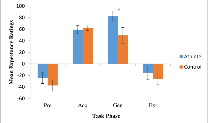
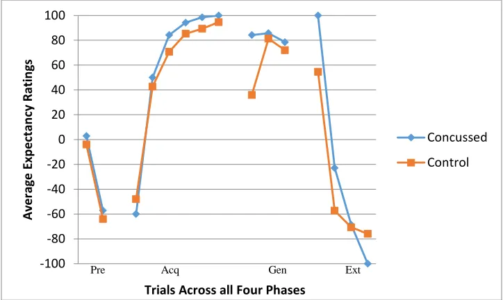
Figure 1. Mean Expectancy Ratings across Phases of the Aversive
Conditioning Task 87
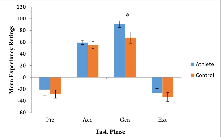
Figure 2. Mean Expectancy Ratings across Phases of the Pleasant
Conditioning Task 89
Figure 3. Mean Expectancy Ratings to the Conditioned Stimulus by
Presentation across all Phases of the Aversive Task 92
Figure 4. Mean Expectancy Ratings to the Conditioned Stimulus by
Presentation across all Phases of the Pleasant Task 92
Figure 5. Mean Reaction Times (ms) to the Conditioned Stimulus across
all Phases of the Aversive Conditioning Task 101
Figure 6. Mean Reaction Times (ms) to the Conditioned Stimulus across
all Phases of the Pleasant Conditioning Task 103
Figure 7. Mediational Analysis of Personal Control, Problem-focused
Coping, and PCSS 106
Figure 8. Mediational Analysis of Timeline-cyclical, Problem-focused
CHAPTER 1
INTRODUCTION
Although the significance of moderate and severe brain injuries has long been
recognized, the impact of milder brain injuries, including concussions, has only recently
been appreciated. Interest in mild traumatic brain injury (mild TBI; mTBI) has recently
been piqued by news of late-life dysfunction and autopsies of a number of professional
athletes engaged in high-contact sports, particularly football. Whereas the vast majority
of individuals recover fully from concussion, there is a small but significant group that
continues to exhibit cognitive, somatic, and affective symptoms beyond 3-months
post-injury (Kraus & Chu, 2005). Such prolonged symptoms are referred to as post-concussion
syndrome.
Further, evidence suggests that a mild brain injury places individuals at risk for
the acquisition of a variety of anxiety disorders, including posttraumatic stress disorder
(PTSD), generalized anxiety, and social phobia (Mooney & Speed, 2001). Of particular
interest in the present study were symptoms of anxiety occurring in the acute phase of
concussion, presenting as either part of the clinical picture of post-concussion syndrome
or the acquisition of specific anxiety disorders.
One major line of research has focused on the neuroanatomical substrates of
aversive classical (Pavlovian) conditioning as a critical component in the acquisition and
maintenance of anxiety disorders. Normal aversive conditioning relies on a vast network
of brain structures collectively referred to as the “fear circuit,” including the amygdala,
hippocampus, insula, anterior cingulate cortex, and other regions of the prefrontal cortex
(Sehlmeyer et al., 2009). Animal models of mild TBI and limited neuroimaging studies in
microscopic structural changes and dysfunction following injury (Giza & Hovda, 2014).
Further, rodent models of concussion that examined changes in aversive conditioning and
anxiety behaviours revealed increased aversive conditioning and generalization, as well
as increases in a variety of anxiety-like behaviours (e.g. Reger et al., 2012;
Almeida-Suhett et al., 2014). As of yet, these connections have not been established in human mild
TBI samples.
A separate line of research has focused on psychological factors impacting
recovery following mTBI. Recently, a limited number of studies have used the Common
Sense Model (CSM; Leventhal, Leventhal, & Contrada, 1998) as a paradigm for
understanding the impact of illness representations and coping strategies on outcome in
mild TBI samples specifically (Snell, Siegert, Smith, & Surgenor, 2011; Snell,
Hay-Smith, Surgenor, & Siegert, 2013; van Wilgen, Kaptein, & Brink, 2010). These studies
have typically focused on concussion symptoms as outcome variables, with less emphasis
on affective symptoms specifically. Further, these studies have focused solely on the
independent effects of illness representations and coping strategies, and have neglected to
investigate the mediational relationship of illness representations, coping, and outcome
proposed by the CSM.
A review of the literature identifies a gap in our understanding of the risk for
increased anxiety and diagnosed anxiety disorders following mild traumatic brain injury.
It also reveals that previous work has examined either physiological or
psychological/behavioural risk factors in isolation, even though the need to consider both
attempts to extend and integrate previous literature on rodent models of mild TBI and the
CSM to fill this knowledge gap.
CHAPTER 2
LITERATURE REVIEW
Concussion
Definition, Prevalence, and Cause
Traumatic Brain Injury (TBI) is a major public health concern. In the United
States alone, it is estimated that 1.8 million to 3.8 million brain injuries occur annually
(Faul, Xu, Wald, & Coronado, 2010; Langlois, Rutland-Brown, & Wald, 2006), with
75% of those injuries classified as mild (mTBI; Gerberding & Binder, 2003). This
classification of severity is based largely on the fact that these injuries involve only a
brief alteration of mental status, in comparison to the prolonged periods of
unconsciousness and posttraumatic amnesia associated with moderate and severe brain
injuries (Centers for Disease Control and Prevention, 2004). Despite the classification of
these injuries as mild, the economic impact is substantial, with mTBI accounting for
approximately 44% of the $56 billion annual cost of TBI in the United States alone
(Thurman, 2001). In 2010, the economic cost of TBI in the United States was estimated
to be even higher at $76.5 billion (Centers for Disease Control and Prevention, 2016).
Diagnosis of concussion is typically made based on a combination of subjective
report of symptoms and signs, neuroimaging findings, balance testing, and cognitive
testing. The range of symptoms include: somatic symptoms (e.g. headache); cognitive
symptoms (e.g., feeling like in a fog); and/or emotional symptoms (e.g., lability). There
(e.g., irritability), cognitive impairment (e.g., slowed reaction times), and sleep
disturbance (e.g., insomnia; McCrory et al., 2013). Within sports, suspected concussions
are often examined on the side-line using brief neuropsychological measures such as the
Sideline Concussion Assessment Tool (SCAT3) or Standardized Assessment of
Concussion (SAC; McCrory et al., 2013). Concussion is associated with negative
findings on conventional diagnostic imaging. Thus, CT and MRI scans are typically
employed only to test for the presence of hematomas and to rule out complications from
more severe head injuries (Eierud et al., 2014). As a result of the lack of diagnostic
markers on conventional neuroimaging tests and an emphasis on subjective symptoms,
concussions are often undiagnosed or misdiagnosed. This problem may be compounded
in athletes motivated to stay in play, who either under-report or do not report their
symptoms at all (Rabinowitz, Li, & Levin, 2013), or in cases of litigation where
individuals can be motivated to over-report symptoms (Belanger, Curtiss, Demery,
Lebowitz, & Vanderploeg, 2005).
The terms mTBI and concussion are used as labels for mild forms of head injury,
although their use does not have general agreement. These terms are often used
interchangeably, but recently, some researchers have argued for distinction between the
terms "mTBI" and "concussion" (McCrory et al., 2013) based on arguments that the
general public does not see the terms as synonymous. In the debate regarding mTBI and
concussion terminology, Ehmed and Sullivan (2015) examined 122 contact-sport players
and their reactions to sport-related vignettes that varied only in the diagnostic label
applied to each vignette ( i.e., concussion, mTBI, or no diagnosis). Participants rated their
the injury, as well as their expectations of poor outcome. The results showed that there
were no differences in players’ perceptions or symptom expectations based on the
diagnostic label provided. For the time being, most experts agree that concussion
represents a form of mTBI, with no more than a transient disruption of function
(Rabinowitz et al., 2014). Definitions of concussion/mTBI vary slightly based on the
specific group providing the definition. For example, the Centers for Disease Control
(CDC) and Prevention’s Mild Traumatic Brain Injury Working Group define a
concussion as “the occurrence of injury to the head arising from blunt trauma or
acceleration or deceleration forces with one or more of the following conditions
attributable to the head injury:
• any period of observed or self-reported
o transient confusion, disorientation, or impaired consciousness,
o dysfunction of memory around the time of injury, or
o loss of consciousness lasting less than 30 minutes;
• observed signs or other neurological or neuropsychological dysfunction, such as
o seizures acutely following the injury to the head,
o irritability, lethargy, or vomiting following head injury, or
o headache, dizziness, irritability, fatigue or poor concentration.”
The Mild Brain Injury Special Interest Group of the ACRM (American Congress
of Rehabilitation Medicine) provides a slightly different definition: “a patient with a mild
TBI is a person who has had a traumatically induced physiological disruption of brain
function, as manifested by at least one of the following:
• any loss of memory for events immediately before or after the accident,
• any alteration in mental state at the time of the accident (e.g., feeling dazed,
disoriented, or confused), or
• focal neurological deficits that may or may not be transient.”
The 5th International Conference on Concussion in Sport (McCrory et al., 2017) led to
the release of a consensus statement that defined a sport related concussion (SRC)
specifically as a “traumatic brain injury induced by biomechanical forces. Several
common features that may be utilised in clinically defining the nature of a concussive
head injury include:
• SRC may be caused either by a direct blow to the head, face, neck or elsewhere
on the body with an impulsive force transmitted to the head.
• SRC typically results in the rapid onset of short-lived impairment of neurological
function that resolves spontaneously. However, in some cases, signs and
symptoms evolve over a number of minutes to hours.
• SRC may result in neuropathological changes, but the acute clinical signs and
symptoms largely reflect a functional disturbance rather than a structural injury
and, as such, no abnormality is seen on standard structural neuroimaging studies.
• SRC results in a range of clinical signs and symptoms that may or may not
involve loss of consciousness. Resolution of the clinical and cognitive features
typically follows a sequential course. However, in some cases symptoms may be
The clinical signs and symptoms cannot be explained by drug, alcohol, or medication
use, other injuries (such as cervical injuries, peripheral vestibular dysfunction, etc.) or
other comorbidities (e.g., psychological factors or coexisting medical conditions).”
What is clear from the above definitions is that a concussion is generally
conceptualized as a transient condition with some change in mental state resulting from
trauma to the head, either as a direct force to the head or as the result of
acceleration-deceleration forces. Other aspects of the definitions vary, with differential importance
placed on neurological signs and symptoms. According to Bigler (2008), definitions of
concussion have four dominant features common to all: 1) brief alteration in
consciousness or neurological function with at least acute changes in mentation and speed
of processing, 2) physical symptoms of fatigue, headache, dizziness, and/or vertigo, 3)
impairments in short-term memory, attention, and concentration, and 4) increased
likelihood for changes in mood and affective functioning. A number of groups (e.g., Mild
Brain Injury Special Interest Group) delineate at which point a more severe diagnosis of
brain injury should be given; this includes a period of loss of consciousness longer than
30 minutes, posttraumatic amnesia (PTA) lasting longer than 24 hours, or a Glasgow
Coma Scale (GCS) assessed at less than 13. The GCS assesses motor, verbal, and eye
responses, and ranges from 3-15 with higher scores indicating higher levels of
functioning.
There are a wide range of situations and events that may lead to a head injury.
Common causes of mTBI include motor vehicle accidents (MVAs), falls (especially in
the very young and in older adults), assault, or struck by/against events (Faul et al.,
infantry soldiers reported injuries with loss of consciousness and 10% reported injuries
with altered mental status during a year-long deployment to Iraq (Hoge et al., 2008).
Recent work suggests even higher rates in this population, with up to 23% of army
personnel screening positive for clinician-confirmed TBI history (Terrio et al., 2009.)
Finally, participation in sports can lead to brain injuries; one estimate suggested that
approximately 300,000 sports-related concussions occur annually in the US (Thurman,
Branche, & Sniezek, 1998). It should be noted that this estimate included only
concussions that involved a loss of consciousness. Given that as many as half of
sports-related concussions go unreported (McCrea, Hammeke, Olsen, Leo, & Guskiewicz,
2004), and that only between 8% and 19.2% of these reported injuries are thought to
involve a loss of consciousness (Langlois, Rutland-Brown, & Wald, 2006), this number is
likely an underestimate. Langlois, Rutland-Brown, and Wald (2006) estimate that
anywhere between 1.6 and 3.8 million total sports-related TBIs occur each year. High
contact sports are most likely to cause concussions, with 4-20% annual incidence of
mTBI in football (Mendez, Hurley, Lassonde, Zhang, & Taber, 2005). Among high
school, college, and amateur athletes, ice hockey and rugby have the highest incidence of
concussion. At the recreational level, female taekwondo participants and male boxers
have the highest frequency of concussion (Mendez et al., 2005).
Biomechanics of Concussion
Biomechanics is defined as the study of biological systems, such as the brain, in
response to physical forces. In mTBI, mechanical forces can damage the brain directly
through delayed damage caused by the initiation of physiological processes leading to
cell dysfunction or death (Giza & Hovda, 2014).
Two important types of forces may play a role in the biomechanics of a
concussion: linear acceleration and rotational/angular acceleration. Early estimates of
injury tolerance levels (the likelihood that an individual will sustain a concussion at an
impact of a given magnitude) proposed that impacts of 90 g linear acceleration sustained
for 9 milliseconds or longer were sufficient to produce mTBI (Ono & Kanno, 1996).
Further research presented a more complicated picture. For example, some studies have
shown that linear acceleration as low as 60 g is sufficient to produce concussion
(Guskiewicz et al., 2007b), whereas others have shown that athletes can sustain impacts
greater than 90 g without any neurological dysfunction (McCaffrey, Mihalik, Crowell,
Shields, & Guskewicz, 2007). Thus, the relationship between impact force and injury
appears to not be direct, but rather moderated by other factors. In terms of rotational
acceleration, this is the type of acceleration that is thought to contribute most to
concussion (King, 2003). These rotational forces produce the shearing of axons and
deeper lesions; fronto-subcortical areas have proven particularly vulnerable to this type of
acceleration (Williamson, Heilman, Porges, Lamb, & Porges, 2013).
A combination of variables appears to better predict who will sustain a concussion
than any single variable in isolation. For example, Greenwald, Gwin, Chu, and Crisco
(2008) found that the best predictor of concussion was an algorithmic combination of
linear acceleration, rotational acceleration, Head Injury Criterion (HIC; a formula that
estimates the likelihood of head injury from impact by taking into account both
colleagues (2010) who found that concussion was most likely to occur when linear
acceleration exceeded 96.1 gs, rotational acceleration exceeded 5,582 rad/sec2, and
impact location was r the front, side, or top of the helmet.
Other studies have modelled the impact of acceleration and deceleration forces.
For example, Viano and associates (2005) simulated movement within the cranium
during concussion using finite element analysis and constructing a detailed anatomic
model of the brain and head accelerations based on game impacts from National Football
League (NFL) videotapes of injured players. These models indicated that the largest
strains occurred in the fornix, midbrain, and corpus callosum. In particular, the
hippocampus, caudate, amygdala, anterior commissure, and midbrain showed 4-5mm
displacements. As will be discussed in subsequent sections, structural changes and the
dysfunction of these medial structures may play a role in the acquisition of anxiety
disorders due to their role in classical conditioning, a type of associative learning that has
been implicated in both the acquisition and maintenance of anxiety.
Neuropathology
There appears to be an absence of any clear morphological or functional
abnormalities in the brains of individuals sustaining a mild TBI (Bigler & Maxwell,
2012). Specifically, conventional imaging techniques such as Computerized Tomography
(CT) and Magnetic Resonance Imaging (MRI) are either negative for any abnormal
findings, or show only minimal levels of damage (Bazarian et al., 2013). In the absence
of overt macroscopic brain pathology, however, mTBI is typically associated with
microscopic brain pathology, specifically widespread axonal injury. Known as diffuse
neuroimaging techniques such as Diffusion Tensor Imaging (DTI; Bazarian et al., 2013).
Given that axons are particularly vulnerable to mechanical injury due to their viscoelastic
properties, diffuse axonal injury (DAI) is common after mTBI. For example, Browne,
Chen, Meaney, and Smith’s (2011) porcine models of mTBI revealed multifocal axonal
pathology. Similarly, as discussed in detail later, DTI has been used to examine
neuropathology in humans with concussion, and has revealed evidence for widespread
axonal injury (e.g., Zhang, Heier, Zimmerman, Jordan, & Ulug, 2006).In addition to the
diffuse axonal injury that is incurred by some following concussion, research has
revealed a cascade of neurochemical changes that begin immediately following the
impact or force causing the head injury.
Neurochemical Changes
Giza and Hovda (2014) provide a comprehensive review of the current
understanding of the neuropathology of concussion using rodent models. Based on their
research, they define concussion as a “neurometabolic cascade of events” that includes:
ionic flux and glutamate release, an energy crisis, cytoskeletal damage, axonal
dysfunction, altered neurotransmission, inflammation, and cell death. This metabolic
cascade is initiated by biomechanical forces that lead to the opening of ion channels via
the disruption of neuronal membranes and axonal stretching. The opening of these
channels causes uncontrolled ionic flux with an efflux of potassium and an influx of
sodium and calcium into the cell. Further depolarization is caused by a hyperacute
indiscriminate release of the excitatory neurotransmitters glutamate. In an effort to restore
ionic homeostasis, sodium potassium pumps relying on adenosine triphosphate (ATP)
the cell converts increased amounts of adenosine diphosphate (ADP) into ATP. The
combination of decreased cerebral blood flow, diminished glucose availability, and an
increased need for glucose leads to an energy crisis in the brain. After this initial period
of hyperglycolysis, glucose metabolism becomes impaired for a period of 7-10 days. The
cytoskeletal damage caused by the biomechanical forces of concussion affects the
dendritic arbors and axons of both neurons and glial cells, which provide support and
protection for neurons. Concussion has also been linked to changes in inflammatory
markers, shown by upregulation of cytokine and inflammatory genes (Li, Lee, Cai,
Sutton, & Hovda, 2004; Patterson & Holahan, 2012).
Research employing non-human animal models of mTBI suggests that within 10
days of injury, chemical and metabolic levels return to normal (Hovda, Yoshino,
Kawamata, Katayama, & Becker, 1991; Yoshino, Hovda, Kawamata, & Becker, 1991).
This is consistent with neuropsychological functioning in humans demonstrating a 10-day
recovery curve (Belanger & Vanderploeg, 2005). The pathophysiological changes of
concussion map onto a number of clinical signs and symptoms. For example, the brain’s
increased need for energy within the acute recovery period is consistent with an increased
vulnerability to a second injury during this time for both humans and animals
(Guzkiewicz et al., 2003; Bigler, 2008). Further, symptom exacerbation with physical
exertion is commonly reported during this time, providing additional evidence that the
brain experiences an increased need for energy during this acute phase (Leddy et al.,
2010). Giza and Hovda (2014) proposed a number of additional connections between the
neurobiology of concussion and early clinical symptoms: ionic flux and migraine,
processing, and slowed reaction times; impaired neurotransmission and impaired
cognition, slowed processing, and slowed reaction time; and protease activation, altered
cytoskeletal proteins, and cell death, and chronic atrophy and development of persistent
impairments. In addition to specific symptoms, neurometabolic changes of decreased
glutamate in the acute phase of concussion correlate with self-reported symptom severity
(Henry, Tremblay, Boulanger, Ellemberg, & Lassonde, 2010).
Many of the above findings were based on non-human animal models of brain
injury. Of course, it is also important to validate these mTBI models in human
populations. On this note, Bergsneider and colleagues (2001) found reduced glucose
metabolism for approximately one month following mild to severe TBI in a patient
sample. Zetterberg et al. (2006) examined markers of neuronal and astroglial injury in the
cerebrospinal fluid (CSF) of amateur boxers and found that indicators of neuronal injury
by-products were significantly related to the number of hits taken during a bout. These
acute pathological changes were found despite the fact that the hits were subconcussive
in nature. In a group of professional boxers, Zhang, Heier, Zimmerman, Jordan, and Ulug
(2006) found subtle white matter abnormalities using DTI techniques. These results were
replicated by Chappell and colleagues (2006) in a group of 81 professional boxers.
Finally, Cohen and associates (2007) found subtle brain volume loss in a 20 patient
sample of mild TBI. These studies suggest that the glucose hypometabolism, axonal
damage, and cell death seen in animal models are consistent with the neuropathology of
concussion in humans.
Researchers examining rodent models of brain injury have employed a number of
different injury mechanism procedures including: lateral fluid percussion (LFP) injury
(Lifshitz, Witgen, & Grady, 2007; Reger et al., 2012), blast overpressure (Genovese et
al., 2013; Elder et al., 2012), controlled cortical impact (CCI; Almeida-Suhett et al.,
2014), and weight-drop procedures (Meyer et al., 2012). These procedures differ in a
number of ways, including severity and level of invasiveness. Despite these differences,
these experimental procedures in rodents produce similar pathological features to those
characteristic of brain injury in humans, specifically neuronal loss, gliosis, and metabolic
and ionic perturbations (Lifshitz et al., 2007).
These studies overall have found a lack of gross pathology, consistent with the
mTBI presentation seen in human populations, but have identified axonal degeneration
and loss of neurons as a consequence of mild TBI procedures in rats. For instance, Heldt
and colleagues (2014) observed scattered axonal degeneration in brain sections of mice
3-8 weeks after blast-induced trauma in spite of a lack of gross cerebral pathology. This
pattern of neuronal loss is consistent with the diffuse axonal injury characteristic of mild
TBI in humans. In addition, despite the lack of gross pathology following a single mild
TBI, some functional impairment over multiple injuries suggests that there may be some
chronic long-term structural changes (DeFord et al., 2002).
In addition to diffuse microstructural changes, some research suggests changes in
structural volume in specific regions, including the amygdala and hippocampus. The
amygdala and hippocampus are bilateral structures located deep within the temporal
lobes that, as a result of their location in the brain, may be particularly vulnerable to the
The amygdala is an almond-shaped structure comprising 13 nuclei, which can be divided
into three major groups: deep or basolateral group, superficial or cortical-like group, and
the centromedial group. It plays an important role in emotional processing, particularly
the processing of fear. The hippocampus is a seahorse-shaped structure found adjacent to
the amygdalae that plays an important role in emotional responding and human memory.
More detailed information regarding the function of these structures is discussed in
subsequent sections.
Meyer et al. (2012) discovered neuronal cell loss in the dorsal hippocampus and
increased cell numbers in subregions of the amygdala. Similarly, Lifshitz and colleagues
(2007) found significant neuronal loss in the hippocampus, but not in the amygdala. In
spite of the literature indicating no reduction in the overall number of neurons within the
amygdala, studies focused on Thy1 excitatory projection neurons specifically have found
evidence of decreased neurons. In two separate studies, mice receiving overpressure air
blasts of 50-60 psi showed decreased numbers of Thy1 enriched neurons in the
basolateral amygdala two months after blast, with reductions by 25% (Heldt et al., 2014)
and 20% (Reiner et al., 2015), respectively. The functional implications of these
disturbances on anxiety and fear processes will be described below.
There have been efforts to investigate structural changes in humans using a wide
range of neuroimaging techniques, including Positron Emission Tomography (PET),
Single-Photon Emission Computed Tomography (SPECT), Magnetoencephalography
(MEG), Electroencephalography (EEG), Magnetic Resonance Imaging (MRI), and
subtypes of MRI including DTI, functional MRI (fMRI), and Magnetic Resonance
Curtiss, and Warden (2007) indicated that there are at least some abnormalities associated
with mild TBI across imaging modalities. Their qualitative review of these studies
suggested that many of the results indicate structural changes relating to mild TBI.
Possibly the most sensitive structural imaging technique to the effects of mTBI is
DTI, which allows for the examination of the structural integrity of white matter tracts by
calculating the amount of directional restriction of water movement in the brain. When
water is unrestricted, it diffuses equally in all directions (isotropic), but when it is
restricted, it will not diffuse equally in all directions (anisotropic). The type of diffusion
is dependent largely on the type of tissue present; for example, in CSF the water is
largely unrestricted, but along axons and myelin sheaths the water is generally restricted
to a movement that is parallel to white matter tracts, due to the fact that white matter is
made of lipids and lipids are hydrophobic (Shenton et al., 2012). There are a number of
DTI measures, with functional anisotropy (FA) values most commonly used as a
sensitive, but non-specific, marker of neuropathology and microstructural change
(Alexander, Lee, Lazar, & Field, 2007). This value provides a marker of the shape of the
diffusion; unrestricted diffusion typically creates a spherical shape while restricted
diffusion created an elongated ellipsoid.
Mild TBI research employing DTI measures have found mixed evidence for FA
values in the acute phase of concussion, with some finding decreased FA values (e.g.,
Matsushita et al., 2011) and others findings increased values (e.g., Bazarian et al., 2007).
However, when examining DTI anisotropy values following mTBI across studies, there
appears to be a pattern that is temporal in nature. The meta-analysis by Eierud et al.
and chronic mTBI tended to be correlated with depressed anisotropy levels. Both are
suggestive of white matter changes, with elevated anisotropy levels thought to reflect
axonal swelling processes in the acute phase, and reduced anisotropy levels related to
damage of myelin or axon membranes, reduced axonal packing density, and/or reduced
axonal coherence (Shenton et al., 2012).
Functional Changes
In addition to the structural changes noted above, the mild TBI literature suggests
that there are abnormalities in functioning, even in areas thought to be structurally intact.
Recent studies employing rodent models have examined the effects of mTBI on the
function of various brain structures, particularly mesial temporal lobe structures including
the amygdala (Meyer et al., 2012; Elder et al., 2012; Lifshitz et al., 2007; Reger et al.,
2012) and hippocampus (Meyer et al., 2012; Lifshitz et al., 2007; Reger et al., 2012).
There is evidence of amygdalar dysfunction after brain injury in rodents across a number
of studies (Hovda et al., 1991; Meyer, Davies, Barr, Manzerra, & Forster, 2012). For
example, Ameida-Suhett and colleagues (2014) used a blast-induced mTBI procedure in
rodents and found subsequent bilateral amygdalar hyperactivity. There is also evidence of
prolonged hippocampal dysfunction following mTBI in rodents (Fendt & Fanselow,
1999), including changes in inhibitory neurotransmission (Reger et al., 2012).
A number of studies have also found alterations in protein synthesis and
neurotransmission in the amygdala. Specifically, with regards to protein synthesis, there
is indication of an elevation in the protein stathmin 1 in the amygdala, which is crucial
for the regulation of innate and learned fear (Elder et al., 2012). Research examining
excitatory receptors and processes in the context of decreased inhibitory receptors and
processes, with a sum excitatory effect, particularly within the basolateral amygdala
(BLA). For example, changes in inhibitory neurotransmission were evidenced by
decreased levels of GAD67, a biosynthetic enzyme for gamma-aminobutyric acid
(GABA), in the amygdala (Reger et al., 2012). Further, these authors found a significant
upregulation of excitatory N-methyl-D-aspartate (NMDA) NR1 receptors in the BLA, a
trend for increased NR2A and B NMDA receptors, and a trend toward decreased
GABA-related inhibition in the BLA and hippocampus. Similarly, Almeida-Suhett and
colleagues (2014) found significant loss of GABAergic interneurons and significant
reductions in the frequency and amplitude of spontaneous and miniature GABAA
-receptor mediated inhibitory postsynaptic currents (IPSCs), indicating a significant
reduction of inhibition in the BLA. This was associated with reduced surface expression
of α1, β2, ƴ2 GABAA receptor subunits. Finally, there were significant increases in the
surface expression and current mediated by α7-nAChR, indicating increased excitability
of principal neurons within the BLA.
There have been investigations of functional changes in humans using a wide
range of neuroimaging techniques. A review of these neuroimaging results by Belanger,
Vanderploeg, Curtiss, & Warden (2007) indicated that there are at least some functional
abnormalities associated with mild TBI in areas found to be structurally intact.
Simmons and Matthews (2012) conducted a meta-analysis of fMRI studies for
individuals with heterogenous mTBI performing a variety of tasks, mostly cognitive and
motor in nature. The authors found dysregulation of function in several prefrontal,
gyri, superior and inferior parietal lobules, superior temporal gyrus, and medial frontal
cortex. In a different fMRI meta-analysis, areas of reduced activity included the middle
frontal gyrus, right middle temporal gyrus, right precentral gyrus, and right anterior
cingulate. Areas of increased activity included the right insula, right inferior parietal
lobule, right cerebellar tonsil, right inferior frontal gyrus, and right supramarginal gyrus
(Eierud et al., 2014). These results are generally consistent with an anterior-to-posterior
pattern of activity in which there is reduced activity in anterior regions and increased
activity in posterior regions. Consistent with these results are studies finding evidence of
functional abnormalities in the amygdala following mild TBI. For example, following
blast induced TBI, bilateral amygdalar hyperactivity has been observed in U.S. soldiers
(Matthews et al., 2011).
Overall, research examining the neuropathology of concussion in rodent and
human populations has found an absence of macroscopic brain pathology, in the context
of microscopic brain pathology characterized by widespread axonal injury, with
associated neurochemical and metabolic changes, neuronal loss and decreased volume in
mesial brain structures, and both hyperactivity and hypoactivity of various brain
structures. The pattern of recovery from this neuropathology and variables affecting
typical outcome are discussed below.
Concussion Outcome
The majority of patients recover completely from mTBI (Iverson, 2007). Despite
the minimal overt brain damage in mild TBI, current statistics indicate that about 10-15%
of mTBI patients will develop persistent cognitive, behavioural, and/or emotional
poor outcome ranging from 24% to 55% by 3-months post-injury, 26% to 51% by
6-months post-injury, and 27.3% to 50% by 12-6-months post-injury (Snell, Siegert,
Hay-Smith, & Surgenor, 2011a). These statistics are hotly debated in the literature and it is
suggested that these rates may be largely inflated due to the self-report nature of
measures, motivational biases, and litigation status. A large meta-analysis conducted by
Binder, Rohling, and Larrabee (1997) found no lasting effects of mTBI at 3-months
post-injury. This meta-analysis was updated and corroborated by Frencham, Fox, and
Maybery (2005). Other researchers (Pertab, James, & Bigler, 2009; Bigler, Farrer, Pertab,
James, Petrie, & Hedges, 2013) have examined the same data and concluded that the
methodological flaws associated with meta-analysis hides a “lost minority,” a minority of
mTBI patients that suffer from persistent symptoms.
The acute outcome of concussion tends to be better in athletes, with athletes
tending to show full neuropsychological recovery within 10 days, whereas concussion
symptoms tend to resolve completely within three months post-injury for the general
population of patients (Belanger and Vanderploeg, 2005). This difference in acute
recovery may be due to a number of reasons at biomechanical, physiological, and
psychological levels (Rabinowitz et al., 2014). From a biomechanical perspective, the
forces involved in sports-related injuries tend to be less severe in comparison to other
common injury mechanisms (i.e. motor vehicle accidents, falls) and the physical
attributes of athletes, including well-developed neck musculature, help minimize
rotational acceleration forces present in mTBI injuries. At the physiological level, higher
pre-injury level of fitness may protect from neuronal injury and properly timed physical
perspective, athletes are less likely to have comorbid psychiatric diagnoses and generally
demonstrate lower stress responses, as measured by adrenocortical responses, autonomic
responses, and psychological responses (e.g., Rimmele et al., 2007; Rimmele et al.,
2009). A study by Verner et al. (2010) found that female athletes similarly demonstrated
a lower cortisol stress response to an experimental stressor in comparison to female
non-athletes. In all three of these studies baseline levels were similar across groups. A lower
stress response in athlete groups may act as a protective factor in recovery. Motivation
may also lead to differences in acute recovery, with the higher motivation of athletes to
return to play may lead them to minimize symptoms (Rabinowitz, et al., 2014), whereas
other mTBI patients may have the opposite motivation.
When examining long-term outcome, however, athletes may be at risk for chronic
problems. For example, studies have shown an association between recurrent concussion
and late-life cognitive dysfunction (Guskiewicz et al., 2005) and depression (Guskiewicz
et al., 2007a) in retired professional football players. Furthermore, more than three
decades after injury, athletes who played at university level and incurred a concussion
continued to demonstrate electrophysiological abnormalities and cognitive and motor
impairments when compared to matched controls with no history of concussion;
specifically, they exhibited delayed and attenuated P300 brain signals, reduced movement
velocity, and lower scores on tasks of episodic memory and response inhibition (De
Beaumont et al., 2009).
Within the present study, emotional complaints related to symptoms of anxiety
following mTBI are of interest. Anxiety following mTBI can present as part of the
disorder subsequent to a head injury. Understanding the etiology of affective symptoms
following mTBI will help in the early identification of those at risk for developing PCS
and acquired anxiety disorders. Before PCS and anxiety disorders are discussed in detail,
various factors that may lead to poor outcome are considered.
Factors affecting Outcome
Recovery after mTBI, including the resolution of cognitive, somatic, and affective
symptoms, remains poorly understood. The majority of research suggests an interplay
between psychogenic and physiogenic factors (King & Kirwilliam, 2011). One early
model put forth by Lishman (1988) suggested that neurobiological factors were solely
implicated in the development of symptoms, while psychological factors accounted for
the maintenance of long-lasting symptoms. However, Silverberg and Iverson (2011)
updated this model by reviewing research in the 20 years following Lishman’s original
paper, suggesting that both neurobiological and psychological factors play a role in the
development and maintenance of post-concussion symptoms. Other factors that have
been examined include demographic variables and factors related to injury mechanism
and severity.
Demographic factors related to outcome include age, gender, and education.
Research generally finds that increased age is related to poorer outcome, particularly
being over the age of 40 (Binder, 1986). Female gender has widely been cited as a
predictor of poor outcome, particularly PCS symptoms (Meares et al., 2008; Edna &
Cappelen, 1987). A study found that females reported more PCS symptoms, but did not
differ from males in respect to number of days before returning to normal functioning and
In terms of educational attainment, the majority of research has found that higher levels
of education are associated with better outcomes (Stulemeijer, van der Werf, Borm, &
Voss, 2008), although at least one study has found a correlation between higher education
and increased odds of poor outcome (Snell et al., 2011a).
The majority of studies have found that injury mechanism and severity of the
injury are not related to outcome. For example, in a study by Snell and colleagues
(2011a) measures of injury severity, including Glasgow Coma Scale scores, duration of
loss of consciousness, and posttraumatic amnesia duration did not differentiate between
good and poor outcome groups. Concussion biomechanics, similarly, do not appear to
play a predictive role in outcome. Guskiewicz et al. (2007b) found that there was no
significant correlation between concussive impact magnitude and post-injury changes in
symptoms, postural control, and cognitive functioning among collegiate athletes. Broglio,
Eckner, Surma, and Kutcher (2011) replicated these findings in a sample of high school
football players, finding no association between cumulative linear or rotational
acceleration and post-concussive outcomes.
It has been suggested that both pre-morbid and current psychiatric and
psychological variables may mediate persistent symptoms following mTBI (McCrae et
al., 2009). Some of the strongest psychological predictors include prior psychiatric
history (Meares et al., 2008; Carroll et al., 2004), including premorbid anxiety or
depressive disorders (Meares et al., 2011), personality traits such as neuroticism
(Keshavan, Channabasavanna, & Reddy, 1981), and stressful life experiences (Lidvall,
Linderoth, & Norlin, 1974). Further, a history of alcohol or substance misuse is
In terms of motivational factors, compensation-seeking, effort, and motivation are
consistently demonstrated as the strongest predictors of outcome (Carroll et al., 2004).
Meta-analysis of outcome suggested that litigation status was associated with greater
cognitive sequelae that were stable or worsened over time (Belanger et al., 2005). As a
result, understanding the factors that play a role in the etiology of true cases of PCS may
be best achieved through the study of concussed athletes, who are generally motivated to
recover quickly in an effort to return to play. Thus, in the current study, the possible
development of PCS and anxiety symptoms is examined during the acute phase of
concussion when individuals may be in a vulnerable neurometabolic state.
Post-Concussion Syndrome
Post-Concussion Syndrome (PCS) is defined as set of symptoms following mTBI
that typically include: physical symptoms, such as headaches and dizziness; cognitive
symptoms, such as problems with memory and concentration; and emotional symptoms,
including irritability, anxiety, emotional lability, and depression. Cases of concussion that
have not recovered within 3 months generally receive a diagnosis of PCS. Our
understanding of PCS is complicated by inconsistencies in definition. The DSM-IV
provided a diagnostic criteria for post-concussional disorder (PCD) in the Criteria Sets
and Axes Provided for Further Study, and the ICD-10 provided a diagnostic set for PCS;
however, the symptom criteria differ between these two diagnostic systems (Bigler,
2008). As a result, there are differences in prevalence rates between the two criteria sets.
Specifically, prevalence rates are higher when using ICD-10 criteria (64%) than DSM-IV
criteria (11%) three-months post-injury (Boake et al., 2005). PCS as a syndrome has been
uninjured individuals. Up to 88% of healthy individuals report post-concussion
syndrome-type symptoms, despite never having sustained a concussion.
There is some evidence to suggest that there is symptom overlap between PCS
and anxiety disorders, including physiological symptoms such as sleep disturbance,
fatigue, cognitive symptoms such as difficulty concentrating, and emotional symptoms
such as irritability and feelings of anxiety. Exploratory factor analyses of the
Post-Concussion Symptom Scale (PCSS; Lovell, Collins, Podell, Powell, & Maroon, 2000), a
22-item scale examining PCS, suggested a factor solution that included somatic,
cognitive, sleep, and emotional factors in a group of athletes following concussion
(Kontos, Elbin, Schatz, Covassin, Henry, Pardini, & Collings, 2012). Potter, Leigh,
Wade, and Fleminger (2006) found a three-factor structure of cognitive, somatic, and
affective factors when using the Rivermead Post Concussion Symptom Questionnaire
(RPQ; King, Crawford, Wenden, Moss, & Wade, 1995), another commonly used
measure for examining PCS. The presence of emotional factors in these measures
underscores the similarity in symptoms between PCS and affective disorders.
In addition to significant symptom overlap, there also appears to be significant
comorbidity between symptoms of emotional distress and PCS symptoms. In one sample,
a measure of anxiety correlated strongly with concurrent PCS symptoms (King, 1996). In
another, there were large group differences on measures of anxiety between a group with
or without PCS (Meares et al., 2006). In addition to anxiety, depression also predicts PCS
(Lange, Iverson, & Rose, 2011). PCS and emotional distress were highly correlated in a
sample of mTBI and trauma control patients (Landre, Poppe, Davis, Schmaus, & Hobbs,
diagnosis predicts the development of PCS in mTBI patients (Meares et al., 2008). In a
pediatric concussion sample, those with premorbid anxiety disorders scored significantly
higher on all three factors of the PCSS than those without anxiety disorders (Joyce,
LaBella, & Carl, 2014). The overlap in symptoms between PCS and anxiety disorders
and the comorbidity between the two makes differential diagnosis difficult and may lead
to an under-diagnosis of anxiety disorders in this population; despite these difficulties,
however, anxiety is a common affective outcome of mTBI and described in detail below.
Anxiety
The majority of studies examining affective outcomes following mTBI find an
increase in anxiety symptoms and/or anxiety disorders (e.g., Epstein & Ursano, 1994;
Mooney & Speed, 2001). However, some early studies did not find any evidence of
anxiety following mTBI. For example, a study by Schoenhuber and Gentilini (1988)
screened 35 head injured patients with the State Trait Anxiety Inventory at 5-17 months
post-injury and found that there were no differences in either the state or trait subscale
between patients and healthy controls. This lack of finding may be due to the small
sample and the long post-injury periods seen in this study; it may be the case that any
acute changes in anxiety resolve over a period of time. Some recent studies have found
elevated levels of anxiety symptoms and disorders following mTBI. According to Rao
and Lyketsos (2002), the most common post-TBI anxiety symptoms include
“free-floating anxiety, fearfulness, intense worry, generalized uneasiness, social withdrawal,
interpersonal sensitivity, and anxiety dreams.” In a meta-analysis, Epstein and Ursano
(1994) found a prevalence rate of 23% for anxiety disorders in mild TBI across three
of TBI across twelve studies. These statistics are consistent with those found by Mooney
and Speed (2001) in which 24% of their participants with mild TBI were classified as
having developed an acquired anxiety disorder. Within the general population, the pooled
lifetime prevalence rate of anxiety disorders across 46 studies from 1980 to 2004 was
16.6% (Somers, Goldner, Waraich, & Hsu, 2006), suggesting that mTBI confers an
increase in the likelihood of developing an anxiety disorder. As noted by Mainwaring,
Hutchison, Camper, and Richards (2012) in a comprehensive review of the emotional
sequelae of sports concussion, anxiety has not been a focus of study for investigators in
this field. For example, while Erlanger and colleagues (2003) identified clinical reports of
post-concussive irritability and nervousness following sports concussion, they did not
examine symptoms of anxiety or the possible presence of anxiety disorders more
specifically.
The prevalence of different types of anxiety disorders following TBI differ.
Across all severity of TBI, 3-28% met the criteria for generalized anxiety disorder
(GAD), 4-17% met criteria for Panic Disorder, 1-10% met criteria for phobic disorders,
2-15% met criteria for obsessive-compulsive disorder (OCD), and 3-27% met criteria for
posttraumatic stress disorder (PTSD; Koponen et al., 2002; Hiott & Labbate, 2002).
According to these data, the most prevalent forms of anxiety disorders following TBI are
GAD, PTSD, and panic disorder. Within the general population, community lifetime
prevalence rates for various disorders are estimated to be: 5% for GAD, 8% for PTSD,
1-2% for Panic Disorder, 4-8.8% for Specific Phobia, and 2.5% for OCD (DSM-IV-TR).
When community prevalence rates are compared with prevalence rates for individuals
and Panic Disorder. Specific phobias and OCD do not appear to be significantly elevated
following TBI.
With regard to mTBI in particular, research with military samples suggests that
PTSD is particularly common after mTBI sustained during combat events. In a group of
2525 soldiers, 43.9% of those reporting a loss of consciousness met criteria for
posttraumatic stress disorder (PTSD). In comparison, 27.3% of those reporting altered
mental status, 16.2% of those reporting other injuries, and 9.1% of those denying any
injury met the criteria for PTSD (Hoge et al., 2008). Other work has found similarly high
rates of PTSD after mTBI (Bryant, 2001), and some studies suggest that combat-induced
TBI approximately doubles the risk for PTSD (Schneiderman, Braver, & Kang, 2008).
The high degree of comorbidity that exists between PTSD and mTBI (Stein &
McAllister, 2009) presents a number of difficulties for understanding their influence on
one another. To begin, the differentiation between mTBI and PTSD is difficult due in part
to the many overlapping and self-reported symptoms including fatigue, irritability, poor
sleep, and a number of cognitive deficits. Differential diagnosis is therefore based largely
on the predominant symptoms. Mild TBI is typically diagnosed on the basis of injury
characteristics, including loss of consciousness, and posttraumatic amnesia and
confusion, details of the injury itself (i.e. self and witness reports; Ruff, Iverson, Barth,
Bush, & Broshek, 2009), and physical symptoms such as headache, difficulties
concentrating, and photophobia and phonophobia. A diagnosis of PTSD is typically made
on the basis of symptoms of re-experiencing, avoidance, negative mood and cognitions,
and arousal (DSM-V). As a result, much controversy exists regarding the differentiation
mTBI and PTSD dates back to WWI and the concept of shell shock; at this time, there
was much debate as to whether these symptoms represented psychic or physiological
causes (Elder et al., 2012). Hoge and colleagues (2008) found a nonsignificant
relationship between mTBI and outcome after controlling for PTSD, suggesting that poor
outcome is the result of psychological variables and not lasting neurotrauma. In contrast,
in a sample of trauma survivors, moderate to severe head injury and PTSD independently
predicted symptom reporting and interacted such that the relationship between head
injury and number of health complaints was stronger when posttraumatic stress disorder
symptoms were more severe (Keatley, d’Alfonso, Abeare, Keller, & Bertelsen, 2015).
Other studies suggest that mTBI puts individuals at risk for developing PTSD. For
example, Mora and colleagues (2009) studied 333 burn victims with or without primary
blast injury/mTBI and found a greater prevalence of PTSD in those with mTBI than those
with other injury mechanisms. Similarly, Walilko and colleagues (2009) examined 124
survivors of Oklahoma City Bombing and explored the relationship between PTSD and
physical injuries; in this sample PTSD and head/brain injuries were significantly
associated, while PTSD was not highly correlated with other injuries. These findings of
Mora (2009) and Walilko (2009) suggest that TBI may predispose individuals to the
development of PTSD. Bryant (2008) expanded on this idea and suggested that mild TBI
may diminish the capacity to employ cognitive resources that would normally be engaged
in problem solving and regulating emotions after trauma, thereby leaving individuals
more susceptible to PTSD and related problems. Similarly, Elder and colleagues (2012)
proposed that TBI might predispose individuals to PTSD if TBI damages brain structures
identify frontal and limbic areas including the prefrontal cortex, amygdala, and
hippocampus (Etkin & Wager, 2007). This is in keeping with neuroimaging findings in
anxiety disorders that identify dysfunction in similar areas, including the amygdala,
insula, and anterior cingulate cortex (Holzschneider & Mulert, 2011). Given that these
medial brain regions are vulnerable to the effects of concussion, their dysfunction
provides a possible biological mechanism for the increased risk for anxiety following
concussion. One possible mechanism for this impact is through their influence on the
hypothalamic-pituitary-adrenal (HPA) axis, which plays an important role in stress
responses and anxiety. Another possible mechanism is through their purported role in
classical conditioning, particularly fear conditioning.
Section I. Impact of Biological Variables: Aversive Conditioning and the HPA
The Role of the Hypothalamic-Pituitary-Adrenal (HPA) Axis
At a biological level, structures that play an important role in the regulation of
responses to stress include the hypothalamic-pituitary-adrenal axis (HPA axis), brain
stem noradrenergic neurons, sympathetic adrenomedullary circuits, and parasympathetic
systems. The HPA axis is the neuroendocrine component of this stress response system.
When a human perceives a stressful situation, the hypothalamus releases
corticotropin-releasing factor (CRF)/corticotropin/corticotropin-releasing hormone (CRH). This then binds to
CRF/CRH receptors on the anterior pituitary gland, signalling the release of
adrenocorticotropic hormone (ACTH). Finally, ACTH binds to receptors in the adrenal
gland to stimulate the release of glucocorticoids, including cortisol (Smith & Vale, 2006).
The hypothalamus is regulated by glucocorticoid feedback, as well as by afferent