ABSTRACT
KIM, MIN JUNG. Characterization of agrin function in chicken and zebrafish
embryogenesis. (Under the direction of Dr. Gregory J.Cole)
Agrin is an extracellular matrix heparan sulfate proteoglycan that plays a
key role in the development of the neuromuscular junction (NMJ) by inducing the
clustering of acetylcholine receptors at synaptic sites of the NMJ. Although recent
studies have extended our understanding of agrin’s function in the nervous system,
its function in the CNS is not clearly understood.
The present study was undertaken to assess the role of agrin in neurite
outgrowth mediated by the basic fibroblast growth factor (FGF-2), using both PC12
cells, and chick retina neuronal cultures. Agrin increases the efficacy of FGF-2
stimulation of neurite outgrowth, as an inhibitor of the FGF receptor abolished
neurite outgrowth in the presence of agrin and FGF-2. Agrin augments and sustains
FGF-2.
To overcome the lethality of agrin gene disruption and the difficulty of
embryonic manipulation of agrin function in mice, a gene encoding zebrafish agrin
was identified and characterized. Zebrafish agrin is expressed in the developing
CNS, the NMJ, and non-neural structures such as the pronephric duct, and
endodermal tissues. A morpholino-based gene targeting against agrin significantly
impair development of tail and the NMJ, and cause severe defects in motor neuron
axon outgrowth and formation of the midbrain-hindbrain boundary, eye, and otic
vesicles. Morphants subsequently develop paralysis, and die at larvae stages.
Knockdown of agrin in zebrafish strikingly resembles phenotypes of
zebrafish FGF-related mutants, such as disruption of the MHB, optic and otic
vesicles during zebrafish development. Inhibition of FGFR synergizes defects from
agrin knockdown resulting in MHB disruption, a shortened tail, small eyes and otic
vesicles, which suggest that agrin modulates the activity of FGF signaling pathways.
In conclusion, my studies show that agrin is essential for NMJ formation as
CHARACTERIZATION OF AGRIN FUNCTION IN CHICKEN
AND ZEBRAFISH EMBRYOGENESIS
by
MIN JUNG KIM
A dissertation submitted to the Graduate Faculty of North Carolina State University
in partial fulfillment of the requirements for the Degree of Doctor of Philasophy
COMPARATIVE BIOMEDICAL SCIENCES
Raleigh 2004
APPROVED BY:
_______________________________ _______________________________ Dr. Philip L. Sannes Dr. Robert R.H. Anholt
____________________________________ Dr. Brenda J. Brizuela
Minor Representative
____________________________________
DEDICATION
To my parents,
Han-Bae Kim and Soon Jin Oh, Who made all of this possible,
With their endless encouragement and support, I could follow my heart’s decision and passion.
also to My lovely husband,
Younghoon Baek,
BIOGRAPHY
Min Jung Kim was born on November 15, 1973 and raised in Seoul, Korea.
She received B.S. degree in Biology at Sook-Myung women’s University in 1996. During her undergraduate studies, she participated in research project on Functional analysis of cytochrome c oxidase and worked with Dr. Eunsook Song and Dr. Bernard Kadenbach at Universitat of Marburg, Germany. From 1996-1998, she pursued a master degree in Zoology from Sook-Myung women’s university , Korea. Her master’s thesis was entitled “Effect of aging on mitochondrial transcription and translation of cytochrome c oxidase”. In 1999, she worked as a researcher at St. Mary’s
hospital, Catholic University, Korea. During this period, she participated in research project involving stem cell biology and identification of leukemia.
ACKNOWLEDGMENTS
The heart decides, and what it decides is all that really matters.
When I started my PhD program, I tried to find what I really want to devote myself for future. I followed my instinct and had a lot of trial and error. During this short journey, I had enormous influence from many people. Without help, support, and encouragement from several persons, I would never have been able to finish this work.
I would like to devotedly thank my advisor, Gregory J. Cole, for his inspiring and enthusiastic encouraging way to guide me to a deeper understanding of neuroscience world, and his deliberate mentoring during the whole work with this dissertation. I would never forget his endless support, patience and concern, which was essential to the completion of all my work.
I would give my special thanks to Dr. Brenda Brizuela for fruitful discussion and help for developmental biology. I am very grateful to my research advisory committee members: Robert Anholt and Phill Sannes.
My thanks also go to Dr. Christine Beattie, Dr. Ava Udvadia, Michelle for a big help to set up zebrafish system. Without their help, I could not reach this goal.
I would like to extend my gratitude to some special people who are always there for me in spite of:
To my beloved husband, Younghoon, for being there whenever I felt down towards my work: To my parents, for always supporting and believing in me, without them, I could not be here at all: To my brother for cheering me up: To my parents-in-law, for their support and concern: To my friends and relatives for supporting me with love and understanding.
TABLE OF CONTENTS
LIST OF TABLES……….………xi
LIST OF FIGURES……….……xii
INTRODUCTION………..1
CHAPTER I THE HEPARAN SULFATE PROTEOGLYCAN AGRIN MODULATES NEURITE OUTGROWTH MEDIATED BY FGF-2………...22
ABSTRACT……….…..23
INTRODUCTION……….….25
MATERIALS AND METHODS……….…..30
MATERALS………..……….………30
PURIFICATION OF AGRIN………..…...30
RESULTS………..….35 AGRIN SPECIFICALLY STIMULATES NEURITE OUTGROWTH
MEDIATED BY FGF-2 IN PC12 CELLS………..………..……….35 AGRIN EXERTS ITS EFFECTS ON FGF-2 MEDIATED NEURITE
OUTGROWTH THROUGH THE FGF RECEPTOR…………..………….39 AGTIN EXERTS ITS EFFECTS ON FGF-2 MEDIATED NEURITE
OUTGROWTH BY MODULATING ERK ACTIVITY IN PC12 CELLS...40 ROLE OF AGRIN IN MODULATING FGF-2 MEDIATED NEURITE
OUTGROWTH IN RETINAL NEURONAL CULTURES…..…….……...44 AGRIN EXERTS ITS EFFECTS ON RETINAL NEURITE OUTGROWTH
VIA AN FGF-2 DEPENDENT ERK SIGNALING MECHANISM………54 AGRIN INCREASES AND SUSTAINS FGF-2 MEDIATED C-FOS
PHOSPHORYLATION……….………....56 DISCUSSION……….59
CHAPTER II
AGRIN IS REQUIRED FOR POSTERIOR DEVELOPMENT AND AXON
PATHWAY FORMATION IN EMBRYONIC ZEBRAFISH………...68
INTRODUCTION……….……….70
MATERIALS AND METHODS………...74
FISH MAINTENANCE………..…..….74
MOLECULAR CLONING………..………..……74
ANTISENSE MORPHOLINO INJECTION……….…75
NORTHERN BLOT ANALYSIS……….……….75
WHOLE MOUNT IN SITU HYBRIDIZATION………..76
ANTIBODY PRODUCTION……….…..………….…77
WESTERN BLOT ANALYSIS………...………..78
IMMUNOHISTOCHEMISTRY……….….…..78
ANALYSIS OF NEUROMUSCULAR JUNCTION DEVELOPMENT..………79
BODIPY STAINING……….…80
ANALYSIS OF APOPTOSIS IN ZEBRAFISH EMBRYOS…………..……….80
RESULTS………..………….81
CLONING AND EXPRESSION PATTERN OF AGRIN mRNA AND PROTEIN DURING EMBRYONIC DEVELOPMENT IN ZEBRAFISH………..…..….81
GUIDANCE AND OUTGROWTH….……….95
DORSAL PROJECTING SECONDARY MOTOR NERVES ARE AFFECTED BY AGRIN KNOCK-DOWN………97
AGRIN KNOCK-DOWN DISRUPTS TRIGEMINAL MOTOR NEURON AXONAL PROJECTION………..………99
AGRIN KNOCK-DOWN LEADS TO IMPAIRED AXON OUTGROWTH BY ROHON-BEARD SENSORY NEURONS………..………100
MUSCLE DEVELOPMENT APPEARS NORMAL IN AGRIN KNOCKDOWN EMBRYOS………...………102
DISCUSSION……….……….107
CHAPTER III AGRIN REGULATES THE DEVELOPMENT OF CNS STRUCTURES THAT REQUIRE FGF SIGNALING PATHWAYS. ………..116
ABSTRACT……….………117
INTRODUCTION………..……..118
MATERIALS AND METHODS……….……122
ANTISENSE MORPHOLINO INJECTION………....…122
SU5402 TREATMENT……….…123
SOLID PHASE BINDING ASSAY……….….123
RESULTS……….…125
AGRIN KNOCK DOWN DISRUPTS NORMAL FORMATION OF THE MIDBRAIN-HINDBRAIN BOUNDARY, EYES AND OTIC VESICLES…..125
AGRIN BINDS TO FGF-8 IN VITRO………..…...………...,127
INHIBITION OF FGF SIGNALING PATHWAYS MIMICS AGRIN KNOCKDOWN PHENOTYPE IN ZEBRAFISH EMBRYOS……….……..127
PERTURBATION OF CNS DEVELOPMENT DUE TO AGRIN KNOCKDOWN IS VIA DISRUPTED FGF SIGNALING………...……….……131
DISCUSSION………..135
GENERAL SUMMARY AND CONCLUSIONS ……….…….139
LIST OF TABLES
1. Phenotypes of embryos injected with agrin morpholino oligonucleotides………....93
2. The average distance between head and tail of early stage zebrafish embryos…….96
LIST OF FIGURES
Figure 1. The structure of glycosaminoglycans in proteoglycans………..2 Figure 2. The primary domain structure of agrin………..12 Figure 3. Agrin potentiates the ability of FGF-2 to promote neurite outgrowth from
PC12 cells………...………37 Figure 4. Quantitation of neurite outgrowth from PC12 cultures treated with FGF-2
and/or agrin………...…..41 Figure 5. Phosphorylation of p42/44ERK in PC12 cells in response to FGF-2 treatment (2.5 ng/ml) in the presence or absence of 200 ng/ml agrin………43 Figure 6. Agrin promotes a rapid initiation of retinal neuronal neurite outgrowth in the presence of FGF-2……….46 Figure 7. Effect of agrin on FGF-2 mediated neurite outgrowth from retinal neurons.. Figure 8. Effect of agrin on FGF-2 mediated retinal neurite outgrowth… …………48 Figure 9. Quantitation of neurite outgrowth from retinal cultures treated with FGF-2
and/or agrin……….. 51 Figure 10. Effect of elimination of cell surface HS-GAGs on retinal neurite outgrowth
in response to FGF-2 treatment………53 Figure 11. Phosphorylation of P-ERK in retinal neurons in response to treatment with
Figure 14. Localization of agrin mRNA in the zebrafish embryo……….84 Figure 15. Localization of agrin protein in zebrafish embryo……….. 87 Figure 16. Immunohistochemistry confirms the efficient inhibition of agrin expression and splice-site-targeted agrin morpholino oligonucleotide alters splicing of agrin in zebrafish………89 Figure 17. Knock-down of agrin by morpholino oligonucleotide injection leads to gross
abnormalities in zebrafish posterior development……….92 Figure 18. Defects of anterior-posterior development occurred during early
development………...95 Figure 19. Effects of agrin morpholinos on primary and secondary motoneuron axonal
growth………98 Figure 20. Agrin knock-down disrupts the axonal projection of trigeminal motor
neurons throughout development……….……101 Figure 21. Truncation of axonal outgrowth by spinal cord sensory neurons during
INTRODUCTION
Proteoglycans are glycosylated proteins that have covalently linked sulfated
glycosaminoglycans, which are attached to serine residues of the protein core (Kjellen
and Lindahl, 1991). Glycosaminoglycans (GAGs) are different disaccharide repeated
polymers that are generated by the structural modification of deacetylation, sulfation
and epimerization that provide diverse biological activities (Lindahl, 1990). These
various structural modified GAG chains are critical for developmental processes as
well as in pathogenesis. Proteoglycans are present in the extracellular matrix, such as
cartilage, basement membrane and connective tissues, as well as at the cell surface. The
types of GAGs can be divided into several groups, which are chondroitin sulfate (CS),
dermatan sulfate (DS), heparan sulfate (HS), heparin, and keratan sulfate (KS) (Figure
1). The structure of GAGs associated with proteoglycans exhibit distinct differences in
the amino sugars found in CS-GAGs and HS-GAGs (acetylgalactosamine and
N-acetylglucosamine, respectively) and in the pattern of sulfation in the individual GAGs.
CS-GAGs typically contain higher levels of sulfate residues than HS-GAGs, with one
sulfate per repeating disaccharide. HS-GAG contains sulfate on both the uronic acid
group and amino sugar group, which gives more variability in sulfation, including the
higher sulfated domains serve as HS-GAG binding domains of HSPGs to
heparin-binding proteins to provide discrete functions (Aviezet et al, 1994; Sanderson et al.,
1994; Cotman et al., 1999; Herndon et al., 1999; Knox et al., 2002). The change of
sulfation pattern of HS-GAGs also has been shown to modulate the binding activity of
specific HSPGs (Nurcombe et al., 1993; Brickman et al., 1998).
Among proteoglycans, heparan sulfate proteoglycans show the highest structural
variability. Heparan sulfate proteoglycans are extracellular and cell surface
macromolecules that consist of a core protein to which heparan sulfate
glycosaminoglycan (HS-GAG) chains are attached. The major HSPGs can be divided
into families based on structure of core protein, as well as cellular localization.
Syndecans and glypicans are cell surface HSPGs (Sanderson and Bernfield., 1988;
David et al., 1990; Filmus et al., 1995; Karthikeyan et al., 1992; Litwack et al., 1994;
Stipp et al.,1994; Watanabe et al., 1995), and perlecan, agrin and collagen XVII are
associated with the extracellular matrix (Lozzo., 1998; Olsen., 1999; Cole and Halfter.,
1996; Halfter et al., 1998).
The most important roles for heparan sulfate proteoglycans have been
demonstrated in cell adhesion, cell migration, modulation of growth factor function,
organization of the extracellular matrix, inflammation, metastasis, and synapse
biological roles in nervous system development. Development of the nervous system
can be largely divided into three stages. First, neurons are generated from neural
precursors and differentiate into specific cell types, such as neurons or glia, followed
by cell migration to their appropriate CNS destination. Second, axons and dendrites
extend from neurons toward target cells, and synaptic contacts are formed and mature.
Third, a functional CNS requires maintenance and plasticity of synaptic arrangements
in order to allow learning, memory, and other important brain functions to occur.
During the process of neural development, various types of proteoglycans have been
shown to have diverse functions and to interact with molecules essential to
development, such as growth factors and morphogens.
In particular, the fibroblast growth factors (FGFs) have well demonstrated
interactions with HSPGs in both embryonic development as well as adulthood. FGFs
are secreted molecules that are involved in the regulation of cell survival,
proliferation, migration and differentiation, as well as the patterning of body axes
(Brickman et al., 1998; Vaccarino et al., 2001; Molteni et al., 2001; Perrone-Capano
and Di Porzio., 2000; Mufson et al., 1999, Andersn et al., 1993; Grothe and
Wewetzer., 1996). At early stages of embryogenesis, FGFs are required to induce
mesoderm and establish the anteroposterior and dorsoventral body axis (Draper et al.,
especially growth and patterning of the brain, the initiation of limb buds, and tooth
morphogenesis (Liu et al., 2003, Scholpp et al., 2003; Walshe and Mason, 2003,
Wiellette and Sive, 2004).
24 FGF family members and 4 FGF receptors have been identified. The
molecular weights of vertebrate FGF proteins are 17 to 34 kDa. FGFs consist of 28
highly conserved domains and six identical amino-acid residues (Otnitz., 2000). The
core protein of FGF-1 and FGF-2 is composed of 12 antiparallel β-strands, which serve
as the heparin-binding domain (Moy et al., 1996; Li and Seddon., 1994). Most FGFs
have N-terminal signal peptides, which allows FGFs to be readily secreted from cells
into the extracellular space. However, FGF-1, -2, -9, -16 and -20 lack conventional
signal peptides, and rather are secreted molecules which may be released from cells by
a mechanism independent of the endoplasmic reticulum-Golgi pathway (Mignatti et al.,
1992; Friesel and Maciag.,1999).
Transgenic mice for most FGFs have been generated and display the various
phenotypes expected for each FGF mutation, from no phenotypic change to embryonic
lethality. Among various FGF family members, FGF-2 deficient mice are viable, but
show failure of neural regulation of blood pressure, decrease of vascular smooth
muscle contractility, and defects and impairment of the cerebral cortex and cervical
due to gastrulation defects, with these mice displaying defects in cardiac, craniofacial,
forebrain, midbrain and cerebellar development (Meyers et al., 1998). Although FGFs
are essential molecules in embryonic development as well as adulthood, mild
phenotypic changes in several FGF transgenic mice may imply substitution from
redundancy of different FGF members. In fact, FGF-8 and FGF-17 can compensate
each other with respect to defects of midbrain-hindbrain boundary development (Xu
and Ornitz., 2000). Moreover, knock down of different FGF molecules synergize the
mild defects of single gene disruption, leading to more severe phenotypic changes. For
example, a similar phenotype of smaller otic placodes was shown in FGF-3 knock out
mice (Mansour et al., 1994) and the ace zebrafish, which is a hypomorphic mutation in
the FGF-8 gene (Reifers et al., 1998). Knock down of both genes resulted in a failure of
otic placode development in zebrafish embryos (Maroon et al., 2002; Leger and Brand,
2002; Phillips et al., 2004).
FGFs have two different possible receptors: cell-surface bound tyrosine kinase
receptors (FGFRs) as high affinity receptors (Coughlin et al., 1988) and heparin-like
glycosaminoglycans, such as HSPGs, as low-affinity receptors (Venkatamaran et al.,
1999; Stauber et al., 2000). Two FGF molecules can form a complex bound to one FGF
receptor, which is connected by a heparan sulfate proteoglycan. The receptor complex
vitro and in vivo (Rapraeger et al., 1991; Yayon et al., 1991; Ornitz et al., 1992; Lin et
al., 1999). In the absence of HS, FGF cannot bind FGFRs, on the other hand, addition
of HS can reconstitute FGF-FGFR complex formation (Yayon et al., 1991). The
HS-chains play a key role in orchestrating the formation and stabilization of the FGF:FGFR
signaling complex (Guimond and Turnbull., 1999; Guimond et al., 2001).
During embryonic development, HSPGs may play a crucial role to regulate FGF
signaling pathways. FGFs are involved in cell proliferation, differentiation and
migration during embryonic development and HSPGs can regulate ternary complex
formation and FGFR oligomerization to regulate FGF related signal transduction. The
relationship between HSPGs and FGFs provides an important clue to their functions
during neural development. The central nervous system is generated from
neuroectoderm, with neural precursors being localized in the ventricular zone of the
neural tube. Neurons and glial cells arise from neural precursor cells in the ventricular
layer and the subventricular layer from embryonic stages into adulthood. During
neurogenesis, many growth factors and morphogens are required to regulate
proliferation and differentiation of neural precursor cells. FGF-2 is a strong candidate
for an essential growth factor for neurogenesis in the developing cerebral cortex. The
spatio-temporal expression of FGF-2 in the embryonic brain coincides with
neurogenesis in mouse (Qian et al., 1997). Moreover, FGF-2 and FGFRs are highly
precursor cells, and promotes self-renewal activity of precursor cells in rat (Qian et al.,
1997; Ghosh and Greenberg, 1995; Vicario-Abejón et al., 1995). In addition, low and
high concentrations of FGF-2 can regulate cell fate from neuronal to oligodendroglial
progeny, respectively (Qian et al., 1997). Since distinct HSPGs have different binding
affinities for FGFs in a spatio-temporal manner, differential glycosylation of HSPGs
may regulate the specificity of FGF binding during neurogenesis. After neurogenesis
occurs, neurons migrate into target regions and extend axons and dendrites to form a
network with other neurons. Recent studies have demonstrated the important role of
proteoglycans in axon guidance. Wang and Denburg found that exogenously added
heparin and heparan sulfates, or elimination of them by heparitinase treatment caused
pathfinding errors in pioneer axons of cultured cockroach embryos (Wang and Denburg,
1992). Recent studies have shown that exogenous addition of heparan sulfate resulted
in failure of retinal axons to enter the optic tectum in Xenopus (Irie et al., 2002). This
failure of axon guidance was dependent on FGF-2 binding of HSPGs and only
FGF-2-binding HSPGs could cause aberrant axonal targeting. In Xenopus, FGF-2 was shown
to stimulate axonal outgrowth and also caused a mistargeting of retinal axons following
addition of exogenous FGF-2 to optic nerve explants (Walz et al., 1997). Explants
treated with a dominant-negative form of FGFR extend axons, but axons failed in target
recognition (McFarlane et al., 1996). Since FGF-2 has been suggested to be involved in
elements for the process of learning and memory. Addition of FGF-2 can improve
long-term potentiation (LTP) after stimulation of subcortical afferents (Abe et al.,
1992). Since enzymatic cleavage of HS by heparitinase, as well as addition of soluble
heparin-type carbohydrates, prevented expression of LTP in rat hippocampal slices,
HSPGs and FGFs may act as important regulators in the modulation of neuronal
connectivity (Lauri et al., 1999).
To understand the role of HSPGs in brain development, we must learn how
synapse formation occurs in CNS as well as how cell proliferation, differentiation and
migration of neurons is regulated during development. A well-known molecule
essential to NMJ formation, agrin, is a strong candidate molecule for an important
element for neurosynapse formation/maintenance and neurogenesis.
In the 1970s, reformation of synapses after muscle denervation was mediated by
extracellular matrix from the synaptic cleft at the neuromuscular junction (Sanes et al.,
1978 and Burden et al., 1979). This implied that an essential factor for synapse
formation existed in the ECM at the NMJ and could organize the induction of pre- and
post-synaptic differentiation. Smith et al purified agrin from basal lamina extracts of
the synapse rich electric organ of Torpedo California (Smith et al., 1984). They
suggested in the “agrin hypothesis” that agrin is the nerve-derived factor required to
This hypothesis was confirmed by the following studies. Agrin knock out mice die
perinatally from a breathing failure and do not form functional synapses at the NMJ
(Gautam et al., 1996). Moreover, overexpression of agrin can restore the reconstruction
of a functional mature synaptic apparatus into non-synaptic region of rodent muscle
(Meier et al., 1997).
The agrin gene encodes a core protein of over 2,000 amino acids with a
predicted molecular mass of approximately 250 kDa. Tsen et al had identified that
agrin belongs to the family of heparan sulfate proteoglycans (Tsen et al., 1995). Agrin
contains at least three O-linked carbohydrate attachment sites which serve as binding
sites for not only heparan sulfate glycosaminoglycan side chains, but also a hybrid
HSPG/CSPG (Winzen et al., 2003) and increase the total mass of the protein to over
600 kDa. Agrin is expressed abundantly in the developing brain and the basal laminae
of lung and kidney (Tsen et al., 1995, Burgess et al., 2002 and Burg et al., 1995).
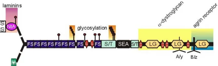
Agrin has multiple domains that can interact with a number of molecules.
Agrin’s primary structure shows a multimodular composition which contains a globular
NtA laminin-binding domain, a central rod-like domain with nine follistatin-like
protease inhibitor domains, two laminin-like epidermal growthfactor (EGF) repeats,
like domains (Figure 2). Theglobules are connected by flexible rod-like structures. The
agrin protein contains threeHS favorable attachment sites in the two domains, both
located in the central part of the molecule:the first regionis located just upstream from
the eighth follistatin-like domain and the second, that contains two possible HS
attachment sites, directlyprecedes the SEA module (Winzen et al., 2003).
Alternative splicing is an important mechanism for functional diversity in the
molecules controlling neuronal activity(Grabowski et al., 2001). Alternative splicing is
also central to the development of the nervous system, the differentiation of neurons
and the formation of their connection patterns. Alternative splicing is extremely well
suited to produce large numbers of subtly different protein functions. Agrin is
alternatively spliced in a highly time and tissue specific manner (Campanelli et al.,
1996;O’Connor et al., 1992; Burgess et al., 1999; Kroger et al., 1997; Daggett et al.,
1996; Gessmann et al., 1996; Thomas et al., 1993; Stone et al., 1995; McMahan et al.,
1992). These multiple isoforms of agrin provide diverse possible functional roles
during development (Deyst et al., 1998; Rupp et al., 1991; Rupp et al., 1992; Annies et
al., 2002). The best-known function of agrin is the aggregating activity of AChRs at the
neuromuscular junction (NMJ), which is dependent on the z insert agrin splicing form
in rodent.
Figure 2. The primary domain structure of agrin (Bezakova, 2002). The signal sequence (SS)
determines whether agrin is to be secreted and the NtA domain provides the binding domain to laminin.
At the N-terminus, 9 follistatin-like domains (FS), laminin EGF-like domains (LE) and
serine/threonine-rich domains (S/T) can be postranslationally modified by glycosylation and contains 3 possible GAG
attachment sites. At the C-terminus, the sea urchin sperm domain (SEA), EGF-like domains (EG),
laminin globular domains (LG) and the A/y and B/z alternative splice sites shows agrin's
at different sites. The COOH-terminal of agrin contains three different splicing sites
which are x, y/A, z/B in mammals/chicken, respectively (Ferns et al., 1992). Agrin’s
C-terminal region plays an essential role in regulating its binding to cell surface proteins
and AChR aggregating activity (Rupp et al., 1991; O’Connor et al., 1992; Lakso et al.,
1992). Isoforms containing 4 amino acids at y/A and either one or both 8 and 11 amino
acids inserts at z/B sites are synthesized by motor neurons and highly active in AChR
clustering activity (Ferns et al., 1992). Isoforms without inserts are formed by muscle
and Schwann cells and are inactive in clustering. The z exons confer the AChR
clustering activity on the protein, whereas the Y exon encodes a heparin-binding site
(Campanelli et al., 1996; Gessmann et al., 1996; Bowen et al., 1996; Rossant et al.,
1995) (Figure 2). Moreover, z insert agrin is expressed only in neurons, whereas
nonneural cells, including glia and myotubes, express only B/z insert-negative forms.
Gautam et al have generated agrin-deficient mutant mice where neuromuscular
differentiation was grossly defective in these mice, and no severe phenotypic defect
was detected in their brain (Gautam et al., 1996). In addition, the disruption of z
inserted isoforms blocked neuromuscular synapse formation and resulted in death after
birth. However, the phenotype in the CNS did not show significant change except for a
slightly smaller size of brain when compared to wild-type littermates (Serpinskaya et
al., 1999). Although y insert of agrin is known for its binding to heparin, the disruption
mean a redundant or dispensable function of the y insert isoforms in mammals.
However, z-negative forms were still expressed in these mutants, and compensation of
z-negative isoforms cannot be ruled out.
The 5’ end of the agrin gene exhibits heterogeneity and gives rise to the
diversity of agrin’s localization, tissue distribution, and function. Mice express twin
distinct short NH2-terminal (SN) and long NH2-terminal (LN) isoforms of agrin
(Neumann et al., 2001; Bixby et al., 2002; Ji et al., 1998; Burgess et al., 2000). The
existence of these distinct isoforms explains the previously noted lack of homology
between the NH2 termini of agrin isolated from rats and chicks (Neumann et al., 2001).
For chicken, SN-agrin is homologous to the transmembrane (TM) - domain and
LN-agrin is equivalent to the secreted signal (SS) -NtA-domain. SN- and LN-LN-agrin are
likely to be transcribed from distinct promoters, and they are expressed in different
patterns throughout development (Burgess et al., 2000; Shigemoto et al., 2000).
SN-agrin is largely confined to the nervous system, whereas LN-SN-agrin is broadly
distributed in neuronal and nonneuronal tissues. Moreover, analysis of native and
recombinant protein indicate that SN and LN-agrin exhibit distinct subcellular
localizations, determined by their NH2 termini: LN-agrin associates with the ECM
while SN-agrin remains attached to cell surfaces (Denzer et al., 1995). LN-agrin
transmembrane protein. The SN-agrin terminus can mediate externalization and
membrane anchoring of heterologous proteins. The perturbation of LN-agrin leads to
neonatal lethality caused by a failure of neuromuscular junction formation that is the
same phenotype as z insert negative isoforms (Burgess et al., 1999; Burgess et al.,
2000). However, both z-/- and LN-/- agrin mice did not show any significant
phenotypic change in the CNS. Thus, basal lamina-associated LN-agrin is required for
neuromuscular synaptogenesis. Even though agrin mutant analysis proved the
importance of agrin in NMJ formation, none of the mutants showed a phenotypic defect
during CNS development as a result of loss of agrin.
Besides alternative splicing mechanism contributing to agrin function, different
regions of agrin’s modular organization provide distinct functional properties, allowing
agrin to interact with a number of distinct molecules. The NtA domain is required for
binding to laminin, which can localize agrin to the basal lamina (Denzer et al., 1995;
Denzer et al., 1997). The laminin-G1 and G2-like domains can bind to α-dystroglycan
which can form the syntrophin-associated glycoprotein complex at the muscle cell
surface (Gesemann et al., 1996; Hopf and Hoch, 1996). The activity of rod-like central
domains has been identified for NCAM, HB-GAM, FGF-2, merosin, thrombospondin,
β-amyloid binding (Cole and Halfter, 1996; Cotman et al., 1999; Burg et al., 1995;
as well as a binding site for heparin and integrin (Martin and Sanes., 1997; Gesemann
et al., 1996; Campanelli et al., 1996).
From studies on agrin’s association with multiple molecules, Musk
(muscle-specific receptor tyrosine kinase) and α-dystroglycan were identified as strong
candidates for agrin’s cellular receptor, which mediates signaling for inducing AChR
aggregation at the NMJ (Sanes et al., 1998; Ruegg and Bixby., 1998; Montanaro et al.,
1998). α-dystroglycan was considered as a direct receptor of agrin due to its activity in
AChR aggregation. However, its absence does not interfere with the formation of
NMJs and it binds agrin’s inactive alternative splicing form B/z-, instead of B/z+ form
(Companelli et al., 1996). Therefore, it is believed dystroglycan might be involved in
the consolidation rather than formation of the synaptic apparatus. On the other hand,
MuSK can be activated by agrin at the NMJ. MuSK-deficient mice die perinatally and
lack postsynaptic specialization at the NMJ much like agrin-deficient mice (Gautam et
al., 1999; Sanes., 1997; DeChiara et al., 1996). Absence of MuSK in myotube cultures
results in a failure to aggregate AChR in response to agrin, and addition of MuSK
allows recovery of the aggregation of AChR (Meier et al., 1997). In addition, the agrin
B/z+ form, but not B/z- form, can induce MuSK phosphorylation (Herbst and Burden.,
2000). Although MuSK can induce the aggregation of AChRs and post-synaptic
possess another agrin receptor, which may be a co-receptor of MuSK.
Outstanding progress has been made toward understanding synaptogenesis at
the NMJ (Bezakove et al., 2001; Terrado et al., 2001; Nitkin et al., 1987; Reist et al.,
1992), but synapses in the CNS have been much less accessible to experimentation. For
instance, the gap between CNS synapses is much narrower than that of neuromuscular
synapses and CNS synaptic clefts do not contain a basal lamina. The diversity of both
their functional and molecular properties such as ion channel/receptor complements,
(Sharp et al., 1996) also renders characterization difficult. Because of easy access to
study mechanisms of synaptogenesis, most studies regarding agrin have focused on
NMJ synaptogenesis. However, high expression of agrin in the developing brain
implies important roles for agrin in CNS, and the existence of a transmembrane form of
agrin could provide a possible explanation that agrin may be expressed at the surface
membranes of CNS neurons in the absence of laminin. Even though the role of agrin in
synaptogenesis in the CNS is controversial, recent studies have been supportive
evidence about this hypothesis (Ma et al., 2000; Borges et al., 2001; Bezakove et al.,
2001; Cotman et al., 1999; Halfter et al., 1997; Hering et al., 1999; Ruegg et al., 2001;
Uhm et al., 2001; Biroc et al., 1993). Gingras et al showed that synaptogenesis is
impaired in neural agrin-deficient superior cervical ganglion (SCG) cultures, with
significantly fewer synaptophysin-labeled nerve terminals and synaptic aggregates of
studies showed that agrin differentially regulates axonal and dendritic growth which
induced both dendritic elongation and dendritic branching (Mantych et al., 2001).
Agrin is specifically localized at sympathetic synapses in vitro, and is consistent with it
playing a role in interneuronal synapse formation (Gingras et al., 2001). The lethality of
agrin mutant mice has been limited the study of agrin’s role in the CNS. Since
neuron-neuron synaptogenesis is progressing even after birth, one cannot rule out the
possibility of agrin’s function in synaptogenesis in CNS beyond previous studies. Even
though the understanding of synaptogenesis in the CNS has been enigmatic,
identification of agrin’s role in synapse formation, maintenance and maturation will
further our understanding of mechanisms of CNS development and agrin’s role in CNS
development.
Based on evidence that agrin is expressed prior to synapse formation, and its
expression is not limited only in neuronal cells, this has suggested a potential role of
agrin throughout development and not just in synaptogenesis. These data imply various
broader roles of agrin for neurogenesis, axonogenesis, blood-brain barrier formation etc.
The generation of distinct classes of neurons at defined positions in the rostro-caudal
and dorso-ventral axis of the neural tube is a fundamental step in the establishment of
the functional complexity of the vertebrate CNS. CNS neurons are generated through a
and undergo final differentiation. Research over the past two decades has elucidated
many of the genetic pathways underlying these biological processes.
Even before the onset of synapse formation, agrin is highly localized in the
ventricular and subventricular zone of the developing brain and spinal cord, and
declines during development before largely disappearing in the adult brain (Stone et al.,
1995; Cohen et al., 1997). Interestingly, the expression pattern of SN-agrin is quite
unique in its temporal and spatial manner during development. SN-agrin is highly
expressed in postmitotic neurons of the cortical plate, moderate in migrating
neuroblasts of the intermediate zone, and lowest in progenitors of the ventricular zone
in forebrain. This pattern is opposite that seen for LN-agrin. Also SN-agrin is abundant
in midbrain, hindbrain, spinal cord, retina, olfactory epithelium, trigeminal ganglion,
and sympathetic ganglia. SN-agrin is selectively expressed by neurons (Neumann et al.,
2001; Bixby et al., 2002; Ji et al., 1998; Burgess et al., 2000). Even though the
involvement of agrin in neurogenesis is not clear, recent results from agrin null mutant
have evoked the question of its role in neurogenesis. Mice lacking z forms of agrin,
display a reduced the number of synapses in the CNS (Serpinskaya et al., 1999).
Secreted axon guidance factors play a major role in the formation of neuronal
networks in the CNS. Even though in vivo the axon guidance roles of HSPGs are
Agrin is expressed by glial cells and highly localized in axonal pathways like the
stratum opticum and the tectobulbar tract in the early developing optic tectum (Cotman
et al., 1999), and even olfactory axons in mature adulthood (O’Connor et al., 1994).
These expression patterns strongly suggest that agrin could be involved in
axonogenesis. Whether agrin could regulate axonogenesis in vivo has not been
examined, however, in vitro studies and agrin’s binding activity to other
axonogenesis-related molecules has been shown. Agrin itself can serve as a transmembrane form and
also binds axon growth related molecules like FGF-2, NCAM, HB-GAM, laminin,
merosin and thrombospondin (Cotman et al., 1999; Dagget et al., 1996; Halfter et al.,
1997; Bixby et al., 2002). Even though it is presently unclear whether agrin inhibits or
promotes axon growth, agrin can exert different activity depending on functional
domains used and the type of neuronal cell examined. The C-terminus of agrin is
known to inhibit axon elongation, but promote dendrite growth in hippocampal
neuronal cultures (Mantych and Ferreira., 2001). Using retinal cell cultures agrin,
when used as a substratum, is inhibitory for axonal growth (Bixby et al., 2002). In
contrast, axonal growth mediated by FGF-2 (Morrison et al., 1986; Walicke et al.,
1986) is modulated by agrin in both PC-12 cells and retinal neurons (Kim et al., 2003),
with agrin promoting FGF-2 mediated axonal growth.
non-synapse-like junction formation has been revealed agrin’s role in the immune system.
Agrin is expressed in lymphocytes and involved in membrane lipid microdomain
organization and T-cell signaling (Trautmann and Vivier., 2001; Shaw et al., 2001).
Agrin can induce the aggregation of immune-related signaling protein, much like the
interaction with AChRs at the NMJ, and can create signaling domains for immune
system through a common lipid raft pathway (Khan et al., 2001). Moreover, agrin is a
key component for basal lamina formation. Agrin is highly intercalated in the brain
microvasculature and the blood brain barrier, where it may provide support as a charge
and molecular size filter (Barber and Lieth., 1997; Rascher et al., 2002). Agrin is also
present in other microvessels like testis, thymus and kidney glomeruli. Since
microvessel maturation for the blood-brain barrier occurs after birth, agrin mutant mice
do not allow analysis of agrin’s role in processes such as blood-brain barrier formation.
Therefore, a detailed analysis of agrin’s function in neurogenesis, axonogenesis and
non-neuronal basal lamina function is critical, to better understand the contribution of
CHAPTER I
The Heparan Sulfate Proteoglycan Agrin Modulates Neurite Outgrowth Mediated
ABSTRACT
While the role of agrin in the formation of the neuromuscular junction is well
established, other functions for agrin have remained elusive. The present study was
undertaken to assess the role of agrin in neurite outgrowth mediated by the
heparin-binding growth factor basic fibroblast growth factor (FGF-2), which we have shown
previously to bind to agrin with high affinity and that has been shown to mediate
neurite outgrowth from a number of neuronal cell types. Using both an established
neuronal cell line, PC12 cells, and primary chick retina neuronal cultures, we find that
agrin potentiates the ability of FGF-2 to stimulate neurite outgrowth. In PC12 cells
and retinal neurons agrin increases the efficacy of FGF-2 stimulation of neurite
outgrowth mediated by the FGF receptor, as an inhibitor of the FGF receptor abolished
neurite outgrowth in the presence of agrin and FGF-2. We also examined possible
mechanisms by which agrin may modulate neurite outgrowth, analyzing ERK
phosphorylation and c-fos phosphorylation These studies indicate that agrin augments
a transient early phosphorylation of ERK in the presence of FGF-2, and augments and
sustains FGF-2 mediated increases in c-fos phosphorylation. These data are consistent
with established mechanisms where heparan sulfate proteoglycans such as agrin may
increase the affinity between FGF-2 and the FGF receptor. In summary, our studies
suggest that neural agrin contributes to the establishment of axon pathways by
INTRODUCTION
Heparan sulfate proteoglycans (HSPGs) are extracellular matrix (ECM) and cell
surface macromolecules characterized by heparan sulfate glycosaminoglycan
(HS-GAG) chains attached to a protein core (Gallagher et al, 1986, Hardingham et al, 1992).
It is becoming increasingly apparent that HSPGs mediate a diverse array of biological
functions that are usually attributed to their GAG chains (Ruoslahti and Yamaguchi,
1991). Specific examples of important functions carried out by HSPGs include
regulating cell adhesion (Cole et al, 1986, Hantaz-Ambroise et al, 1987; Bernfield et al,
1992), cellular growth and differentiation (Castellot et al, 1986, Ruoslahti and
Yamaguchi, 1991), wound healing (Andriessen et al, 1997), and tumorigenesis
(Tumova et al, 2000, Derksen et al, 2002, Tapanadechopone et al, 2001). The
structural heterogeneity of heparan sulfate chains results in specific HSPGs being
capable of mediating distinct functions (Aviezer et al, 1994; Herndon et al, 1999; Knox
et al, 2002) and modulating binding to specific classes of heparin-binding molecules
(Sanderson et al, 1994; Cotman et al, 1999; Knox et al, 2002), resulting in the same
HSPG being capable of eliciting different functions in different cell types (Sanderson et
al, 1994). In addition, structural modification of a HSPG’s heparan sulfate chains
during processes such as development can alter the function of the HSPG (Nurcombe et
information in their HS-GAG chains, which can impinge on a variety of cellular
processes particularly during nervous system development.
The importance of HSPGs to nervous system development has been suggested
by their abundance in basal laminae, which are known to provide a supportive
environment for neurite outgrowth (Halfter et al, 1987; Davies et al, 1987; Condic and
Bentley, 1989; Bovolenta and Fernaud-Espinosa, 2000; Yamaguchi, 2001).
Accordingly, a number of HSPG-binding ECM proteins serve as excellent substrates
for neurite outgrowth, including fibronectin (Rogers et al, 1983, 1985), laminin (Rogers
et al, 1983, Lander et al, 1985) and thrombospondin (O’Shea et al, 1991). In addition,
heparin-binding growth factors such as the fibroblast growth factors (FGFs) are potent
neurite-outgrowth promoting molecules (Rydel and Greene, 1987; McFarlane et al,
1995; Chai and Morris, 1999), and the binding of HSPGs to FGFs has been shown to
potentiate their neurite-outgrowth promoting activity (Walz et al, 1997). FGF-2 has
been shown to be localized to developing axon pathways, and in particular has been
shown to play an integral role in the establishment of the retinotectal pathway
(McFarlane et al, 1995; Walz et al, 1997). HSPGs have also been shown to co-localize
with FGF-2 in the developing retinotectal pathway (Ford et al, 1994; Joseph et al,
1996; Cotman et al, 1999), implicating these HSPGs in FGF-2 function. This is
sulfate as a co-factor that may modulate FGF-FGFR signaling (Kan et al, 1993; Zhang
et al, 2001). Thus, identification of the HSPGs that modulate FGF activity would
extend our understanding of the role of these molecules in FGF-mediated functions
such as axonal growth.
Agrin is a large, ECM HSPG (Tsen et al, 1995) that was originally identified in
the electric ray Torpedo californica neuromuscular junction based on its ability to
promote the aggregation of acetylcholine receptors (Nitkin et al, 1987). Agrin is
abundantly expressed in basal laminae in the kidney, lung and brain, and in the kidney
glomerular basement membrane agrin has been shown to regulate glomerular filtration
(Groffen et al, 1999; Yard et al, 2001). However, in brain the function of agrin has
remained elusive. Agrin has been suggested to be required for synaptogenesis during
CNS development (Ferreira, 1999; Bose et al, 2000), and recent studies have shown
that synapse formation between sympathetic neurons is also impaired in agrin-deficient
mice (Gingras et al, 2002). However, in vitro studies using CNS neurons from agrin
knock-out mice indicates normal synapse formation between neurons (Li et al, 1999).
Immunohistochemical studies have shown that agrin expression is developmentally
regulated, is strongly correlated with periods of formation of axon pathways (Halfter et
al, 1997), and that agrin colocalizes with known neurite-outgrowth promoting
molecules, such as merosin and FGF-2, during periods of axonal growth (Halfter et al,
agrin binds with high affinity to a number of neurite-outgrowth promoting molecules
that include laminins, tenascin, thrombospondin, and FGF-2 (Cotman et al, 1999).
Numerous studies have begun to implicate agrin as contributing to cell adhesion
processes in the developing nervous system, as agrin has been shown to mediate cell
adhesion in vitro (Burg et al, 1995; Martin and Sanes, 1997; Bixby et al, 2002).
Extending these observations to ascertain whether agrin’s role in cell adhesion may be
related to modulating neurite outgrowth have been less informative. Agrin has been
shown to promote axonal branching and dendritic branching and elongation in
hippocampal neurons, while inhibiting elongation of the main axon in these neurons
(Mantych and Ferreira, 2001). Agrin also has been suggested to be inhibitory to neurite
outgrowth using other neuronal types (Campagna et al, 1995; Chang et al, 1997;
Halfter et al, 1997; Bixby et al, 2002). The ability of agrin to inhibit axonal growth in
vitro would seem to run counter to its abundant expression in developing axon
pathways such as the optic nerve (Halfter et al, 1997; Cotman et al, 2000), suggesting
that agrin may not express inhibitory activity in such developing axon pathways.
Accordingly, agrin co-localizes with FGF-2 in the developing optic nerve (Cotman et al,
2000), and FGF-2 has been shown to promote axon growth in the optic tract that is
dependent on the presence of HSPGs (McFarlane et al, 1995; Walz et al, 1997),
To test our hypothesis that agrin may act as a co-receptor for FGF-2 and is
involved in FGF signaling, we tested whether agrin modulates the ability of a neurite
outgrowth-promoting molecule such as FGF-2 to promote neurite outgrowth, using
either primary retinal neuronal cultures or the established neuronal PC-12 cell line.
Our studies demonstrate that agrin potentiates neurite outgrowth mediated by FGF-2,
and that agrin’s modulation of FGF-2 mediated neurite outgrowth is dependent on
agrin’s HS-GAG chains. Our studies therefore suggest that agrin’s localization to
developing axonal pathways in association with binding ligands such as FGFs is of
functional significance, allowing agrin to modulate the function of these neurite
outgrowth-promoting molecules and possibly fine tune axonal responses to
METHODS
Materials
Fertilized eggs from white horn leg chickens were purchased from the North
Carolina State University Department of Poultry Science, and were incubated at 38°C
until use. Culture media, sera, and human laminin-1 were purchased from Invitrogen,
recombinant human basic FGF (FGF-2) was obtained from Chemicon International.
The FGFR-1 inhibitor SU5402 was purchased from Calbiochem. All other chemicals
and reagents were purchased from Sigma Aldrich or Bio-Rad. Nitrocellulose
membrane was obtained from Schleicher & Schuell. Antibodies were obtained from
the following companies: Phospho-ERK kinase (Thr202/Tyr204) antibody,
ERK1(K-23) from Santa Cruz Biotechnology, phospho-c-fos (Ab-2) from Oncogene Research
Products, and horseradish peroxidase-conjugated secondary antibodies and Supersignal
West Pico chemiluminescent substrate kits from Pierce.
Purification of agrin
purified agrin was treated with nitrous acid as previously described (Burg et al, 1995)
or treated with heparitinase as previously described (Cotman et al, 2000).
Primary retina neuronal and PC12 cell cultures
E6 chick retinas were isolated, incubated with 0.25% Trypsin-EDTA for 15
minutes, and then triturated with a fire-polished Pasteur pipet as previously described
(Burg et al, 1995). Cell suspensions were cultured on a laminin-1 substratum in L15
medium containing 1% BSA and penicillin-strptomycin. The laminin-1 substratum
was prepared by coating 35 mm tissue culture plates with nitrocellulose (Lagenaur and
Lemmon, 1987) and incubating with 5 µg/ml of laminin-1 (Invitrogen). The
substratum was then incubated with 2% BSA in PBS and washed with PBS. Cells (3 X
106) were then plated on the laminin-1 substratum and grown at 37oC in a 5% CO2
atmosphere.
PC12 rat adrenal pheochromocytoma-derived cells (PC12 cells) were grown in
Dulbecco’s modified Eagle’s medium containing 5% fetal bovine serum, 10% horse
serum and antibiotics at 37°C in a 5% CO2 atmosphere. Tissue culture plates (6-well)
were coated with poly-L-lysine and washed with PBS prior to plating of PC12 cells.
Cells were plated at 5 X 10⁵ cells / well to analyze for neurite outgrowth, and 3 X 10⁶
medium prior to addition of FGF-2 and/or agrin.
Neurite Outgrowth Assays
Retinal neuronal cultures and PC12 cells were treated with FGF-2 (2.5 or 10
ng/ml), agrin(200 ng/ml), HS-GAG free agrin(200 ng/ml) and the FGFR inhibitor
SU5402 (20µM) for 1 day and 6 days, respectively. Cultures were fixed in 4%
paraformaldehyde/PBS and coverslipped in 30% glycerol/PBS prior to quantitation of
neurite outgrowth. Cells with neurites of a length at least twice the diameter of the
neuronal cell body were scored as positive. The lengths of the longest neurites of 10
cells (10 cells/dish), from random fields, were measured from the photographs of the
PC 12 cultures using SPOT software for each individual experiment, and was repeated
for 10 fields from each culture dish. For retinal cultures the 25 longest neurites were
measured from these cultures, for four independent experiments.
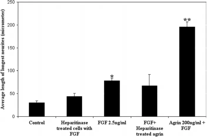
To assess of the role of endogenous HSPGs in FGF-2 mediated neurite outgrowth,
freshly plated retinal cell cultures were treated with 1 U/ml heparitinase for 1 h in
serum-free medium. Cells were then washed with fresh medium and treated with
To assess the effects of agrin on neurite outgrowth, agrin from embryonic chick
brain or vitreous were employed. In initial studies identical effects were observed for
both sources of agrin, and therefore in subsequent experiments only agrin purified from
vitreous body was employed.
Immunoblotting analysis
Retinal neuronal and PC12 cells were treated with FGF-2 (2.5 or 10 ng/ml), agrin
(200ng/ml), HS-GAG free agrin (200ng/ml) and SU5402 (20 µM) for 10 minutes, 30
minutes, 1h and 3 h. Cells were then washed with ice-cold PBS at the appropriate time
period and incubated in lysis buffer (20mM Tris-HCl(pH 8.0), 137mM NaCl , 0.5mM
EDTA, 1% Triton X-100, 10% glycerol, 10mM Na2P2O7, 10mM NaF, 1µg/ml aprotinin,
10µg /ml leupeptin, 1mM sodium orthovandate, 1mM PMSF). Cell lysates were
centrifuged and protein concentrations were determined by the Bradford assay using
the Bio-Rad reagent and immunoglobulin as a standard protein. 30µg of supernatant
proteins were separated by SDS-polyacrylamide gel electrophoresis and transferred to
nitrocellulose membranes. Immunoblots were blocked with 20mM Tris-HCl (pH7.5),
137mM NaCl, 1% BSA, 0.1% Tween-20 and then incubated with polyclonal antibodies
specific for either ERK1/2(1:1000), phospho-ERK1/2(1:1000), or
anti-phospho-c-fos (1 µg/ml) at 4°C overnight. Immunoblots were washed with 20 mM
anti-rabbit, goat, or mouse antibodies (1:250,000). Membranes were incubated with
Supersignal West Pico chemiluminescent Substrate kits (Pierce) and exposed to X-ray
film (Kodak). Membranes were stripped with 2% SDS, 62.5mM Tris-Cl (pH6.8),
100mM β-mercaptoethanol at 70°C for 30 minutes prior to reblotting with other
RESULTS
Agrin specifically stimulates neurite outgrowth mediated by FGF-2 in PC12 cells.
To begin to investigate a role for agrin in neurite outgrowth mediated by
heparin-binding neurite-promoting proteins, we used PC12 cells as a model system. PC12 cells
are an established cell line that exhibit the properties of neurons in the presence of NGF
or FGF-2 (Greene and Tischler, 1976). HS-GAGs have been shown to potentiate the
neurite promoting effects of NGF and FGF-2 in PC-12 cells (Damon et al, 1988; Lesma
et al, 1996), with greater effects observed when presented in combination with FGF-2
than with NGF (Damon et al, 1988). For our analyses of agrin in modulating
FGF-mediated neurite outgrowth, we employed agrin that was immunopurified from either
embryonic chick brain or vitreous body. Our initial studies demonstrated identical
effects for both brain and vitreous agrin, and therefore the experiments described here
employed only vitreous agrin due to the ease of purifying vitreous body agrin.
To examine the role of full-length vitreous agrin on neurite outgrowth, PC12 cells
were incubated for 6 days with FGF-2 in the presence or absence of various
concentrations of agrin that ranged from 100-250 ng/ml. We found that optimal effects
employed for the experiments described here. Previous studies examining the effect of
FGF-2 on neurite outgrowth from PC12 cells have routinely used FGF-2 concentrations
ranging from 20-50 ng/ml, so our initial experiments employed 25 or 50 ng/ml of
FGF-2 in the absence or presence of agrin. Under these conditions a pronounced neurite
growth was elicited from PC-12 cells, but in the presence of agrin there was no
difference in the extent of neurite outgrowth (data not shown). In view of recent
studies showing that in the developing optic tract FGF-2 mediated axon growth is
abolished by elimination of HS-GAGs (Walz et al, 1997), and restored by the addition
of exogenous FGF-2, we reasoned that in order to assess the potential role of agrin in
modulating neurite outgrowth suboptimal doses of FGF-2 need to be employed in order
to mimic the in vivo process.
We therefore carried out studies using 2.5 ng/ml of FGF-2, to determine the
efficacy of this concentration of FGF-2 in promoting neurite outgrowth from PC12
cells. PC12 cells remained undifferentiated when untreated or incubated with agrin
alone (Figure 3 A,D), or incubated with agrin treated with nitrous acid to remove
HS-GAG chains (Figure 3E), indicating that agrin does not promote the differentiation of
PC12 cells. We have shown previously that agrin’s protein core retains full binding
activity (based on ability to bind tenascin) when treated with nitrous acid to remove
by neurite outgrowth, was observed following treatment of PC12 cells with 2.5 ng/ml
of FGF-2 (Figure 3B). The addition of nanomolar concentrations of agrin (200 ng/ml)
to FGF-2 treated PC12 cells resulted in a significant enhancement of neurite outgrowth
(Figure 3C), which was for the most part eliminated when agrin’s HS-GAG chains
were removed (Figure 3F).
The extent of neurite outgrowth in response to FGF-2 or FGF-2 and agrin was
quantified by measuring the average length of the longest neurites from randomly
selected neurons from cultures. For each experiment 10 random microscope fields
were selected, and the 10 longest neurites from neurons in each field were measured.
As shown in Figure 4A, treatment of PC12 cells with low concentrations of FGF-2 (2.5
ng/ml) resulted in a promotion of neurite outgrowth when compared to untreated PC12
cells. With these lower doses of FGF-2 the extent of neurite outgrowth was
significantly less than the neurite outgrowth resulting from exposure of PC12 cells to
both FGF-2 and agrin (Figure 4A). For example, the average length of the longest
neurites in FGF-2 treated (2.5 ng/ml) PC12 cells is 240.3 µm, while for PC12 cells
treated with FGF-2 and agrin the average of the longest neurites is 390.2 µm. At 10
ng/ml doses of FGF-2 a similar pattern is observed, with the average length of these
100 longest neurites being 238.9 µm in 2 treated cells and 476.1 µm in
effects on neurite outgrowth in PC12 cells, with the average neurite length being 297.5
µm. This remaining neurite outgrowth promoting activity for nitrous acid treated agrin
may result from incomplete removal of HS-GAG chains. The distribution of the longest
neurites observed in PC12 cells exposed to the various protein treatments was also
calculated from 100 neurons, and is shown in Figure 4B. It can be seen that
significantly longer neurites are observed in PC12 cells treated with both FGF-2 and
agrin, and that a higher percentage of these PC12 cells have longer neurites.
Agrin exerts its effects on FGF-2 mediated neurite outgrowth through the FGF
receptor.
To determine the extent to which agrin enhances FGF-2 dependent PC12 cell
differentiation through the FGFR and it’s associated ERK signaling pathway, we tested
the effects of the FGFR-1 inhibitor SU5402 on PC12 cell neurite outgrowth. SU5402
interacts with the catalytic domain of the FGFR1 to inhibit its tyrosine kinase activity,
and only acts as a weak inhibitor of the PDGF receptor and does not inhibit the insulin
and EGF receptors (Mohammadi et al, 1997). PC12 cells were exposed to FGF-2
and/or agrin and SU5402 for six days, and neurite outgrowth was then quantified. As
shown in Figure 3 and 4A, the majority of PC12 cells treated with FGF-2 and agrin,
neurite outgrowth. The majority of cells lacked neurites (Figure 3G-I and Figure 4A),
indicating the requirement of a functional FGFR for neurite outgrowth in our assays.
Thus, these data indicate that agrin’s effects on FGF-2 mediated neurite outgrowth in
PC12 cells are dependent on a functional FGFR-1.
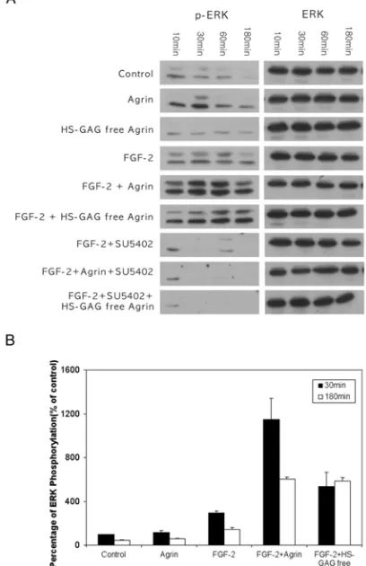
Agrin exerts its effects on FGF-2 mediated neurite outgrowth by modulating ERK
activity in PC12 cells.
Our data showing that the FGFR-1 inhibitor SU5402 eliminates agrin’s effects on
FGF-2 mediated neurite outgrowth in PC12 cells suggests that agrin must be acting
through the FGFR1 and its signaling pathway to modulate FGF-2 activity. To test this
hypothesis directly, we have examined the effect of agrin on FGF-2 mediated changes
in ERK signaling in PC12 cells. Previous studies have shown that FGFs activate ERK
in various cell types, including retinal neurons (Perron and Bixby, 1999), and that
sustained activation of ERK was necessary for neurite outgrowth in retinal neurons
(Dimitropoulou et al, 2000). Thus, we addressed whether agrin modulates the degree
of induction and duration of induction of ERK phosphorylation in PC12 cells. In PC12
cells, FGF-2 stimulated phosphorylation of both the p42 and p44 isoforms of ERK
diminished (Figure 5A and 5B). In contrast, PC12 cells exposed to FGF-2 and agrin
exhibited stimulation of ERK phosphorylation within 10 minutes, which was still
elevated when compared to basal levels at 3 h post-exposure to FGF-2 and agrin
(Figure 5A and 5B). Treatment of PC12 cells with agrin alone, or HS-GAG free agrin,
did not produce a significant, reproducible elevation of ERK phosphorylation (Figure 5
A and 5B), indicating that agrin’s effects on ERK phosphorylation are mediated via
FGF-2 and the FGFR-1. Densitometric analysis of ERK phosphorylation shows that
the level of ERK phosphorylation, when compared to control levels, increased 3.8-fold
after 30 minutes, and even increased 4.2-fold after 3 h in the presence of both FGF-2
and agrin, when compared to FGF-2 alone (Figure 5B). In addition, even though
activation of ERK by FGF-2 and agrin decreased after 1 h, the level of activation of
ERK at 3 h post-exposure was higher than FGF-2 treated cells at 1 h post-exposure.
These data provide strong evidence for agrin augmenting the efficacy of FGF-2
activation of the FGFR-1, and maintaining this activation for longer periods than for
cells exposed to FGF-2 alone. Interestingly, ERK phosphorylation by FGF-2 and agrin
lacking HS-GAG chains was not completely reduced to FGF-2 levels, although the
extent of neurite outgrowth under these conditions is similar between the two
treatments. Importantly, phosphorylation of ERK was completely blocked by SU 5402,
whether agrin or HS-GAG free agrin was used in combination with FGF-2. These data
agrin can significantly augment and maintain for longer periods the high level of
ERK phosphorylation in the presence of FGF-2, providing a mechanism for agrin’s
ability to enhance neurite outgrowth in the presence of concentrations of FGF-2 that are
suboptimal for rapid and complete PC12 cell differentiation.
Role of agrin in modulating FGF-2 mediated neurite outgrowth in retinal
neuronal cultures.
In light of our demonstration that agrin can potentiate the effects of FGF-2 on
PC12 cell neurite outgrowth, we thought it important to analyze the role of agrin on
FGF-2 mediated neurite outgrowth in a CNS neuronal cell type known to be regulated
by FGF-2. Thus, we selected chick retinal neurons, as it has been demonstrated that
retinal neuronal axonal growth is regulated by FGF-2 (McFarlane et al, 1995; Walz et
al, 1997; Perron and Bixby, 1999) that is dependent on the presence of HSPGs (Walz et
al, 1997). In addition, FGF-2 and HSPGs such as agrin co-localize to the retinotectal
pathway during the developmental period associated with establishment of this axon
pathway (Ford et al, 1995; Joseph et al, 1996; Cotman et al, 1999). To facilitate
survival and neurite outgrowth of primary cultures of retinal neurons these cells were
2), did not permit outgrowth of retinal neurites (Halfter et al, 1997). Thus, in the
experiments described here we employed a laminin-1 substratum, but added agrin
exogenously to the culture medium. Furthermore, the concentrations of agrin added to
culture medium (200 ng/ml) were substantially lower than the amounts of agrin (50
µg/ml) that were adsorbed as a substratum in our previous studies (Halfter et al, 1997).
The addition of agrin was in either the presence or absence of FGF-2. Interestingly,
when E6 retinal neuronal cultures were exposed to FGF-2 and agrin, sprouting of
neurites was observed within 1 h of plating, in contrast to the absence of neuronal
sprouting from retinal neurons grown on laminin-1 alone or exposed to FGF-2 alone
(Figure 6). Thus, agrin is able to stimulate a rapid neuronal response to FGF-2.
Importantly, our data suggest that the ability of ECM proteins and growth factors to
modulate neurite outgrowth is highly dependent on the levels of these proteins
presented to cells, as previous studies by Perron and Bixby (1999) demonstrated that
retinal neurons exhibit rapid neurite outgrowth within 30 minutes on a substratum
comprised only of 10 µg/ml of laminin. Thus, on a substratum of 5 µg/ml of laminin-1
as used in our experiments, neurons only initiate a rapid neurite sprouting when
exposed to both 2.5 ng/ml FGF-2 and 200 ng/ml agrin (Figure 6).
To assess a dose-response relationship for FGF-2 on chick retinal neurons, we
exposed retinal neurons to varying concentrations of FGF-2. Previous studies in chick
outgrowth 2-fold when compared to cells plated on laminin-1 only (Perron and Bixby,
1999). We therefore tested 25 or 50 µg/ml of FGF-2 for its affect on retinal cell neurite
outgrowth, and the effect of agrin on this neurite outgrowth. As shown for PC12 cells,
we observed extensive neurite outgrowth from chick retinal neurons using 25 or 50
ng/ml of FGF-2, and agrin was unable to augment this neurite outgrowth (data not
shown). Thus, retinal cell cultures were exposed to lower concentrations of FGF-2,
using 2.5 or 10 ng/ml of FGF-2, to assess the role of agrin in the modulation of FGF-2
mediated neurite outgrowth.
When primary E6 retina cultures are examined at 1 day after treatment with 2.5
ng/ml FGF-2 in the presence or absence of agrin, it can again be seen that agrin
modulates the ability of FGF-2 to regulate neurite outgrowth. Compared to retinal
neurons grown on a laminin-1 substratum (Figure 7A), the addition of FGF-2 augments
neurite outgrowth, and FGF-2 plus agrin further augments outgrowth (Figure 7B and
7C). When 10 ng/ml FGF-2 is employed, the effects of agrin on neurite outgrowth are
more pronounced, indicating the potent activity of agrin as a regulator of FGF-2
function (Figure 8A and 8B). When retinal cultures are treated with agrin alone, by
adding agrin to the culture medium, we do not observe any change in the extent of
neurite outgrowth on laminin-1, indicating that under these conditions agrin does not