156
Brain Tumor Detection and Segmentation Based On
Component Analysis Method Using Discrete Wavelet
Transform
1
Mrs.D.Sherlin,
2Dr.D.Murugan
1Research Scholar, Manonmaniam Sundaranar University, Abishekapatti, Tirunelveli 627 012, Tamil Nadu, India,
2Professor and Head, Department of Computer Science & Engineering,Manonmaniam
Sundaranar University, Abishekapatti, Tirunelveli 627 012, Tamil Nadu, India 1[email protected], 2[email protected]
Abstract- Manual Identification of tumors from MRI brain images is tedious because of the large number of dataset images generated. But the early identification of brain tumors helps to save life of many patients. Medical Image processing booms up with recent technology innovation as the new technique brings out the solution for this major problem with automatic brain tumor detection and segmentation.The proposed work involves more technical challenges which majorly involves segregation of large image dataset, Identification of tumor location, Classification of tumor type and so on. To implement this the entire process is divided into four main procedures. The stages are Preprocessing, Feature extraction, Segmentation and Post processing. The methodology followed here in the process of segmentation involves various process at each stages. The main algorithm used in the process of segmentation is implementing the Component Analysis method using Discrete Wavelet transform. Component Analysis method works with the statistical procedures for the variables that are collected from the image and transform them into linear uncorrelated variables. Based on these principal components the segregated regions of variables that match the tumor affected areas can be identified and classified. This proposed methodology uses the sampled images and information that are obtained from the Discrete Wavelet Transform (DWT) technique. Thus the obtained results shows the good accuracy percentage in identifying the tumor location.
Keywords: Brain Tumor, MRI, PCA, DWT, Segmentation, Component Analysis, Discrete Wavelet Transform.
I. INTRODUCTION
Cancer is a life killing disease which happens to occur in anytime in our lifetime. One cannot predict the occurrence of the cancer as it does not show its symptoms in the body immediately. Cancer is nothing but the abnormal growth of any cells or tissues within the body. These abnormal cells grows as day by day there by making these cells larger and larger. These abnormal cells can spread over other parts of the body as well. These cancer cells are very dangerous as these effect in risking the life of the patient. These type of abnormal and uncontrolled cell growth that occur in brain tissues are called Brain Tumors. Medically all the brain tumors are not cancerous. Brain tumors are basically classified into two major categories called Benign and Malignant. Benign tumors are less aggressive tumors which does not have the ability to invade and enter the neighboring tissues or cells. These type of tumors
generally do not spread but they can become large. Once these tumors are removed, the risk of re-occurrence is very less. The most common benign tumors in the brain are called “Meningioma”. These are usually covered by a protective “SAC” which is generated by the human immune system. These sac helps to segregate the tumor from the rest of the body and so these can be easily identified and removed. Under repeated treatment, there is very little risk for the life of the individual.
157 detection of a painless lump. These types of tumors
are “elastic,” which grow fairly large before they can be detected. As they grow further, symptoms of pain and soreness may occur as these tumors coerce against organs, blood vessels and nerves.
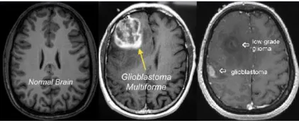
Figure 1. MRI Brain Image comparing normal and Glioblastoma and Benign Tumors Glioblastoma Tumor and Left shows the normal brain image.
Glioblastoma, these are the most common malignant tumors in the brain. These are deadliest of primary brain tumors that occur in adults and it is of a kind of tumors which are termed as “Gliomas”. These are classified as the most dangerous (Grade – IV) type which develops from the lineage of star shaped glial cells which support nerve cells called astrocytes. These gets developed primarily in the cerebral hemispheres but can blowout in other parts of the brain, brainstem, or spinal cord. Because of its lethalness, this was selected as the first brain tumor to be sequenced as part of The Cancer Genome Atlas (TCGA) [1], a national effort to map the genomes of the many types of cancer. Based on this effort, researchers invented that these has four distinct genetic subtypes that respond in a different way to antagonistic therapies, making treatment extremely difficult and challenging.
Automatic detection of these type of tumors are the main aim of this work and the forthcoming paper is sectioned as below. Section II covers the related work of various other researchers. Section III explains on the various methodologies implemented in this research along with the flowchart. Section IV analyze the simulation results and discussion on the work done. Section V ended with the conclusion of the research work with future directions.
II. RELATED WORK
Y. Zang in his paper, “AnMR Brain Images Classifier via Principal Component Analysis and Kernel Support Vector Machine”, 2012 [2], proposed a novel method to classify a tumor from a given MR brain image whether it normal or abnormal.Y.Zang implemented the Principal component Analysis method for the detection of tumor and for the classification purpose whether normal or abnormal is done using Kernel Support Vector Machine technique. According to him, both these techniques helps to easily classify the tumor from theMR brain images. SwapnaliSawakare, in her paper, “Classification of Brain Tumor Using Discrete Wavelet Transform, Principal Component Analysis and Probabilistic Neural Network”, 2014 [3], described about an efficient method for automatic brain tumor segmentation. Also the author suggested on the different methodologies for the extraction of tumor tissues from MR images. In her research activity, the author carried out the segmentation technique using K-means clustering algorithm. According to the author, this K-means based segmentation works with better performance. Final decision making was implemented in two stages: feature extraction using GLCM and PCA and the classification using PNN-RBF network. Robert H. Caverly, “MRI Fundamentals: RF Aspects of Magnetic Resonance Imaging”, 2015 [4], described in his work about the MRI. According to the author, Magnetic resonance imaging (MRI) is an imaging technique based on the nuclear magnetic resonance (NMR) phenomenon and provides the methods of obtaining detailed, high-contrast images for use by the medical community for diagnostic purposes.
III. PROPOSED METHODOLOGY
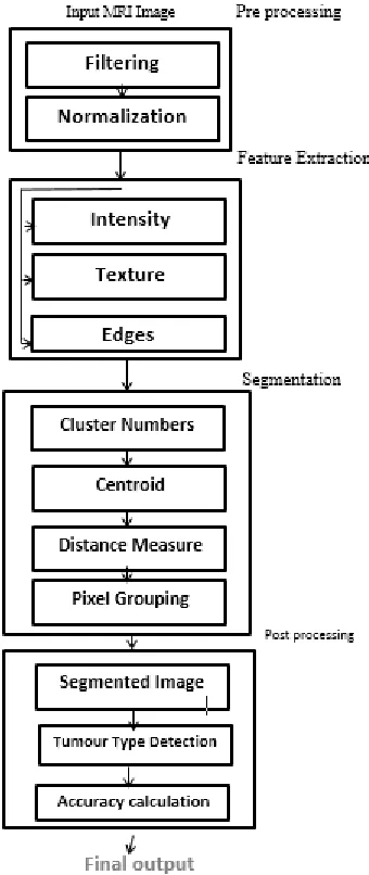
158 Figure 2. Proposed Block diagram
During the Pre-processing stage, this mainly involves two variety of processing, Filtering and Normalization. MRI images when captured are prone to noise disturbances. These MRI images generally have Gaussian noise. The noise occurrence in modern MRI have become very less now-a-day, but due to the thermal effect, noise do occur. The input MRI image is processed with Filters for removal of any unwanted noise. The filters used in this process are Median filters as these filters performs better than Gaussian Filter [5].The image is normalized for intensity value normalization and also to enhance the image quality.
The second stage of the work is the Feature extraction. During this stage intensity, features and edges are extracted. The entire extraction process uses Discrete Wavelet Transform (DWT) method. The wavelet transform is a powerful and best mathematical tool for feature extraction. This has been used to extract the feature coefficient from brain MRI images. The main advantage of wavelets is that for the benefit of classification, for a function of a signal they provide localized frequency information which helps to extract the information. The important feature of this wavelet is multi scale representation of function. By using the wavelets, one can analyze the various levels of resolution for a given function. DWT of image signal helps to produce a non-redundant representation for an image, which provides better image information for spatial and spectral localization when compared with other multi scale representations.Numerous features are possible and they are generallycategorized into texture and statistical features. Featureextraction may depend on applications and they are used totrain the classifier [6].
Third and important stage is Segmentation. The segmentation process is carried out using two important techniques, threshold segmentation and Principal component analysis method. The part of the image containing the tumor generally have more intensity when compared to the rest of the region. Segmentation process is carried out on the basis of threshold value. In order to implement this the extracted image is converted into binary image. Thresholding technique is nothing but the segmenting of an MRI image by their intensities with the help of binary partition.
I (p) = 1, if gray level > T
I (p) = 0, if gray level < T ……. (1)
A grayscale image is turned into a binary (black and white) image by first choosing a grey level Tin the originalimage. Then every pixel is turned black or white according to their grey value whether it isgreater than or less than T.
159 which is often used in signal and image processing
[7, 8]as a technique for data compression, data dimension reduction or their decorrelation as well.
Figure 3. Graphical illustration of Principal component Analysis
PCA is mathematically explained as an orthogonal linear transformation which transforms the data to a new coordinate system in such a way that the greatest variance from the projection of some data lie on the first coordinate(called the first principal component), the second greatest variance on thesecond coordinate, and the series continues.In statistics, PCA is a method to simplify a multidimensional dataset to lower dimensions which helps for analysis, visualization and data compression.Thus, new basis vectors are calculated for the particular data set.From Figure 3, we can combine the observed images with their identified basis vectors to represent a PCA image with the data in the new coordinate system where the basis vector follow the modes of greater variance in the data.
Then the final stage of segmentation is carried out by clustering the vectors based on the threshold intensity and tumors can be segmented easily by combining the cluster of high intensity and top gradient clusters.Final stage is the post processing stage where the segmented tumors are classified based on their severity. Linear kernel functional classifier is used as this is the simplest way to predict the input patterns in a linearly detectable way. This is independent of dimensionality and feature space.
IV. RESULTS AND DISCUSSION
Our algorithm was implemented in MATLAB R 2014a version on a PC with Intel Pentium Core Duo 1.9GHz processor and 4GBRAM. The computationtime for tumor segmentation ranged from
less than a minute depending on the size of the image.
Figure 4. Input Benign MRI brain image Only one input image can be loaded at a time where the image type must satisfy any one of the following extension like Bit Map File(BMP), Portable Network Graphics (PNG), Tagged Image File (TIF), Graphics Interchange Format(GIF).The coding is organized in such a way that the while running the program the user can select the input image by browsing the image dataset. Here the input image has been taken from the MRI dataset images for benign type of tumor.
Figure 5. Threshold Image
160 image.The command “im2bw” converts the MR
image into binary image.
Figure 6. Clustering Image
In output image, it replaces all the pixels in theinput image with luminance greater than the defined threshold level with value 1(white) and all others with value0(black). In this way in the segmented image we can distinguish the portion of tumor from the image.
Figure 7. Tumor Segmented Image
Figure 7 shows the segmented tumor image. The same process has been implemented for the malignant MRI image whose input image is taken as the below figure 8.
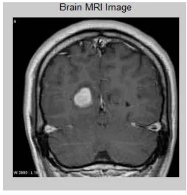
Figure 8. Malignant MRI image
Figure 8 below shows the segmented tumor for the malignant type. The process follows the similar
procedure as the previous benign tumor and the segmented tumor results is shown in the below figure 9.
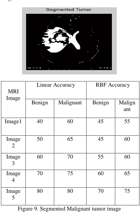
Figure 9. Segmented Malignant tumor image The accuracy tabulation for different set of images are tabulated below
MRI Image
Linear Accuracy RBF Accuracy
Benign Malignant Benign Malign ant Image1 40 60 45 55
Image 2
50 65 45 60
Image 3
60 70 55 60
Image 4
70 75 60 65
Image 5
161 Thus
by using this algorithm, an efficient Brain Tumor Classification method is constructed with maximum recognition rate of 100% .This method could serve as an accurate classification of Brain Tumor diagnosis.
V. CONCLUSION
Automatic brain tumor segmentation is the important modern innovation which ease the process of medical diagnosis. In this paper, we come over the Severity of Brain tumor and the need for automatic segmentation, the process of segmenting the tumors, methods involved in the process. The simulation results shows that the tumor segmentation is accurate and the accuracy results are tabulated. The Linear accuracy and RBF accuracy both are calculated for different samples of benign and malignant tumors with good accuracy. Thus the proposed work shows that the brain tumor segmentation can be done in very less time and hence ease the process of medical specialist.
REFERENCES
[1] https://cancergenome.nih.gov/cancersselected/glio blastomamultiforme
[2] Y. Zhang and L. Wu, “MR Brain Images Classifier Via Principal Component Analysis and Kernel Support Vector Machine”, Progress In Electromagnetics Research, Vol. 130, 2012. [3] .SwapnaliSawakare and Dimple Chaudhari,
“Classification of Brain Tumor Using Discrete Wavelet Transform, Principal Component Analysis and Probabilistic Neural Network”, International Journal for Research in Emerging Science and Technology, Volume-1, November 2014.
[4] Robert H. Caverly, “MRI Fundamentals: RF Aspects of Magnetic Resonance Imaging”, IEEE Microwave Magazine, Volume 6, July 2015 [5] Min, Aye & Mar Kyu, Zin. (2017). MRI Images
Enhancement and Tumor Segmentation for Brain. 270-275. 10.1109/PDCAT.2017.00051.
[6] P. Nagaraja, T. Kalaiselvi, P. Sriramakrishnan, "Brain Tumor Segmentation from MRI Head Scans through GSO based FCM Clustering and Region Growing Technique", International Journal of Computer Sciences and Engineering, Vol.06, Issue.04, pp.348-354, 2018.
[7] H. H. Barret. Foundations of Image Science. John Wiley & Sons, New Jersey, U.K., third edition, 2004.
[8] R. C. Gonzales and R. E. Woods. Digital Image Processing. Prentice Hall, second edition, 2002. 795 pages, ISBN 0-201-18075-8.
[9] http://people.ciirc.cvut.cz/~hlavac/TeachPresEn/1 1ImageProc/15PCA.pdf