Candida albicans
HIBERT RACHUONYO OPINDE (BSc) I56/20520/2012
A Thesis Submitted in Partial Fulfillment of the Requirements for the Award of the Degree of Master of Science (Microbiology) in the School of Pure and
Applied Sciences of Kenyatta University
DECLARATION
This thesis is my original work and has not been presented for a degree in any other University.
Signature ………..……… Date ……..…….……
Hibert Rachuonyo Opinde Department of Microbiology Kenyatta University
This thesis has been submitted for examination with our approval as the University supervisors.
Signature ………... Date.……...……...….
Dr. Anthony Kebira Department of Microbiology
Kenyatta University
Signature ……….. Date.……...……...…..
Dr. Grace W. Gatheri Department of Plant Sciences
DEDICATION
ACKNOWLEDGEMENTS
Thanks to Almighty God for this far He has brought us, his blessings and the gift of a healthy life. Despite the many challenges we have been through He has been good to us and we have been able to overcome them. I sincerely thank my supervisors; Dr. Anthony Kebira and Dr. Grace W. Gatheri for their relentless support, encouragement, relentless support and guidance during my research work. Special appreciation Professor L.E. Newton for the help and guidance offered in the process of identification of the plants. I would also like to dearly appreciate the support and input from my fellow colleagues; Philip Ogola, Japheth Wambani, Nathan Kiboi, James Kimani, Wycliffe Arika, Majorie Oruru, Christine Nakhumincha and Gilbert Koskey. May almighty God give you good health and continue blessing you in your endeavor’s throughout your lives. I
TABLE OF CONTENTS
DECLARATION ... i
DEDICATION ... ii
ACKNOWLEDGEMENTS ... iii
TABLE OF CONTENTS ... iv
LIST OF TABLES ... viii
ABBREVIATIONS AND ACRONYMS ... ix
ABSTRACT ...x
CHAPTER ONE ...1
INTRODUCTION ...1
1.1 Background information ...1
1.2 Problem statement ...4
1.3 Justification ...5
1.4 Research questions ...6
1.5 Hypotheses ...7
1.6 Objectives of the study...7
1.6.1 General objective...7
1.6.2 Specific objectives...8
1.7 Significance of the study ...8
CHAPTER TWO ...9
LITERATURE REVIEW ...9
2.1.1 Tagetes minuta (Southern cone marigold) ...9
2.1.2 Aloe secundiflora...11
2.1.3 Bulbine frutescens ...12
2.1.4 Vernonia lasiopus ...13
2.2 Test microorganisms ...14
2.2.1 Escherichia coli ...14
2.2.2 Salmonella typhi ...15
2.2.3 Staphylococcus aureus ...15
2.2.4 Candida albicans...16
2.2.5 Enterococcus faecalis ...17
2.2.6 Shigella flexineri ...17
2.3 Phytochemicals in medicinal plants ...18
2.3.1 Tannins ...19
2.3.2 Flavonoids ...20
2.3.3 Alkaloids ...21
2.3.4 Saponins ...23
CHAPTER THREE ...25
MATERIALS AND METHODS ...25
3.1 Plants sampling ...25
3.2 Preparation of plant extract ...25
3.3 Preparation of Media ...26
3.3.2 Preparation of Muller-Hinton agar ...26
3.4 Preparation of susceptibility test discs ...27
3.5 Test bacterial organisms ...28
3.5.1 Isolation and Morphological identification ...29
3.5.2 Biochemical identification ...31
3.5.3 Maintenance of the bacterial and fungal cultures...33
3.6 Antimicrobial susceptibility testing ...33
3.7 Combined effect test ...36
3.8 Qualitative phytochemical analysis ...37
3.8.1Tannins ...37
3.8.2 Flavonoids ...37
3.8.3 Alkaloids ...37
3.8.4 Saponins ...38
3.9 Statistical analysis ...38
CHAPTER FOUR ...39
RESULTS ...39
4.1 Efficacy test of the plant extracts on the bacterial pathogens and Candida albicans ...39
4.2 Combined effect of the plant extracts on the bacterial pathogens and Candida albicans ...47
4.3 Qualitative phytochemical analysis ...52
5.1 Discussion ...53
5.1.1 Antimicrobial activity ...54
5.1.2 Combined effect of the plant extracts ...58
5.1.3 Phytochemicals...62
5.2 Conclusion ...65
5.3 Recommendations ...65
5.4 Suggestions for further studies ...66
LIST OF TABLES
Table 3.1:Standard for biochemical tests on isolated microorganisms ... 32 Table 4.1:Average zones of inhibition in millimetres when plant extracts
are used singly... 42 Table 4.2:Interactions between the plants extract and test microorganism
when used singly ... 43 Table 4.3:Minimum inhibitory concentration in micrograms/milliliter ... 44 Table 4.4:Minimum bactericidal concentration and minimum fungicidal
concentration in micrograms/milliliter ... 46 Table 4.5:Average zone of inhibition in millimetres when extracts are
combined ... 50 Table 4.6: Interactions between the plants extract and test microorganism
ABBREVIATIONS AND ACRONYMS
AB Aloe secundiflora and Bulbine frutescens plant extract combination ANOVA Analysis of variance
AT Aloe secundiflora and Tagetes minuta plant extract combination AV Aloe secundiflora and Vernonia lasiopus plant extract combination BT Bulbine frutescens and Tagetes minuta plant extract combination BV Bulbine frutescens and Vernonia lasiopus plant extract combination DMSO4 Dimethyl sulphoxide
FeCl3 Iron (III) chloride HCl Hydrochloric acid
INT 2-(4-iodophenyl)-3-(4-nitrophenyl)-5-phenyl-2H-tetrazolium chloride MBC Minimum bactericidal concentration
MIC Minimum inhibitory concentration SAS Statistical analysis software
ABSTRACT
Plant extracts with medicinal value have been used to treat many diseases that can either be bacterial, fungal or parasitic among many others. Plants with medicinal value produce certain chemical elements known as phytochemicals that have antimicrobial activity. Enteric bacterial pathogens are disease-causing microorganisms that are usually located in the intestinal tracts of either animal or human beings. The pathogenic members are usually associated with infections that are characterized by; enteric fevers, abdominal pain, diarrhoea, and vomiting.
Candida albicans is a yeast fungus that is mainly found in the mucosal cavity of
the vagina and intestinal tract as a normal microbiota but it can cause systematic infections in immunocompromised individuals. This study was aimed at determining the antimicrobial activity, combined effects of the selected plant leaf extracts of Tagetes minuta, Aloe secundiflora, Vernonia lasiopus and Bulbine
frutescens against selected clinical isolates of Escherichia coli, Salmonella typhi,
Staphylococcus aureus, Shigella flexineri, Enterococcus faecalis and Candida
albicans obtained from Kenyatta University Health Centre; using the Kirby-Bauer
method. In addition, qualitative analysis of the phytochemicals present in the extracts was also determined. The plants materials were obtained from Kenyatta University arboretum and identified by Taxonomist; Prof L.E. Newton and voucher specimen deposited in the University herbarium. The collected data was then analyzed in SAS version 9.1 using ANOVA and further subjected to a post hoc test with P<0.05 being considered significant. When used singly and in combinations against the test microorganisms the average zones of inhibition were found to be significant at P<0.05. When the plant extracts were used in low concentrations against the test microorganisms; Vernonia lasiopus was more active against Shigella flexineri (MIC 3.3µg/ml, MBC 7.1µg/ml), Bulbine
frutescens against Shigella flexineri (MIC 3.2µg/ml, MBC 6.2µg/ml), Aloe
secundiflora against Shigella flexineri (MIC 3.7µg/ml, MBC 8.0µg/ml) and
Tagetes minuta against Enterococcus faecalis (MIC 5.1µg/ml, MBC 6.3µg/ml)*.
CHAPTER ONE INTRODUCTION
1.1 Background information
Medicinal plants are used by almost 80% of the world’s population for their basic
health care because of their low cost and ease in availability (Shazadi et al., 2010). From the dawn of civilization, people have developed a great interest in plant-based drugs and pharmaceutical products (Shazadi et al., 2010). In the last few decades, many bacterial organisms have continued to show increasing resistance against current antimicrobial agents (Nascimento et al., 2000). Herbal drugs made from medicinal plants have been used from ancient times to treat various diseases and their antimicrobial properties make them a rich source of many potent drugs (Srivastava et al., 2005).
The use of herbal medicinal plants has always played a positive role in the control or prevention of diseases such as diabetes, heart disorders and various cancers (Mohanta et al., 2003). Some medicinal plants have been used in the production of various drugs singly or in combination and even as principal raw material for the production of other conventional medicines (Tahir and Khan, 2012). Tagetes
minuta L. is also known as Southern Cone Marigold, Stinking Roger or black
either annually or perennially and mostly herbaceous plants (Soule, 1996). Other common species of the family include Tagetes erecta, Tagetes patula and Tagetes
tenuifolia (Tereschuk et al., 1997). The herbaceous plants were mostly found in
North and South America but some species have become naturalized around the world (Soule, 1996). Tagetes minuta extracts have been used as medicinal tea in some areas (Soule, 1993). The total extracts from leaves, flowers, stem and other parts of the plant have shown antibacterial activity against Gram positive and Gram negative bacteria (Tereschuk et al., 1997). Extracts from the other common species have also been used as medicine in treating various illnesses such as stomach problems and intestinal disorders (Broussalis et al., 1999).
Some of the components contained in Tagetes minuta extracts such as flavonoids have been tested and proved to have antimicrobial activity against not only bacteria but also fungi and some nematodes (Cushnie and Lamb, 2005). The genus Aloe belongs to Aloeaceae (Liliaceae) family which has around 360 to 400 different species (Newall et al., 1996). Aloe species have antibacterial, antifungal, anticancer, antiviral and immunomodulatory properties (Holzmuller et al., 2002).
Aloe secundiflora Engl. is also known as Aloe floramaculata, Aloe engleri and
Aloe marsabitensis (Kaingu et al., 2013). Other common species include Aloe
arborescens, Aloe chabaudii, Aloe turkanensis, Aloe greatheadii, Aloe cameronii
and Aloe excels. Aloe secundiflora leaf components have been credited for
(Mwale et al., 2005). Bulbine is a genus of plants in the family Xanthorrhoeaceae and subfamily asphodeloideae and its members are well known for their medicinal value (Acock, 1988). They are succulent plants with most species having yellow flowers while others are white, orange or pink flowers. The most common species is Bulbine frutescens wild which is popularly grown in flower gardens (Van Wyk, 2008). Many species have bulb shaped tuber and they are chiefly found in South Africa with a few species extending to the tropics of Africa and Australia (Coopoosamy et al., 2000). Vernonia lasiopus O. Hoffman belongs to the tribe Vernonieae in the family Asteraceae which mostly contains herbaceous plants (Keeley, 2007). Vernonia is a shrub that grows in tropical Africa and has a height of about 2-5 metres, elliptical leaves of up to 20 centimetres and a rough bark (Ijeh and Ejike, 2011).
1.2 Problem statement
The increase in resistance to many commercially produced synthetic antimicrobial agents by microorganisms has been increasing with time hence the need of searching for new antimicrobial agents (Ramesh and Okigbo, 2008). There has been a need to increase alternative antimicrobial agents leading interests of evaluating extracts from plants known to have medicinal value for the manufacturing of herbal antimicrobial agents by Pharmaceuticals Company. Most of the bacterial and fungal pathogens have shown the capability of developing resistance to some of the available commercially available antimicrobial agents.
1.3 Justification
The use of herbal plant extracts with medicinal value has increasingly been advocated and incorporated in the production of antimicrobial agents. This is due to the need of increasing the base of already available antimicrobial agents that are commercially produced. Plants with medicinal value have been used to make homemade concoctions that have not been scientifically validated to have antimicrobial activity. Furthermore, different parts of the same medicinal plants have been used in making antimicrobial agents giving contrasting antimicrobial activity against pathogenic microorganisms. In this study, the antimicrobial activity of crude extracts from the leaves of Tagetes minuta, Aloe secundiflora,
Bulbine frutescens and Vernonia lasiopus were tested for antimicrobial activity
against pathogenic bacteria and fungus Candida albicans.
The plants leaves were selected because in some other similar studies carried out showed extracts from the other plants parts; Tagetes minuta seeds (Shazadi et al., 2010), Aloesecundiflora roots (Induli et al., 2012), Bulbine frutescens roots (Van
Wyk et al., 1997) and Vernonia lasiopus roots (Toyang and Verpoorte, 2013).
This is because phytochemicals have been known to be secondary metabolites that are usually produced by plants and accumulated in different parts of the plant. Some of these phytochemicals have been thought to be responsible for the antimicrobial activity shown by extracts from various herbal plants. The finding in the study could provide an insight into the antimicrobial activity of the plant leaf extracts against the test pathogenic bacteria and fungus Candida albicans. Furthermore, the combination of the plant extracts will provide an insight if there is any combined effect of the plant extracts against the test microorganism. This may lead to the future formulation of herbal concoctions that are scientifically validated not only at home but also for commercial production hence increasing the base of antibacterial agents available. This may provide an effective and alternative available antimicrobial agent for people who cannot afford conventionally synthetically produced medicine.
1.4 Research questions
i) Do the extracts from Tagetes minuta, Aloe secundiflora, Vernonia
lasiopus and Bulbinefrutescens have any antimicrobial activity against the
bacterial pathogens and fungus Candida albicans when used separately? ii) Combined extracts of Tagetes minuta, Aloe secundiflora, Vernonia
lasiopus and Bulbine frutescens have synergistic antimicrobial activity
iii) What are the phytochemicals present in the extracts of Tagetes minuta,
Aloe secundiflora, Vernonia lasiopus and Bulbinefrutescens?
1.5 Hypotheses
i) Extracts from Tagetes minuta, Aloe secundiflora, Vernonia lasiopus and
Bulbine frutescens do not have a bactericidal effect on bacterial pathogens
and fungicidal effect on Candida albicans.
ii) The combinations of extracts from Tagetes minuta, Aloe secundiflora,
Vernonia lasiopus and Bulbine frutescens does not have different
antimicrobial activity against bacterial pathogens and fungus Candida
albicans than when used separately.
iii) Phytochemicals present in the crude extracts from Tagetes minuta, Aloe
secundiflora, Vernonia lasiopus and Bulbinefrutescens are not known.
1.6 Objectives of the study 1.6.1 General objective
To determine the antimicrobial effect of Tagetes minuta, Aloe secundiflora,
Vernonia lasiopus, and Bulbine frutescens extracts on bacterial pathogen and
1.6.2 Specific objectives
i) To determine the antimicrobial activity of plant extracts from Tagetes
minuta, Aloe secundiflora, Vernonia lasiopus and Bulbine frutescens
against bacterial pathogens and Candida albicans.
ii) To determine the impact of combined plants extracts against the bacterial pathogens and Candida albicans.
iii) To determine the qualitative phytochemical present in the plant extracts obtained from Tagetes minuta, Aloe secundiflora, Vernonia lasiopus and
Bulbinefrutescens.
1.7 Significance of the study
The main aim of the study was to determine if the plant extracts from Tagetes
minuta, Aloe secundiflora, Vernonia lasiopus and Bulbine frutescens have
CHAPTER TWO LITERATURE REVIEW 2.1 Medicinal plants
Medicinal plants have been identified and used throughout human history (Lichterman, 2004). The use of medicinal plants to treat diseases is almost universal among non-industrialized societies and is often more affordable than purchasing expensive conventional drugs (Fabricant and Farnsworth, 2001). The World Health Organization (WHO) estimates that 80% of the world population especially Asian and African countries use herbal medicine for some aspect of primary health care (http://www.traffic.org/medicinal-plants, 30th march 2014). Over 120 active compounds currently isolated from the higher plants are widely used in modern medicine and 80% of these show a positive correlation between their modern therapeutic use and the traditional use of the plants from which they are derived (Fabricant and Farnsworth, 2001).
2.1.1Tagetes minuta (Southern cone marigold)
The genus Tagetes belongs to the Asteraceae family which presently comprises of 56 species, 27 biennials, and 29 perennials. Tagetes species are grown all over the world as multipurpose plants. The most common species are Tagetes minuta,
Tagetes patula, Tagetes erecta and Tagetes tenuifolia (Soule, 1993). Tagetes
species and chemotypes from its genus have been largely examined for biologically active metabolites that can be used in industry and medicine (Green
plant are said to be accumulated in the organs of the plant and their essential oils have not only antimicrobial effect but also insecticidal properties (Piccaglia et al., 1997). Plant parts such as flowers and leaves have been known to contain flavonoids that are scavengers for free radicals which enhances the antimicrobial activity of the Tagetes minuta extracts (Rivas, 1991). Phytochemicals from the plant such as carotenoids have also been used in pharmacological preparations and they have been found to contain anti-aging and anticancer effects (Bennish et al., 1992). The plant extracts have been used in treating intestinal and stomach problems (Tereschuk etal., 1997; Broussalis et al., 1999). Tagetes minuta extracts such as its volatile oil and other components have been used in the flavoring of food products and as perfumes.
The plant has also shown inhibitory activity against some pathogens and insects (Green et al., 1991). Studies carried out have shown that leaf extracts from most of the Tagetes species including Tagetes minuta contain flavonoids that have shown antimicrobial potential against both Gram positive and Gram negative bacteria (Tereschuk et al., 1997). Extracts from Tagetes minuta leaf flowers and stem extracted using methanol have shown to contain secondary metabolites including terpenes which are thought to be responsible for antibacterial activities (Lopez et al., 2008). Components of some of the essential oils extracted from
camphene which has been found to be effective against Erwinia amylovora and
Staphylococcus aureus (Facon, 1996).
2.1.2 Aloe secundiflora
Aloes are perennial succulent xerophytes which develop water storage tissues in leaves to survive in areas with low or erratic rainfall (Talmadge et al., 2004). The plant is mainly found in cultivation, having no naturally occurring population although closely related Aloes do grow in northern parts of Africa (Akinyele and Odiyi, 2007). The plant is an almost sessile perennial herb that has leaves 30-50 centimetres long and 10 centimetres broad at the base, bright yellow tubular flowers 25-35centimetres in length arranged in a slender loose spike (WHO, 1999). The genus Aloe is common in Kenya; with about 60 taxa recognized (Hendricks and Wright, 1979).
Aloe secundiflora has been used in treating ailments including; chest problems,
polio, malaria and stomach ache by herbalists in the Lake Victoria region (Kigondu et al., 2009). Aloe secundiflora leaf components have been credited for antibacterial, antifungal and antiviral and antihelmintic medicinal properties (Kaingu et al., 2013). Aloe extracts have been used for many centuries for their curative and therapeutic properties (Habeeb et al., 2007). Aloe products have also been used in pharmaceuticals, cosmetic and food industries (Eshun and He, 2004).
Aloes contain over 75 nutrients and 200 active compounds including enzymes,
salicylic acid (Park and Jo, 2006). Extracts of Aloes especially its leaf gel have shown antibacterial activity by inhibiting the growth of both Gram-negative bacteria and Gram-positive bacteria (Ferro et al., 2003). Specific compounds isolated from Aloe vera such as anthraquinones (Garcia et al., 2006) and dihydroxyanthraquinones (Wu et al., 2006) as well as saponins (Reynolds and Dweck, 1999) have been proposed to have antibacterial activity. Studies carried out have also shown that Gram-negative bacteria are more susceptible to most Aloes extracts (Cock, 2008). Crude extracts of Aloes show a broad range of antimicrobial activity because it contains all the constituent of the sap (Atherton, 1997). Previous studies have shown some antibacterial activity of crude extract of
Aloe secundiflora (Waihenya et al., 2002).
2.1.3 Bulbine frutescens
Bulbine is a genus of plants in the family Xanthorrhoeaceae and subfamily
asphodeloideae and its members are well known for their medicinal value (Acock, 1988). Bulbine frutescens wild and Bulbine natalensis baker are the most common species known (Van Wyk and Gericke, 2000). Bulbine plant has been used for medicinal purposes in the early stages of the eighteen century by Dutch and British settlers of South Africa in treating various ailments (Coopoosamy et al., 2000). Many species have bulb shaped tuber. It’s chiefly found in South Africa with a few species extending to the tropics of Africa and Australia (Coopoosamy
et al., 2000). They are succulent plants with most of the species having yellow
frutescens is mostly grown as an ornamental plant in the flower garden at homes in South Africa (Van Wyk and Gericke, 2000). The leaves of the plant have been used in the treatment of wound thought to be infected with bacterial pathogens and it has shown antibacterial properties (Kelmanson et al., 2000). Some of the species of the plant found in South Africa have been used for blood cleansing, treatment of ringworms and gravel rush by some local communities such as the Xhosa (Coopoosamy et al., 2000). A decoction of bulbs and roots of some of the species has been used in the treatment of some of the venereal diseases in women and stomach upsets (Van Wyk, 2008).
2.1.4 Vernonia lasiopus
Vernonieae is a tribe which has about 1300 species and in the family Asteraceae
some of the phytochemical components found in its extracts have the antimicrobial capability (Koul et al., 2003). Its extracts have also been used in treating some of the sexually transmitted diseases in southern parts of Africa (Kambizi and Afolayan, 2001). In North America some of the species of the genus Vernonia such as Vernonia altissima, Vernonia fasciculata, and Vernonia
flaccidifolia have been found to contain effective properties for them to be used as
blood purifiers, uterus toner and also contain sesquiterpene lactone which can also help in preventing atherosclerosis (Crellin et al., 1989). In Brazil Vernonia
condesata commonly known to locals as necroton or figatil has been used in
traditional medicine to treat analgesic, anti thermal, anemic, anti-inflammatory and as an antibacterial agent (Da Silva et al., 2013).
2.2 Test microorganisms 2.2.1 Escherichia coli
Escherichia coli are normal flora in the body of human beings and they can be
resistant Escherichia coli isolated from water (Rahman et al., 2011). Other studies carried out on plants with a medicinal value such as Allium sativum has shown antimicrobial activity against Escherichia coli (Ziarlarimi et al., 2011).
2.2.2 Salmonella typhi
Salmonella typhi is a Gram-negative bacterial pathogen that causes gastroenteritis
in humans (Ibarra and Steele, 2009). In developing countries, it is mainly associated with causing typhoid fever (Watson and Holden, 2010). Typhoid fever is a major cause of death around the world in a limited setting and globally remains as one of the most infectious diseases (Buckle et al., 2012). The disease is estimated to be responsible for about 26.9 million infections and 269,000 deaths in 2010 (Buckle et al., 2012). Studies carried out have shown that herbal extracts and dietary spices from medicinal plants have antimicrobial activity against
Salmonella typhi (Shan et al., 2007). Other studies have shown that herbal
extracts from medicinal plants not only have antimicrobial activity on Salmonella
typhi found in vegetables but also against other disease-causing bacteria
pathogens such as enteropathogenic Escherichia coli and Listeriamonocytogenes (Cutter, 2000).
2.2.3 Staphylococcus aureus
Staphylococcus aureus is Gram-positive bacteria that cause diseases such as skin
world exceeds one of the developed countries (Nickerson et al., 2009). The increasing use of antimicrobials against Staphylococcus aureus has led to the development of resistance hence need to develop new antimicrobial agents (Kwon
et al., 2007). Medicinal plant extracts have shown a wide range of antimicrobial
activity against both bacterial and fungal pathogens (Manvi et al., 2010). Studies carried out have shown that some edible plants extracts also have antimicrobial activity against Staphylococcus aureus (Alzoreky et al., 2003). Other studies carried out have shown a great synergistic activity of plant extracts and spices when used against not only pathogenic, probiotic and food spoilage pathogens such as Staphylococcus aureus, Salmonella typhi, Escherichia coli and other bacteria organisms, both Gram positive and Gram negative (Das et al., 2012).
2.2.4 Candida albicans
Candida albicans is a normal microbiota mainly found in the mucosal cavity,
developing nations (Geyid et al., 2005). Other studies carried out have shown that herbal extracts are generally effective against Candida albicans (Pramila et al., 2012; Hussein et al., 2014).
2.2.5 Enterococcus faecalis
Enterococcus faecalis is a Gram-positive bacteria that is a commensal as well as
an opportunistic pathogen (Murray, 1990). The bacteria belongs to the genus of lactic acid bacteria of phylum firmicutes and when observed under the microscope they are seen to occur in pairs or in short chains (Maurya etal., 2012). They have been increasingly becoming nosocomial pathogens over the past decade (Dupre et al., 2003; Van – Kerchaven et al., 2004). They are known to cause clinical infections such as urinary tract infection, bacteremia, bacterial endocarditis, diverticulitis and even meningitis (Ryan et al., 2004). Herbal plant extracts from some of the previous studies carried out have shown antimicrobial activity against that Enterococcus faecalis.
2.2.6 Shigella flexineri
Shigella is a genus of Gram-negative, rod-shaped facultative bacteria responsible
due to water or food contamination (Qu et al., 2012). Shigellosis is responsible for most of the diarrhoea episodes and causes approximately over one million deaths annually (Kotloff et al., 1999). Over time the increase in the use of conventionally produced antimicrobials against Shigella flexineri has led to the development of resistance (Bennish et al., 1992). The increase in resistance has led to the need for an alternative to the antimicrobials produced (Palpasa et al., 2011). This has led to the extensive study of extracts from medicinal plants with antimicrobial activities (Palpasa et al., 2011). Some of the medicinal plants extracts have been known to contain polyphenols such as flavonoids that have shown antimicrobial activity against pathogenic bacterial organisms (Cowan, 1999).
2.3 Phytochemicals in medicinal plants
and plant products for antimicrobial activity has shown that higher plants represent a potential source of novel antibiotic prototypes (Afolayan, 2003). Numerous studies have identified compounds within herbal plants that are effective antibiotics (Afolayan, 2003). Some of the commonly used traditional remedies have already produced compounds that are effective against antibiotic-resistant strains of bacteria (Kone et al., 2004).
2.3.1 Tannins
Tannin is astringent vegetable product found in a wide range of plants parts ranging from the barks, roots, seeds, fruits, leaves, galls and roots (Ramakrishnan, 2006). They occur naturally in plants and are water soluble phenolic compounds of the higher molecular weight of about 500 - 3000 containing phenolic hydroxyl groups that make them to effectively cross-link with proteins and other macromolecules (Ramakrishnan, 2006).
(Lopes et al., 1999). Tannins have been used in inhibiting the growth of many fungi, yeasts, bacteria and viruses (Chung et al., 1998). Studies carried out have shown that tannins have antibacterial activity (Akiyama et al., 2001). Some of the bioactive compounds of tannins such as catechin and pyrogallol found in vegetable tannins have been found to be toxic to microorganisms (Cowan, 1999). Tannins have been found not only effective against pathogenic microbes but also have a significant value as a cytotoxic and an antitumor agent (Josh et al., 2013).
2.3.2 Flavonoids
metabolism process (Cushnie and Lamb, 2005). Flavonoids from some medicinal plants have been found to inhibit the synthesis of the nucleic acids, cause permeability of the inner bacterial membrane and a dissipation of the membrane potential of Gram negative and Gram positive bacteria (Cushnie and Lamb, 2005). Some of the bioactive components that have been isolated from flavonoids have been found to contain antifungal, antibacterial and insecticidal activities (Abdel et al., 2013). Previous studies carried out have shown that when mixed with antibiotics they have synergistic activity and suppress many pathogenic microorganisms in numerous in vitro and in vivo studies (Cushnie and Lamb, 2011; Manner et al., 2013). Additional in vivo studies have shown that flavonoids can be used as pharmaceutical drugs for bacterial infections or through the dietary intake to offer protection against infection (Zamora et al., 2012).
2.3.3 Alkaloids
and cyclopeptides alkaloids and pseudoalkaloids (Faulkner et al., 2006). They have a wide range of pharmacological activities such as antiasthma, antimalarial, anticancer, cholinomimetic, vasodilatory, antiamyhyrithic, analgesic, antibacterial and antihyperglycemic activities (Cushnie and Lamb, 2014). Some alkaloids have been known to possess psychotropic and stimulant activities and have been used as recreational drugs and entheogenic rituals (Blankenship et al., 2005). Alkaloids have great antimicrobial activity against bacterial pathogens such as
Escherichia coli, Klebsiella pneumonia, Staphylococcus aureus and Pseudomonas
aureginosa (Maatalah et al., 2012).
2.3.4 Saponins
They are a class of chemical compounds found in various plant species and they are amphipathic glycoside grouped structurally by having one or more hydrophilic glycosides moieties combined with lipophilic triterpene (Hostettmann and Martson, 1995). In plants, saponins are known to provide protection against microbes and fungi (Riguera, 1997). Saponins have been used by a wide range of commercial therapeutic claims for natural products whereby in organismal or human benefit are often based on preliminary biochemical and cell biology studies (Skene and Phillip, 2006). Saponins are also considered as one of the natural antimicrobial products that make up the defense system of the plants and some can be beneficial rather than harmful to animals (Rupasighe et al., 2003; Hubert et al., 2005).
There has been evidence of the presence of saponins in traditional medicine preparations where the administration is through oral means that is expected to lead to the hydrolysis of glycosides from terpenoids (Asl et al., 2008). Studies carried out have shown medicinal plant extracts fractions rich in saponins are effective against microorganisms such as Escherichia coli, Salmonella typhi,
Aeromonas hydrophilia and other fungal pathogens such as Candida albicans
CHAPTER THREE MATERIALS AND METHODS 3.1 Plants sampling
The plants were randomly collected in densely populated areas in the Kenyatta University arboretum. The plants were then placed on a table and the leaves selected from the plants using the following criteria: those with no dead leaves, those with no flowers and of the same height. The plant material was randomly sampled in densely populated areas where; twenty samples of Tagetes minuta, ten samples of Vernonia lasiopus, five samples of Aloe secundiflora and five samples
of Bulbine frutescens plants based on the criteria were collected. Voucher
specimens were prepared and deposited in the University herbarium in Plant Sciences Department for future reference. The plants were brought to the laboratory and thoroughly washed in running water to remove debris and dust particles and then rinsed using distilled water and finally air dried.
3.2 Preparation of plant extract
The air dried leaves from the plants were ground into powder and 500 grams were soaked in 750 millilitres of Analar grade (AR) methanol in a conical flask for 72 hours, placed in a Gallenkamp shaker rotating at 65 revolutions per minute. The contents were homogenized and filtered using Whatman filter paper no. 1. The filtrate was poured into a round bottom flask and concentrated using a Buchi Rotavapor R-200 yielding 2.8 grams of Bulbine frutescens, 3.1 grams of Aloe
The extracts were then stored in a labelled amber glasses bottle slightly opened where they were further left to dry bottle at room temperature away from light and heat in a laminar flow before being used for antimicrobial efficacy test.
3.3 Preparation of Media
The media used were Muller Hinton agar and Potato dextrose agar (Sharau®) were
prepared according to commercially given instructions.
3.3.1 Preparation of Potato dextrose agar
39 milligram of potato dextrose agar powder was added into one litre of distilled water in a flat-bottomed conical flask. Boiling while mixing was done so as to dissolve the potato dextrose agar powder. The flask was then tightly closed using cotton wool and further covered with aluminum foil. The mixture was autoclaved for 15 minutes at 121 degree celsius after which it was left to cool down to room temperature. 40mg of tetracycline was added to inhibit bacterial growth and the media stirred before dispensing in Petri dishes. The media was poured in the Petri dishes in a laminar flow to give uniform depth of 3-4 millimetres. The Petri dishes were left to cool and after which they were placed in a sterile plastic bags and stored at a temperature of 2-8 degree celsius before use.
3.3.2 Preparation of Muller-Hinton agar
agitation and boiled for one minute to completely dissolve the media. The flask was then tightly closed using cotton wool and further covered with aluminum foil. The mixture was autoclaved for 15 minutes at 121 degree celsius after which it was left to cool down to room temperature. The media was poured in the Petri dishes in a laminar flow to give uniform depth of 3-4 millimetres. The Petri dishes containing the media were then placed in a sterile plastic bags and stored at a temperature of 2-8 degree celsius before use.
3.4 Preparation of susceptibility test discs
Discs of 6 milliliters were prepared from Whatman no.1 filter paper. This was done by punching the filter papers using a paper punch. The discs prepared filled four McCartney bottles. The discs were then sterilized by autoclaving at 121-degree celsius for 15 minutes after which the autoclave was left to cool before removing the McCartney bottles containing the discs. The discs were dried in hot air oven at 50 degrees celsius to remove moisture (Arunkumar et al., 2009).
The discs used for the antimicrobial activity were impregnated with formulated stock solution of the plant leaf extracts of 1000µg/ml of Tagetes minuta, Aloe
secundiflora, Bulbinefrutescens and Vernonia lasiopus. This was done by; taking
pick. The disc was then left to stay in the plant leaf extracts stock solution for two hours. The discs were then removed and placed in sterile Petri dish in a laminar flow and left to dry for 30 minutes. The discs impregnated with leaf extracts from each plant were then picked by sterilized forceps and stored in a sterilized McCartney bottle and stored in a refrigerator at a temperature of 4-8 degree celsius before being used for the antimicrobial susceptibility test.
3.5 Test bacterial organisms
The test microorganisms used in the study were clinical isolates of Escherichia
coli, Salmonella typhi, Enterococcus faecalis, Staphylococcus aureus, Shigella
flexineri, Enterococcus faecalis and Candida albicans. The microorganisms were
isolated from samples collected from first-time patients at Kenyatta University Health Centre Laboratory, Nairobi. The samples were collected from patients who showed symptoms associated with enteric bacterial infections such as fever, abdominal pain, diarrhoea, vomiting and have no known drug resistance.
formed. The morphologically identified microorganisms were then subjected to a biochemical test for identification up to biochemical level.
3.5.1 Isolation and Morphological identification
The faecal material collected for the isolation and morphological identification of enteric bacteria was first mixed with distilled sterile water. The mixture was serially diluted up to 10-6. A wire loop was sterilized by heating and the left to cool. It was then dipped into the serially diluted sample of 10-6 and streaked on selective and differential media in Petri dishes known to support the growth of each of the test microorganism used. The Petri dishes were then tightly closed using a parafilm and incubated for 24 hours at 37 degrees celsius. The plates were then removed and the bacteria`s identified according to their morphological characteristics.
Escherichia coli was isolated by streaking the sample on Eosin methylene blue
Salmonella typhi was isolated by streaking sample on Salmonella-Shigella agar and Hektoen agar. The Petri dish tightly closed used parafilm and incubated at 37 degrees celsius for 24 hours. The plate was then later observed for growth. The growth of colonies with or without black centres on Salmonella-Shigella agar and blue-green colonies with black centres on Hektoen agar is a morphological growth characteristic of Salmonella typhi. Shigella flexineri was isolated by streaking sample on Salmonella-Shigella agar. The Petri dish tightly closed used parafilm and incubated at 37 degrees celsius for 24 hours. The plate was then later observed for growth. The growth of transparent and translucent colonies on
Salmonella-Shigella agar is a morphological growth characteristic of Shigella
flexineri.
Enterococcus faecalis was isolated by streaking the sample on Columbia agar
Staphylococcus aureus was isolated by streaking the diluted sample from clean infected woundson Mannitol agar. The Petri dish tightly closed used parafilm and incubated at 37 degrees celsius for 24 hours. The plate was then later observed for growth. The growth of yellow colonies with yellow zones on Mannitol salt agar is a morphological growth characteristic of Staphylococcus aureus.
Candida albicans was isolated by streaking the diluted sample from high vaginal
swabs on Sabourauds agar containing tetracycline to prevent bacterial growth. The Petri dish tightly closed used parafilm and incubated at 37 degrees celsius for 24 hours. The plate was then later observed for growth. The growth of white to cream colonies, smooth, glabrous, yeast-like colonies on Sabourauds agar is a morphological growth characteristic of Candida albicans.
3.5.2 Biochemical identification
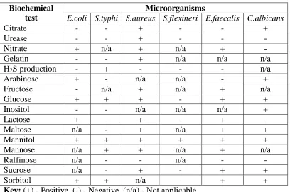
Durham tubes were inserted to media containing broth and later observed for gas production. If there was gas production the result was termed as positive and no gas production as negative. The tubes were then closed and autoclaved at 121-degree Celsius for 15 minutes and left to cool before being stored in a refrigerator before use. One millilitre inoculum of the isolated test microbes from a diluted sample of 10-6 was introduced to the media and incubated at 37 degrees celsius and observation did after 24 hours. The tubes containing solid media were observed for colour change. Where colour change occurred the results were termed as positive and no colour change as negative using standards (http://www.microbiologyinfo.com/biochemical test, 15th June 2014).
Table 3.1: Standard for biochemical tests on isolated microorganisms Biochemical
test
Microorganisms
E.coli S.typhi S.aureus S.flexineri E.faecalis C.albicans
Citrate - - + - - +
Urease - - + - - -
Nitrate + n/a + n/a + -
Gelatin - - + n/a n/a n/a
H2S production - + - - - n/a
Arabinose + - n/a n/a - +
Fructose - n/a + n/a + n/a
Glucose + + + - + +
Inositol - - n/a n/a n/a +
Lactose + - + - + -
Maltose n/a - + n/a + +
Mannitol + + + + + +
Mannose n/a + + n/a + n/a
Raffinose n/a - - n/a - -
Sucrose n/a - + - + +
Sorbitol + + n/a - + +
3.5.3 Maintenance of the bacterial and fungal cultures
The cultures of the clinical isolates of the test microorganisms were maintained on agar slants. The agar slants were prepared by making 250 millilitres of nutrient agar and potato Dextrose agar prepared according to their commercial instruction. The solution of the Medias was divided into bijou bottles at equal amount of 10 millilitres. The bijou bottles were then closed and placed in an autoclave and sterilized for about 15 minutes at 121 degrees celsius. The autoclave was left to cool before the bijou bottles containing the media are removed. The bottle was then placed on a wedge in a laminar flow slanting at around 45 degrees and left to cool forming agar slants. The bacterial microorganisms were streaked on nutrient agar slant whereas Candida albicans was streaked on potato dextrose agar slant. The streaked agar slants were placed in an incubator at 37 degrees celsius and observed for growth after every 24 hours. The test microorganism was subsequently subcultured after every 48 hours to maintain their viability.
3.6 Antimicrobial susceptibility testing
The microorganisms used (Escherichia coli, Salmonella typhi, Enterococcus
faecalis, Staphylococcus aureus, Shigella flexineri, Enterococcus faecalis and
Candida albicans) were concentrated by comparing it with a 0.5 McFarland
The stock solution concentration of each extract was formulated using the formula below:
S×M = C Q
Where: S - Solute weight (milligrams), Q - Dilution solvent (milliliters), M - Conversion units to micrograms, C - Concentration (µg/ml).
The discs prepared from the Whatman filter paper no.1 were impregnated with different the plant leaf extracts (Tagetes minuta, Aloe secundiflora, Vernonia
lasiopus and Bulbine frutescens) from the highest concentration of 1000µg/ml to
the lowest concentration of 1µg/ml by serially halving the concentration in subsequent dilutions using and stored in McCartney bottles away from light (Mangoma et al., 2010).
The antimicrobial efficacy test was carried out using Kirby-Bauer method (Newall et al., 1996). Muller Hinton agar (Sharau®) was used for bacterial test microorganisms and potato dextrose agar for Candida albicans in the spread plate technique where the clinical isolates were spread using sterilized cotton wool swabs. They were exposed to extracts impregnated discs in milligrams per microliter from Aloe secundiflora, Tagetes minuta, Vernonia lasiopus and Bulbine
frutescens. The discs were placed with equal distance between them on agar
Positive control standard discs containing ciprofloxacin (5µg/ml) was used for the bacteria’s Escherichia coli, Salmonella typhi, Enterococcus faecalis, Shigella
flexineri; vancomycin (3µg/ml) for Staphylococcus aureus, and fluconazole
(15µg/ml) for Candida albicans. A negative control of discs impregnated with 4% Dimethyl sulphoxide and 100% Analar (AR) methanol were also used. The Petri dishes were incubated at 37-degree celsius for 24 hours, after which they were removed and observed to see if there was the formation of any zones of inhibition by the extracts from the plant against the test microbes. The experiment was carried in duplicates and the diameter of zones of inhibition formed was measured and their average determined.
Minimal inhibitory concentration (MIC) was determine using the broth tube method according to Eloff et al., 1998) whereby: 100µl of 250mg/ml of methanol extract was added to 100µl of sterile bacteriological peptone in the first well of the 96 well microplate and mixed well with a micropipette. 100µl of this dilution was transferred subsequently to wells two folding each dilution of the original extract. This was done to the extracts of Aloe secundiflora, Bulbine frutescens,
Vernonia lasiopus, and Tagetes minuta. An inoculum of 100µl (0.5 McFarland
standard ) of overnight clinical cultures of ; Escherichia coli, Salmonella typhi,
Staphylococcus aureus, Shigella flexineri, Enterococcus faecalis and fungus
Candida albicans were added in each of the wells. Triplicate of each microplate
were then incubated at 37-degree celsius for 24 hours. After incubation 40µl of 0.2 mg/µl of INT was added in each of the wells and the plates examined after an additional 60 minutes of incubation. Growth was indicated by a red colour (conversion of INT to formazan). The lowest concentration at which the colour was apparently invisible as compared to the next dilution was taken as the minimum inhibitory concentration (Rabe et al., 2002). Minimum bactericidal concentration (MBC) and minimum fungicidal concentration (MFC) was determined according to Rabe et al., 2002 whereby: 100µl of the suspension was taken from microplate wells that demonstrated no growth and inoculated on agar plates. The plates were incubated at 37 degrees celsius for 24 hours. In the case where there was no bacterial growth and also not greater than the minimum inhibitory concentration was used to determine the maximum bacterial concentration and maximum fungicidal concentration.
3.7 Combined effect test
The combinations were determined using the permutation formula of P=N! / (N-R)! Producing six combinations namely: Aloe secundiflora and Tagetes minuta (AT), Aloe secundiflora and Vernonia lasiopus (AV), Bulbine frutescens and
Tagetes minuta (BT), Bulbine frutescens and Vernonia lasiopus (BV), Aloe
secundiflora and Bulbine frutescens (AB) and Vernonia lasiopus and Tagetes
minuta (VT). The combined effect was determined by using the plants leaf
the test microorganisms in replicates and the zones of inhibition measured. The average zones of inhibition formed when the extracts were used in combinations against the test microbes were compared with the zones of inhibition when they are used singly.
3.8 Qualitative phytochemical analysis
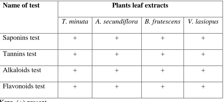
The presence of saponins, tannins, flavonoids and alkaloids in the crude extract were determined according to the method defined by Congesta etal (2005).
3.8.1Tannins
Each of the extracts was weighed to 0.5mg and dissolved in 1 ml of distilled water. Filtration was carried out after 2ml of FeCl3 was added. If there was the presence of a blue or black precipitate then it indicated the presence of tannins.
3.8.2 Flavonoids
Each of the extracts was weighed to 0.5mg and dissolved in 1 ml of ethanol and filtered. 2ml of 1% HCl and magnesium ribbon was added to the filtrate. If there was the formation of a pink or red colour it indicated the presence flavonoids.
3.8.3 Alkaloids
3.8.4 Saponins
Each of the extracts was weighed to 0.5mg and dissolved in 1 ml of methanol and filtered. Distilled water was added and shaking done for a few minutes. If there was persistence frothing then it indicated the presence of saponins.
3.9 Statistical analysis
The data collected was exported to Microsoft excel spreadsheet where descriptive statistics were carried out. The data was analyzed using SAS version 9.1 whereby; ANOVA (one way) was carried out to show statistical difference using the varying zones of inhibition between the test microbes exposed to plant leaf extracts from Tagetes minuta, Aloe secundiflora, Vernonia lasiopus and Bulbine
frutescens. Two-way ANOVA was also carried out to determine if there was any
CHAPTER FOUR RESULTS
All the bacterial pathogens (Escherichia coli, Salmonella typhi, Staphylococcus
aureus, Shigella flexineri and Enterococcus faecalis) and Candida albicans were
tested against plant extracts of the concentration of 1000µg/ml impregnated on discs. The bacterial pathogens and Candida albicans cultures of 0.5 McFarland standard were used for the efficacy test.
4.1 Efficacy test of the plant extracts on the bacterial pathogens and Candida albicans
All the bacterial pathogens (Escherichia coli, Salmonella typhi, Staphylococcus
aureus, Shigella flexineri and Enterococcus faecalis) and fungus Candida
albicans were exposed to the plant extract in replicates and they showed varied
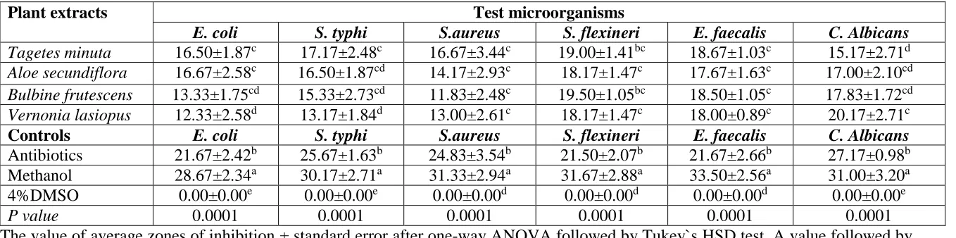
antimicrobial activity (Table 4.1). Aloe secundiflora extract produced the largest average zone of inhibition of 16.67±2.58mm against Escherichia coli when compared to the other plant extracts (Table 4.1).
produced the largest average zone of inhibition of 17.17±2.48mm against
Salmonella typhi when compared to the other plant extracts (Table 4.1). The
average zone of inhibition formed by an extract from Vernonia lasiopus was significantly different to those of Tagetes minuta (P˂0.05; Table 4.1). Moreover, the average zones of inhibition formed by ciprofloxacin, methanol and DMSO (negative control) were significantly different to those formed by the plant extract (P˂0.05; Table 4.1). Tagetes minuta extractproducedthe largest average zone of
inhibition of 16.67±3.44mm against Staphylococcus aureus when compared to other plant extracts (Table 4.1). The average zones of inhibition formed by the plant extracts were not significantly different from each other (P˃0.05; Table 4.1).
However, the average zone of inhibition formed by the plant extracts were significantly different to those formed by vancomycin, methanol and DMSO (negative control) were significantly different to those formed by the plant extract (P˂0.05; Table 4.1). Bulbine frutesens extract produced the largest average zone
of inhibition of 19.50±1.05mm against Shigella flexineri when compared to other plant extracts (Table 4.1). The average zones of inhibition formed by the plant extracts were not significantly different from each other (P˃0.05; Table 4.1). However, the average zones of inhibition formed by ciprofloxacin, methanol and DMSO (negative control) were significantly different to those formed by the plant extract (P˂0.05, Table 4.1). Tagetes minuta extract produced the largest average
compared to other plant extracts (Table 4.1). The average zones of inhibition formed by all the plant extracts were not significantly different (P˃0.05; Table 4.1). However, the average zones of inhibition formed by ciprofloxacin (positive control), methanol and DMSO (negative control) were significantly different to those formed by the plant extract (P˂0.05; Table 4.1). Vernonia lasiopus extract produced the largest zone of inhibition of 20.17±2.71mm against Candida
albicans when compared to other plant extracts (Table 4.1). The average zone of
Table 4.1: Average zones of inhibition in millimetres when plant extracts are used singly against bacterial pathogens and Candida albicans
The value of average zones of inhibition ± standard error after one-way ANOVA followed by Tukey`s HSD test. A value followed by the same superscript within the same column are not significantly different (P˃0.05).
Key: DMSO - Dimethyl sulphoxide, Plant extracts concentration (1000µg/ml) Antibiotics standard discs of; Ciprofloxacin (5µg/ml) for Gram negative bacteria, Vancomycin (3µg/ml) for Gram positive bacteria, Fluconazole (15µg/ml) for Candida albicans.
Plant extracts Test microorganisms
E. coli S. typhi S.aureus S. flexineri E. faecalis C. Albicans
Tagetes minuta 16.50±1.87c 17.17±2.48c 16.67±3.44c 19.00±1.41bc 18.67±1.03c 15.17±2.71d
Aloe secundiflora 16.67±2.58c 16.50±1.87cd 14.17±2.93c 18.17±1.47c 17.67±1.63c 17.00±2.10cd
Bulbine frutescens 13.33±1.75cd 15.33±2.73cd 11.83±2.48c 19.50±1.05bc 18.50±1.05c 17.83±1.72cd
Vernonia lasiopus 12.33±2.58d 13.17±1.84d 13.00±2.61c 18.17±1.47c 18.00±0.89c 20.17±2.71c
Controls E. coli S. typhi S.aureus S. flexineri E. faecalis C. Albicans
Antibiotics 21.67±2.42b 25.67±1.63b 24.83±3.54b 21.50±2.07b 21.67±2.66b 27.17±0.98b Methanol 28.67±2.34a 30.17±2.71a 31.33±2.94a 31.67±2.88a 33.50±2.56a 31.00±3.20a
4%DMSO 0.00±0.00e 0.00±0.00e 0.00±0.00d 0.00±0.00d 0.00±0.00d 0.00±0.00e
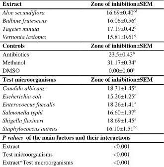
Table 4.2: Interactions between the plants extract and test microorganism when used singly
The value of average zones of inhibition ± standard error of mean (SEM) after two-way ANOVA followed by Tukey`s HSD test. A value followed by the same superscript within the same column are not significantly different (P˃0.05).
Key: Antibiotics (ciprofloxacin, vancomycin, and fluconazole), DMSO - dimethyl sulphoxide
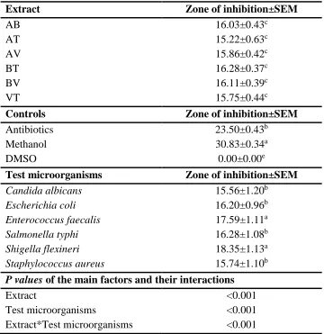
The average zone of inhibition formed by the plants extracts against all the test microorganism were significantly different to those formed by antibiotics, methanol and DMSO (P<0.05; Table 4.2). Moreover, zones formed by Tagetes
minuta extract against all the test microorganism was significantly different to
Extract Zone of inhibition±SEM
Aloe secundiflora 16.69±0.40cd
Bulbine frutescens 16.06±0.56d
Tagetes minuta 17.19±0.42c
Vernonia lasiopus 15.81±0.61d
Controls Zone of inhibition±SEM
Antibiotics 23.5±0.43b
Methanol 31.17±0.34a
DMSO 0.00±0.00e
Test microorganisms Zone of inhibition±SEM
Candida albicans 18.31±1.45a
Escherichia coli 15.26±1.25c
Enterococcus faecalis 18.26±1.41a
Salmonella typhi 16.60±1.37b
Shigella flexineri 18.69±1.45a
Staphylococcus aureus 16.10±1.51bc
P values of the main factors and their interactions
Extract <0.001
Test microorganisms <0.001
those formed by Vernonia lasiopus and Bulbine frutescens (P<0.05; Table 4.2). However, it was not significantly different to zones formed by Aloe secundiflora (P>0.05; Table 4.2). The average zones of inhibition formed by the plant extracts against Enterococcus faecalis, Candida albicans, and Shigella flexineri were not significantly different from each other (P>0.05; Table 4.2). However, they were significantly different to those formed by Escherichia coli, Salmonella typhi and
Staphylococcus aureus (P<0.05; Table 4.2). The interaction between the plant
extracts and test microorganisms were significant (P<0.05; Table 4.2).
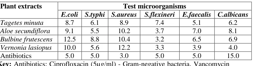
Table 4.3: Minimum inhibitory concentration in micrograms/milliliter of plant extracts against bacterial pathogens and Candida albicans
Plant extracts Test microorganisms
E.coli S.typhi S.aureus S.flexineri E.faecalis C.albicans
Tagetes minuta 8.7 6.1 8.9 7.4 5.1 6.2
Aloe secundiflora 9.1 5.5 10.2 3.7 7.0 8.1
Bulbine frutescens 12.5 8.8 10.4 3.2 6.5 6.9
Vernonia lasiopus 10.0 5.6 12.2 3.3 3.9 4.0
Antibiotics 5.0 5.0 3.0 5.0 5.0 15.0
Key: Antibiotics; Ciprofloxacin (5µg/ml) - Gram-negative bacteria, Vancomycin (3µg/ml) - Gram-positive bacteria, Fluconazole (15µg/ml) - Candida albicans)
Tagetes minuta extract was more active against Escherichia coli when compared
Tagetes minuta extracts were more active against Staphylococcus aureus at low concentration when compared to the other plant extracts. The positive control antibiotic (Vancomycin) used against Staphylococcus aureus, showed to be more active in low concentration when compared to all the extracts (Table 4.3). Bulbine
frutescens was more active against Shigella flexineri at low concentration when
compared to the other plant extracts. Furthermore, the level of concentration of
Bulbine frutescens extract was lower when compared to the used standard
antibiotic (Ciprofloxacin) against Shigella flexineri (Table 4.3).
The plant extract from Vernonia lasiopus was more active at low concentration against Enterococcus faecalis when compared to the other plant extracts. Its concentration was lower when compared to the used standard antibiotic (Ciprofloxacin) (Table 4.3). All the plant extracts were more active against
Candida albicans as compared to the used standard antibiotic (Fluconazole).
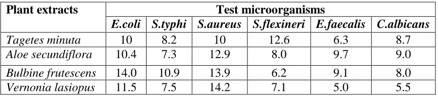
Table 4.4: Minimum bactericidal concentration and minimum fungicidal concentration in micrograms/milliliter of plant extracts against bacterial pathogens and Candida albicans
Plant extracts Test microorganisms
E.coli S.typhi S.aureus S.flexineri E.faecalis C.albicans
Tagetes minuta 10 8.2 10 12.6 6.3 8.7
Aloe secundiflora 10.4 7.3 12.9 8.0 9.7 9.0
Bulbine frutescens 14.0 10.9 13.9 6.2 9.1 8.0
Vernonia lasiopus 11.5 7.5 14.2 7.1 5.0 5.5
Tagetes minuta extract was bactericidal in low concentration against Escherichia
coli when compared to the other extracts (Table 4.4). The plant extract from Aloe
secundiflora was bactericidal against Salmonella typhi at low concentration when
compared to other plant extracts (Table 4.4). When used against Staphylococcus
aureus the extract from Tagetes minuta was bactericidal at low concentration
when compared to the other plant extracts (Table 4.4). Bulbine frutescens plant extract was bactericidal against Shigella flexineri at low concentration when compared to the other plant extracts (Table 4.4). When used against Enterococcus
faecalis and Candida albicans, Vernonia lasiopus was bactericidal and fungicidal
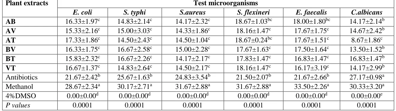
4.2 Combined effect of the plant extracts on the bacterial pathogens and Candida albicans
The plant extracts showed to be both more effective and less effective in some instances against all the test microorganisms when used in combinations.
Vernonia lasiopus and Tagetes minuta plant extract combinations formed the
largest average zone of inhibition of 16.67±1.37mm against Escherichia coli when compared to the other plant extract combinations (Table 4.5). The average zones of inhibition formed by the plant extracts combinations were not significantly different (P>0.05; Table 4.5). However, they were significantly different to those formed by ciprofloxacin, methanol and DMSO (negative control) (P<0.05; Table 4.5).
Bulbine frutescens and Vernonia lasiopus; Bulbine frutescens and Tagetesminuta
plant extract combinations formed the largest average zones of inhibition of 16.67±2.58mm and 16.67±2.26mm against Salmonella typhi when compared to the other plant extract combinations (Table 4.5). The average zones of inhibition formed by all the plant extracts combinations were not significantly different (P>0.05; Table 4.5). However, the average zones of inhibition formed by ciprofloxacin, methanol and DMSO (negative control) were significantly different to those formed by the plant extract (P˂0.05; Table 4.5). Bulbine frutescens and
Vernonia lasiopus plant extract combination formed the largest average zone of
other plant extract combinations (Table 4.5). The average zone of inhibition formed by all the plant extracts combinations were not significantly different (P>0.05; Table 4.5). However, the average zones of inhibition formed by vancomycin, methanol and DMSO (negative control) were significantly different to those formed by the plant extract (P˂0.05; Table 4.5). Aloe secundiflora and
Bulbine frutescens; Aloe secundiflora and Tagetes minuta plant extract
combinations formed the largest average zones of inhibition of 18.67±1.03mm and 18.67±0.24mm against Shigella flexineri when compared to other plant extracts combinations (Table 4.5). The average zone of inhibition formed by the plant extracts combinations was not significantly different to those formed by ciprofloxacin (positive control) (P>0.05; Table 4.5).
However, methanol and DMSO (negative control) were significantly different to those formed by the plant extract and ciprofloxacin (P˂0.05; Table 4.5). Aloe
secundiflora and Bulbine frutescens plant extract combination produced the
largest average zone of inhibition of 18.00±1.80mm against Enterococcus faecalis when compared to the other plant extracts (Table 4.5). The zones formed by the plant extracts were not significantly different (P>0.05; Table 4.5). Moreover, Aloe
secundiflora and Bulbine frutescens plant extract combination formed zones that
extract combinations and ciprofloxacin (P<0.05; Table 4.5). Bulbine frutescens
and Tagetes minuta plant extract combination formed the largest average zone of