Biologics: Targets and Therapy 2018:12 43–60
Biologics: Targets and Therapy
Dovepress
submit your manuscript | www.dovepress.com 43
R E V I E W
open access to scientific and medical research
Open Access Full Text Article
New frontiers in oncolytic viruses: optimizing and
selecting for virus strains with improved efficacy
Kenneth Lundstrom PanTherapeutics, Lutry, Switzerland
Abstract: Oncolytic viruses have demonstrated selective replication and killing of tumor cells. Different types of oncolytic viruses – adenoviruses, alphaviruses, herpes simplex viruses, Newcastle disease viruses, rhabdoviruses, Coxsackie viruses, and vaccinia viruses – have been applied as either naturally occurring or engineered vectors. Numerous studies in animal-tumor models have demonstrated substantial tumor regression and prolonged survival rates. Moreover, clinical trials have confirmed good safety profiles and therapeutic efficacy for oncolytic viruses. Most encouragingly, the first cancer gene-therapy drug – Gendicine, based on oncolytic adeno-virus type 5 – was approved in China. Likewise, a second-generation oncolytic herpes simplex virus-based drug for the treatment of melanoma has been registered in the US and Europe as talimogene laherparepvec.
Keywords: immunotherapy, viral vectors, clinical trials, drug approval
Introduction
Gene-therapy applications were initiated in the 1990s by utilization of both nonviral and viral delivery vectors.1 Although some progress was seen early on, the whole field,
especially the utilization of viral vectors, was severely hampered by some setbacks. Particularly, the death of a young patient treated with adenovirus vectors for the non-life-threatening disease ornithine transcarbamylase2 significantly reduced the interest
in gene therapy and slowed down its progress. Furthermore, retrovirus vectors used for treatment of children suffering from severe combined immunodeficiency (SCID) showed integration of a therapeutic gene into the LMO2 proto-oncogene region, which triggered leukemia development in some patients.3,4 In hindsight, it is obviously easy
to criticize the scientific community for moving too quickly into clinical trials without the proper safety conditions established. The setbacks, however, forced some serious reengineering of viral vectors and clinical protocols to improve delivery and targeting and to meet appropriate safety standards. These modifications include the introduction of elements controlling replication and expression, as well as means of termination of virus propagation by addition of the prodrug ganciclovir after administration of replication-competent Sindbis virus (SINV) carrying a fusion of the herpes simplex virus (HSV) TK gene and the SINV protein nsP3.5 In the long run, vector engineering
has significantly improved the properties of second- and third-generation vectors and enabled their safe applications for the treatment of various diseases.6
In this review, the focus is entirely on viral vectors in cancer therapy. One of the key issues from the birth of gene therapy has been delivery, and it remains the talking
Correspondence: Kenneth Lundstrom PanTherapeutics, 49 Route de Lavaux, Lutry CH1095, Switzerland
Tel +41 79 776 6351
Email [email protected]
Journal name: Biologics: Targets and Therapy Article Designation: REVIEW
Year: 2018 Volume: 12
Running head verso: Lundstrom
Running head recto: New frontiers in oncolytic viruses DOI: http://dx.doi.org/10.2147/BTT.S140114
Biologics: Targets and Therapy downloaded from https://www.dovepress.com/ by 118.70.13.36 on 19-Aug-2020
For personal use only.
Dovepress
Lundstrom
point.1 Intensive vector engineering addressing targeting and
delivery by the introduction of target-specific recognition signals and/or delivery-enhancing molecules, such as poly-mers and liposomes, has contributed to increased efficacy. Furthermore, the design of packaging cell lines has signifi-cantly facilitated the utilization of viral vectors for cancer treatment in experimental animal models. An interesting approach comprises employing oncolytic viruses as both naturally occurring7 and engineered8 vectors can provide
superior therapeutic efficacy, due to their selective tumor cell-killing capacity and potential induction of systemic antitumor immunity.9 Today, a number of different oncolytic
viruses, such as adenoviruses (Ads),10 HSV,11 alphaviruses,
rhabdoviruses,12 Newcastle disease virus (NDV),13 vaccinia
viruses (VVs),14 and others have been evaluated for antitumor
activity in a number of animal models and in clinical trials.
Viral vectors desirable for
therapeutic strategies
Commonly, both nonviral and viral vectors have been applied in cancer therapy.1 The use of nonviral vectors has mainly
been favored by their straightforward application and gener-ally good safety profiles, while the attractive features of viral vectors relate to their ability to provide superior delivery and extreme levels of transgene expression. Viral vectors have in general been characterized by their broad range of host-cell tropism and extreme expression levels of heterologous genes.15 Transient high-level expression is especially
attrac-tive for cancer-therapy applications, as the presence of anti-tumor and/or toxic products is limited in time. Alternatively, expression vectors comprised of regulation and termination signals have been engineered to restrict vector spread and long-term toxicity. Generally, viral vectors carrying either a DNA or RNA genome can accommodate foreign genetic information of different sizes, depending on which type of viral vector is used.15 For instance, vectors based on HSV and
VV are capable of accommodating more than 30 kb of foreign DNA, whereas most vector systems allow packaging of 6–8 kb of inserts, which is sufficient for covering more or less any therapeutic gene. Only adenoassociated viruses (AAVs) show a somewhat-limited packaging capacity in the range of 4 kb, but even that allows accommodation of a wide range of appropriate therapeutic genes. Both replication-deficient and -competent viral particles have been applied in immunization and therapeutic interventions in animal models.15 Moreover,
alphavirus vectors have been utilized in the form of naked RNA and plasmid DNA for the delivery of therapeutic genetic
information for toxic, anticancer, and immunostimulatory genes, as well as for miRNA and shRNA16.
Another issue is the potential immunogenicity triggered by the administration of viral vectors. In this context, the original Ad vectors have demonstrated strong immunogenic-ity, although later-generation versions with gene deletions have proven to be less immunogenic.17 AAV vectors have
also shown strong immunogenicity, especially after virus readministration, which has been circumvented by using different AAV serotypes for subsequent injections.18
The discovery of oncolytic viruses, which can provide specific replication in tumor cells and further induce killing without affecting normal cells, has provided attractive alter-native opportunities for cancer-therapy applications. In this context, naturally occurring oncolytic viruses and genetically engineered vectors have been subjected to cancer therapy and cancer-vaccine studies in animal models (Table 1), as described in detail herein (“Examples of therapeutic applica-tions of oncolytic viruses” section).
Mechanisms of oncolytic activity
Both natural and engineered oncolytic viruses utilize the general routes of recognition of cell-surface receptors and fusion to the plasma membrane with the special capabil-ity of establishing a lytic cycle in malignant cells, while normal tissues remain unaffected.19,20 The mechanism of
action occurs through RAS-pathway activation or by genetic modifications.21,22 In this context, HSV has been demonstrated
to replicate only in tumor cells dependent on TK activity.23
Moreover, in addition to the continuous replication in tumor cells, oncolytic viruses can recruit uninfected cells nearby without resulting in chromosomal integration or causing any major disease.24 One interesting feature of oncolytic
reovi-ruses,25 HSV,26 and VV27 is their ability to induce adaptive
immunoresponses, which can contribute indirectly to tumor cell death. Similarly, oncolytic Ads,28 Coxsackie virus (CV)
B3,29 and measles virus (MV)30 can induce stress of the
endo-plasmic reticulum, which attracts immune cells and results in immunologic cell death.
Other studies have demonstrated that viral infections of tumors can contribute to the immunosuppressive milieu by inducing immunostimulatory cytokines and chemokines.31–33
Although the production of cytokines and chemokines recruits and activates neutrophils, natural killer cells, mac-rophages, and CD4+ and CD8+ T lymphocytes, contributing
to viral clearance, it can also alter immunosuppression and stimulate antitumor responses.34–36 Moreover, various
Biologics: Targets and Therapy downloaded from https://www.dovepress.com/ by 118.70.13.36 on 19-Aug-2020
Dovepress New frontiers in oncolytic viruses
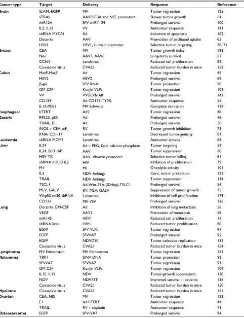
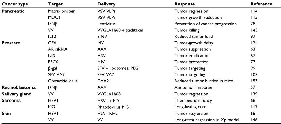
Table 1 Examples of preclinical cancer therapy applications for viral vectors
Cancer type Target Delivery Response Reference
Brain SLAM, EGFR
sTRAIL miR124 IL2, IL12 shRNA MYCN Decorin HSV1 MV
AAV9 CBA and NSE promoters SFV-miRT124
VV Ad AAV
HSV1, survivin promoter
Tumor regression Slower tumor growth Prolonged survival Antitumor response Induction of apoptosis Promotion of paclitaxel uptake Selective tumor targeting
125 64 100 141 162 65 70, 71 Breast CEA Neu CCNY Coxsackie virus MV AAV5, AAV6 Lentivirus CVA21 Tumor-growth delay Long-term survival Reduced cell proliferation Reduced tumor burden in mice
123 62 82 152 Colon MazF-MazE HSV2 β-gal GM-CSF VV CD133 IL12-PDL1 Ad HSV2 SFV RNA Kunjin VLPs VVGLVh168 Ad CD133-TYML MV Schwarz Tumor regression Prolonged survival Tumor protection Tumor regression Prolonged survival Antitumor response Complete remission 49 69 90 109 142 55 129
Esophageal hTERT Ad5 Tumor regression 48
Gastric RPL23, p53
TRAIL, E1 iNOS + CEA scFv RNAi CDH17 Ad Ad RV Lentivirus Prolonged survival Prolonged survival Tumor-growth inhibition Decreased tumorigenicity 46 50 73 81
Leukemia shRNA MCM7 Lentivirus Antitumor activity 83
Liver IL24
IL24, Bcl3 IAP HSV-TK shRNA miR30 IL2 M1 IL2 TRAIL TSCL1 MLV, GALV Wtp53-mi30-shRNA CD133
Ad + PEG, lipid, calcium phosphate AAV
AAV, albumin promoter HIV
M1
NDV Anhinga NDV Anhinga
Ad-Wnt-E1A-(δ24bp)-TSLC1 RV, MLV, GALV
Lentivirus MV VSV
Tumor targeting Tumor suppression Selective tumor killing Inhibition of proliferation Oncolytic activity Cure, tumor protection Tumor suppression Prolonged survival
Suppression of tumor growth Inhibition of cell proliferation Prolonged survival 52 60 61 79 101 133 134 54 75 179 126
Lung Decorin, GM-CSF
VEGF miR145 shRNA livin EGFP EGFP EGFP Coxsackie virus Ad AAV2 HSV1 HIV1 SFV VLPs SFVVA7 NDVD90 CVA21
Inhibition of lung metastasis Prevention of metastasis Reduced cell proliferation Reduced tumor proliferation Tumor regression
Prolonged survival Tumor-selective replication Reduced tumor burden in mice
56 58 11 80 91 95 131 154
Lymphoma MV Edmonston MV Edmonston Tumor regression 121
Melanoma TRP1 SFVVA7 GM-CSF IL15, IL12 NDV Coxsackie virus SINV DNA SFVVA7 Kunjin VLPs NDV NDV73T CVA21 Tumor protection Tumor regression Tumor regression Tumor-growth suppression Improved survival in patients Reduced tumor burden in mice
92 93 109 130 136 150
Myeloma Coxsackie virus CVA21 Reduced tumor burden in mice 151
Ovarian CEA, NIS
E1 TRAIL
MV Ad hTERT RV + cisplatin
Tumor regression Antitumor response Antitumor response 122 44 73
Osteosarcoma EGFP SFV-VA7 Prolonged survival 94
(Continued)
Biologics: Targets and Therapy downloaded from https://www.dovepress.com/ by 118.70.13.36 on 19-Aug-2020
Dovepress
Lundstrom
antiviral immunoresponses have been shown to contribute to the anticancer activity of oncolytic vesicular stomatitis virus (VSV), Maraba virus, VV, HSV, and reovirus by inducing IFN1, leading to the secretion of several immunostimulatory cytokines and chemokines, such as tumor necrosis factor (TNF) and TRAIL.37 Similarly, expression of
proinflamma-tory genes, such as IL12 or IL18, from oncolytic HSV38 and
Ad39 vectors has enhanced tumor-specific immunity.
More-over, coexpression of IL12 and CCL2 from an oncolytic HSV vector accelerates the recruitment of activated macrophages and T cells without affecting virus replication, albeit provid-ing improved survival rates.40
An interesting finding relates to enhanced antitumor activity in the presence of preexisting antiviral immunity. While improved survival has been obtained in immunocom-petent tumor models, the same phenomenon is not present in immunosuppressed mice.41 In contrast, innate immune cells
are capable of rapid clearance of replicating oncolytic HSV particles, which presents a significant limitation of oncolytic virotherapy.42 Furthermore, it was discovered in a Phase IB
clinical trial with the oncolytic HSV1-derived g134.5-deleted
G207 vector that a stronger inflammatory response and IFN-stimulated gene expression were detected in long-term survivors compared to nonresponders.43 In summary, four
phases contribute to oncolytic virotherapy: direct cellular lysis, cytokine-induced apoptosis, innate immune-cell cyto-toxicity, and antigen-specific adaptive T-cell killing.
Examples of therapeutic
applications of oncolytic viruses
A number of oncolytic viruses have been subjected to stud-ies in animal-tumor models (Table 1) and in a few clinical trials (Table 2). Ads represent the most frequently used viral vectors subjected to cancer therapy. For instance, animal models for ovarian,44 prostate,45 gastric,46 and brain cancer47
have been established. Related to gastric cancer, expression of RPL23 and p53 from a bicistronic Ad vector provides sig-nificantly better tumor-suppression activity in gastric cancer cells and antitumor responses in MKN45 cells compared to administration of the Ad-p53 vector alone.46 Furthermore,
administration of the bicistronic Ad-RPL23/p53 shows sur-vival benefits in a human gastric tumor model. In another approach, oncolytic Ad expressing luciferase (VRX007-Luc) was subjected to intratumoral injections in a Syrian hamster model, which provided similar levels of inhibition of tumor growth, as observed for immunosuppressive and chemotherapeutic agents such as cyclophosphamide. As human telomerase activity is present in more than 85% of primary cancers, the human telomerase reverse transcriptase (hTERT) promoter has been inserted into an attenuated Ad5 vector, resulting in significant tumor regression in an esophageal tumor model.48 Furthermore, introduction of the
bacterial MazF–MazE toxin–antitoxin system into an Ad vector has provided dose-dependent killing of KRAS cells and considerable tumor shrinkage in vivo without displaying
Cancer type Target Delivery Response Reference
Pancreatic Matrix protein MUC1 IFNβ
VV IL12
VSV VLPs VSV VLPs Lentivirus
VVGLV1h68 + paclitaxel SINV
Tumor regression Tumor-growth reduction Prevention of cancer progression Tumor killing
Reduced tumor load
114 115 78 145 97
Prostate CEA
AR siRNA NIS PSCA
β-gal SFV-VA7 Coxsackie virus
MV AAV HSV HIV1
SFV + liposomes, PEG SFV-VA7
CVA21
Tumor-growth delay Tumor suppression Tumor eradication Tumor protection Tumor targeting Tumor targeting
Reduced tumor burden in mice
124 63 67 77 99 103 153
Retinoblastoma IFNβ AAV Antitumor response 57
Salivary gland VV VVGLV1h68 Tumor regression 139
Sarcoma HSV1
MG1
HSV1 + PD1 Rhabdovirus MG1
Therapeutic efficacy
Long-lasting cure
68 117
Skin HSV1
VV
HSV1 RH2 VV
Tumor regression
Long-term regression in Xp model 66 146
Abbreviations: AAV, adenoassociated virus; Ad, adenovirus; AR, androgen receptor; β-gal, β-galactosidase; CVA21, Coxsackie virus A21; GALV, gibbon ape leukemia virus; GM-CSF, granulocyte-macrophage colony stimulating factor; HSV-TK, herpes simplex virus thymidine kinase; HSV1, herpes simplex virus 1; hTERT, human telomerase reverse transcriptase; IL2, interleukin 2; iNOS, inducible nitric oxide synthase; MV, measles virus; NDV, Newcastle disease virus; PEG, polyethylene glycol; RV, retrovirus; scFv, single-chain variable fragment; SFV, Semliki Forest virus; SINV, Sindbis virus; VLPs, virus-like particles; VSV, vesicular stomatitis virus; VV, vaccinia virus; Xp, Xeroderma
pigmentosum.
Table 1 (Continued)
Biologics: Targets and Therapy downloaded from https://www.dovepress.com/ by 118.70.13.36 on 19-Aug-2020
Dovepress New frontiers in oncolytic viruses
any side effects.49 Similarly, an oncolytic Ad vector with
a tumor-specific promoter expressing the TRAIL and E1A
genes has induced apoptosis in gastric cancer cell lines, inhibition of peritoneal metastasis, and prolonged survival in tumor-bearing mice.50 In attempts to improve oncolytic
Ads, incorporation of polymers, liposomes, and nanoparticles has extended the circulation time and reduced vector-based immunogenicity.51 In this context, formulation of oncolytic
vectors with polyethylene glycol, lipids, and calcium reduces liver sequestration and systemic toxicity of oncolytic Ads expressing IL24 (PLC-ZD55-IL24) in BALB/c mice.52
Intra-venous injection demonstrates efficient targeting of Huh7 tumors, with no observed toxicity.
Related to neuroblastoma, multidrug resistance has been a major issue hindering successful chemotherapy. It has triggered the engineering of an oncolytic Ad vector carrying shRNAs against the MYCN oncogene (ZD55-shMYCN),
which correlates with the expression of the protein MRP.53
ZD55-shRNA-based downregulation of MYCN inhibited tumor-cell proliferation and induced apoptosis in neuro-blastoma cells. Furthermore, ZD55-shRNA was capable of resensitizing doxorubicin-resistant cells to doxorubicin and resulted in reduced proliferation, increased apoptosis, and inhibited cell migration, which reduced the in vivo growth rate of neuroblastoma xenografts. In another approach, the dual-regulated oncolytic Ad wnt-E1A(δ24bp)-TSLC1 targeting the Wnt- and Rb-signaling pathways and carrying the TSLC1 tumor suppressor was engineered.54 In vivo administration
showed efficient inhibition of growth of transplanted tumors of hepatic cancer stem cells and prolonged survival in mice. Oncolytic Ad vectors targeted to the CD133 (prominin 1) cell-surface marker present on cancer stem cells have been developed by Ad-library screening.55 The engineered vector
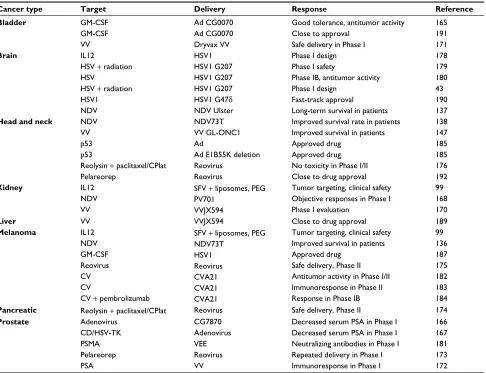
with the CD133-targeting motif (AdML-TYML) showed Table 2 Examples of clinical cancer therapy applications for viral vectors
Cancer type Target Delivery Response Reference
Bladder GM-CSF
GM-CSF VV
Ad CG0070 Ad CG0070 Dryvax VV
Good tolerance, antitumor activity Close to approval
Safe delivery in Phase I
165 191 171
Brain IL12
HSV + radiation HSV
HSV + radiation HSV1 NDV
HSV1 HSV1 G207 HSV1 G207 HSV1 G207 HSV1 G47δ
NDV Ulster
Phase I design Phase I safety
Phase IB, antitumor activity Phase I design
Fast-track approval
Long-term survival in patients
178 179 180 43 190 137
Head and neck NDV
VV p53 p53
Reolysin + paclitaxel/CPlat Pelareorep
NDV73T VV GL-ONC1 Ad
Ad E1B55K deletion Reovirus
Reovirus
Improved survival rate in patients Improved survival in patients Approved drug
Approved drug No toxicity in Phase I/II Close to drug approval
138 147 185 185 176 192
Kidney IL12
NDV VV
SFV + liposomes, PEG PV701
VVJX594
Tumor targeting, clinical safety Objective responses in Phase I Phase I evaluation
99 168 170
Liver VV VVJX594 Close to drug approval 189
Melanoma IL12
NDV GM-CSF Reovirus CV CV
CV + pembrolizumab
SFV + liposomes, PEG NDV73T
HSV1 Reovirus CVA21 CVA21 CVA21
Tumor targeting, clinical safety Improved survival in patients Approved drug
Safe delivery, Phase II Antitumor activity in Phase I/II Immunoresponse in Phase II Response in Phase IB
99 136 187 175 182 183 184
Pancreatic Reolysin + paclitaxel/CPlat Reovirus Safe delivery, Phase II 174
Prostate Adenovirus
CD/HSV-TK PSMA Pelareorep PSA
CG7870 Adenovirus VEE Reovirus VV
Decreased serum PSA in Phase I Decreased serum PSA in Phase I Neutralizing antibodies in Phase I Repeated delivery in Phase I Immunoresponse in Phase I
166 167 181 173 172
Abbreviations: Ad, adenovirus; CD, cytosine deaminase; CPlat, carboplatin; CV, Coxsackie virus; GM-CSF, granulocyte-macrophage colony-stimulating factor; HSV-TK,
herpes simplex virus thymidine kinase; NDV, Newcastle disease virus; PEG, polyethylene glycol; PSMA, prostate-specific membrane antigen; PSA, PS antigen; SFV, Semliki
Forest virus; VEE, Venezuelan equine encephalitis; VV, vaccinia virus.
Biologics: Targets and Therapy downloaded from https://www.dovepress.com/ by 118.70.13.36 on 19-Aug-2020
Dovepress
Lundstrom
selective infection and lysis of CD133+-cultured cells. Nude
mice vaccinated with AdML-TYML were protected against challenges with CD133+ colorectal carcinoma (CRC).
More-over, strong antitumor responses were observed in mice with established CD133+ CRC xenografts after intratumoral
injec-tions of AdML-TYML. In another study on CRC, oncolytic Ads expressing decorin (DCN), a regulator of cancer devel-opment and progression, and the granulocyte-macrophage colony-stimulating factor (GM-CSF) showed significant inhibition of tumor growth and lung metastasis after intra-tumoral administration in mice with implanted CT26 xeno-grafts.56 Furthermore, multiple protumorigenic pathways were
downregulated and antitumor immunoresponses activated. AAV vectors have also been evaluated in a number of cancer-therapy studies. One issue of concern has been the strong immunogenicity presented by readministration of AAV vectors.18 As a special case due to the immunoprivileged
nature of the eye, intravitreal injection of AAV expressing IFNβ has provided a strong antitumor effect in a preclinical retinoblastoma model without any issues of readministration.57
Despite immunogenicity issues, AAV2-based expression of VEGF generates prevention of pulmonary metastases in mice with implanted 4T1 tumors.58 In another approach,
AAV3 targets hepatoblastoma and hepatocellular carcinoma (HCC) cell lines efficiently by using hepatocyte growth fac-tor recepfac-tor (HGFR) as a cellular corecepfac-tor.59 Furthermore,
AAV vectors have been used in combination therapy of the p53-independent Bcl3-insensitive apoptotic protein and IL24 in HepG2 cells and nude mice in vivo.60 In another study on
HCC, HSV-TK expression driven by the albumin promoter and human alpha-fetoprotein (AFP) enhancer from AAV showed selective killing of AFP-positive HCC cells, but not nonhepatocyte tumor cells or AFP- or albumin-negative hepatic tumor cells.61 In the context of oral administration,
AAV5 and AAV6 serotypes expressing a truncated form of the Neu oncogene have shown significantly improved survival and long-lasting protection in 80% of mice implanted with Neu-positive TUBO breast tumors.62
AAV vectors have also been applied in gene silencing. In this context, two unique shRNAs induced apoptotic cell death in androgen receptor-positive prostate cancer cells and suppressed tumor growth after intratumoral injection of mice with implanted xenografts from either androgen-responsive or castration-resistant prostate cancer cells.63 Furthermore,
tail-vein injections provided xenograft elimination within 10 days. Engineering of the ubiquitous chicken beta actin (CBA) and neuron-specific enolase (NSE) promoters into an AAV9 vector was monitored for bioluminescent reporter-gene
expression after intravenous administration.64 The AAV9
vec-tor carrying the NSE promoter showed 100-fold lower expres-sion in the liver. The AAV9-CBA vector targeted astrocytes, neurons, and endothelial cells, while the AAV9-NSE vector provided mainly neuron-specific expression. Moreover, both AAV9-CBA and AVV9-NSE expressing sTRAIL generated slower tumor growth and significantly prolonged survival in mice with intracranial xenografts from glioblastoma patients. Recently, DCN expression from AAV vectors was evaluated in vitro and in vivo.65 It was demonstrated that transduced
neuroblastoma cells expressed DCN and systemic adminis-tration of AAV-DCN in nude mice promoted intratumoral uptake of paclitaxel.
HSV-based cancer therapy has been verified in a syngenetic C3H squamous-cell carcinoma model using the lytic HSV1 RH2 vector.66 In addition to therapeutic
efficacy observed after intratumoral injection, growth in contralateral tumors was also significantly suppressed. In another application, an oncolytic HSV1 vector containing four copies of miR145 targeting the 3'-end untranslated region (UTR) of the essential HSV ICP27 gene was able to decrease cell proliferation and prevention of colony forma-tion of non-small-cell lung cancer (NSCLC) cells, which further enhanced cancer-cell killing when combined with radiotherapy.11 It has also been demonstrated that oncolytic
HSV vectors expressing NIS increased antitumor activity by concentration of radioactive iodine in human prostate LNCaP cells.67 Moreover, intratumoral injection of HSV-NIS resulted
in efficient tumor eradication in nude mice implanted with LNCaP xenografts, and systemic administration provided prolonged survival. Oncolytic HSV vectors have also been tested in syngenetic mouse-rhabdomyosarcoma models in combination therapy with the cell-death-inhibiting ligand PDL1, which might provide a new approach for treatment of childhood soft-tissue sarcomas.68 Moreover, oncolytic
HSV2 vectors show significant inhibition of tumor growth and prolonged survival of BALB/c mice with implanted CT26 tumors.69 Additionally, HSV2 replication contributes
to reduced myeloid-derived suppressor cells and regulatory T cells in the spleen, which also decreases the number of dendritic cells in tumor-draining lymph nodes.
A glioma-specific HSV1 amplicon virus has been engineered to target tumor cells selectively by replacing the HSV1 ICP4 promoter with the tumor-specific survivin promoter.70 Furthermore, incorporation of 5 miR124 target
sequences into the 3'UTR of the ICP4 gene provided trans-lational regulation. The SU4124 HSV1 vector demonstrated enhanced expression of survivin and eIF4E in glioma cells
Biologics: Targets and Therapy downloaded from https://www.dovepress.com/ by 118.70.13.36 on 19-Aug-2020
Dovepress New frontiers in oncolytic viruses
and increased expression of miR124 in normal mouse and human brain tissue. Moreover, a strong antitumor effect was observed in a panel of glioma cell lines. Additionally, signifi-cantly increased antitumor activity was discovered in mice with human U87 glioma tumors after intratumoral injections.
An interesting observation relates to enhanced replication of oncolytic HSV in glioblastoma after short-term nutritional restriction (fasting).71 Glioblastoma cell lines from human
patients subjected to transient fasting for 24 hours increased late HSV expression and improved viral yields. Transient fasting for 48 hours followed by a 24-hour recovery doubled luciferase activity after intratumoral HSV administration in orthotopic glioblastoma xenografts.
Retroviruses have been subjected to a number of cancer-therapy applications, including recombinant bifunctional retrovirus vectors expressing a single-chain variable fragment (scFv) antibody to CEA and the inducible nitric oxide syn-thase (iNOS) gene.72 SCID mice subcutaneously injected with
MKN45 cells expressing CEA showed significant inhibition in tumor growth with 70% reduction in tumor size. The prob-lem of drug resistance has been addressed by demonstrating that retroviruses expressing the TRAIL gene are susceptible to A2780/DDP ovarian cancer cells, which in combination with cisplatin treatment enhanced antitumor activity in nude mice with implanted A2780/DDP xenografts.73 In attempts to
improve the safety of retrovirus-based therapy for hemato-logical malignancies, T cells with chimeric antigen receptors have been engineered.74 Additionally, deletion of oncogenes
and inactivation of oncogenic signaling pathways have been achieved by introduction of Cas9, zinc finger nucleases (ZFNs), or transcription activator-like effector nucleases (TALENs) into retrovirus vectors.
Replicating retrovirus vectors based on murine leukemia virus and gibbon ape leukemia virus (GALV) have proven effective in tumor killing.75 Comparison of murine leukemia
virus and GALV indicated more rapid replication kinetics for the latter in tumors, and in vivo GALV-based suicide-gene therapy demonstrated efficient suppression of HCC-tumor growth. In another study, it was shown that replication competence of retroviruses can provide a powerful tool for generation of novel tumor-specific retrovirus variants, which can be generated by natural selection.76 Moreover, retrovirus
vectors are able to integrate stably into the genome of cancer cells, which can contribute to long-lasting therapeutic effi-cacy, keeping in mind that the integration event is controlled to avoid any unwanted effects, as discussed previously.3
Belonging to the family of retroviruses, lentiviruses have also found a number of applications in cancer therapy. In this context, lentivirus vectors expressing PSCA have
been targeted to DC-SIGN-expressing 293T cells and bone marrow-derived dendritic cells, which provided protection against lethal tumor challenges in the TRAMP-C1 synergic tumor model and reduced tumor growth in animals with pre-existing tumors.77 In another study, self-inactivated lentivirus
vectors expressing human IFNβ achieved 90% transduction efficiency in pancreatic tumor cell lines, leading to inhibition of cell proliferation and induction of cell death.78
Further-more, progression of pancreatic cancer was prevented for 15 days in mice after administration of lentivirus human IFNβ.
Lentivirus vectors have also been employed in gene silencing. For instance, delivery of lentivirus vectors car-rying Wtp53-pPRIME-mi30-shRNA to AFP-positive liver cells resulted in inhibition of proliferation in Hep3B cells and in mice.79 Moreover, lentivirus-based delivery of
shR-NAs for Livin efficiently induced apoptosis in tumor cells, reduced proliferation of tumors, and contributed to cell-cycle arrest.80 Reduced proliferation and increased apoptosis was
also observed in MKN28 gastric cancer cells and in vivo after delivery of lentivirus vectors carrying CDH17 RNAi.81
Related to breast cancer. lentivirus vectors expressing shRNA were used to knock down cyclin Y (CCNY) expression in MCF7 and MDA-MB231 cells, resulting in substantial decrease in cell proliferation and colony formation and inhibition of cancer-cell growth through activation of Bad and GSK3β and cleavage of poly (ADP-ribose) polymerase (PARP) and caspase 3 in a p53-dependent manner.82
Lentivi-rus vectors have also been applied for targeting MCM7 with shRNAs to suppress the endogenous expression in K562 cells as a novel approach for the treatment of leukemia.83
In attempts to enhance lentivirus gene transfer, nanofibrils have been engineered to provide highly versatile and broad delivery profiles and to facilitate lentivirus concentration.84
Additionally, a platform for insertional mutagenesis was estab-lished for lentiviruses to induce HCC efficiently in various mouse models and for the identification of four previously unknown liver cancer-associated genes.85 In another approach,
lentivirus vectors were pseudotyped with truncated MV glycoproteins, which provided targeting of lymphocytes and antigen-presenting cells through signaling lymphocyte activa-tion molecule (SLAM) acting as an entry receptor.86
Reporter-gene expression confirmed the targeting, and administration of pseudotyped lenti-MV glycoproteins showed predominant induction of antigen-specific CD8+ T cells and suitability for
vaccines eliciting antigen-specific immunoresponses. Alphaviruses have been subjected to vector engineering, especially for Semliki Forest virus (SFV),87 SINV,88 and
Venezuelan equine encephalitis (VEE) virus.89 Applications
for cancer therapy have included administration of RNA
Biologics: Targets and Therapy downloaded from https://www.dovepress.com/ by 118.70.13.36 on 19-Aug-2020
Dovepress
Lundstrom
replicons, recombinant alphavirus particles, and layered DNA vectors. For instance, immunization of mice with SFV-LacZ RNA demonstrated tumor regression and provided protection against challenges with tumor cells.90 Likewise, SFV EGFP
particles subjected to intratumoral injections resulted in tumor regression in immunodeficient mice with human lung carcinoma xenografs.91 Additionally, immunization of mice
with SINV plasmid DNA carrying the TRP1 gene showed antitumor activity and immunoprotection in mice.92
Oncolytic alphaviruses occur naturally and have also been engineered from avirulent SFV strains. High infection rates and lysis of cancer cells were observed for the avirulent SFVA7(74) strain (SFVVA7) and a single intraperitoneal or intravenous injection showed significant tumor regression in SCID mice with established melanomas.93 Similarly,
improved survival rates were observed after SFV-VA7-EGFP administration in nude mice with osteosarcoma94
and orthotypic lung-tumor xenografts.95 Furthermore,
SFV-VA7-EGFP particles demonstrated efficient replication and killing of two canine-tumor cell lines, and no adverse events occurred in beagle dogs after intravenous administration of 2×10⁵ particles.96
Adequate attention has been paid to tumor targeting of alphavirus vectors. In this context, it has been shown that SINV particles possess natural tumor targeting after intra-peritoneal administration of mice implanted with tumor xeno-grafts, and subcutaneous SINV-IL12 administration reduced the tumor load to 6.2% of control mice.97 In contrast, studies
on SFV particles showed no tumor targeting.98 For this
rea-son, liposome-encapsulated SFV particles were engineered to provide tumor targeting of β-galactosidase after systemic delivery of SFV-LacZ particles in SCID mice.99 Moreover,
encapsulated SFV-IL12 particles showed good safety pro-files in kidney carcinoma and melanoma patients.99 Another
approach comprises engineering six tandem neuron-specific miR124 sequences between the nsP3 and nsP4 genes in the SFV4 genome, which provided glioma targeting and limited spread in the central nervous system (CNS) in BALB/c mice after intraperitoneal delivery of SFV4-miR124 particles.100
Moreover, the naturally occurring oncolytic M1 alphavirus is capable of selective killing of zinc-finger antiviral protein (ZAP)-deficient cancer cells, providing potent oncolytic efficacy and high tumor tropism in vitro and in vivo.101 In
another study, the safety of M1 was evaluated in nonhuman primates prior to initiation of clinical trials.102 Five macaques
received three rounds of 109 pfu of M1 intravenously and were
monitored for a number of physiological and biochemical parameters, neutralizing antibodies, and clinical symptoms.
No clinical, biochemical, immunological, or medical imaging indicated any evidence of toxicity, suggesting that M1 can be safely used for intravenous administration in cancer patients.
Recently, SFV-VA7 particles were evaluated in human VCaP, LNCaP, and 22Rv1 prostate cancer cell lines and in the nonmalignant RWPE1 prostate epithelial cell line, as well as in subcutaneous and orthotopic mouse LNCaP xenograft models.103 Interestingly, all prostate cancer cell lines,
irre-spective of their hormone-response status, were efficiently killed by SFVVA7, whereas RWPE1 cells were resistant to SFVVA7, indicating tumor targeting of SFV. This result is in contrast to previous findings of lack of tumor targeting of SFV particles.98 In vivo, a single peritoneal dose of SFVVA7
showed eradication of all subcutaneous and orthotopic LNCaP tumors.
Flaviviruses are enveloped ssRNA viruses engineered for recombinant protein expression and cancer therapy.104–108 For
example, Kunjin virus vectors expressing GM-CSF provided cure in more than 50% of mice with established subcutaneous CT26 colon carcinomas after intratumoral administration.109
Moreover, regression of B16-OVA melanoma tumors was obtained after 5 days, with a cure rate of 67%. Subcutane-ous administration of Kunjin GM-CSF particles resulted in tumor regression in CT26 lung metastasis in BALB/c mice.
Rhabdoviruses, such as rabies virus110,111 and VSV,112 have
been engineered as expression vectors applicable for cancer therapy with a special emphasis on oncolytic VSV vectors.113
The attractive features of VSV comprise high susceptibility of cancer cells, lack of antiviral responses induced by type I IFN, ease of manipulation, replication in the cytoplasm, and no preexisting immunity in humans. VSV particles show superior oncolytic activity in 13 relevant human pancreatic cell lines in comparison to Ads, Sendai virus, and respiratory syncytial virus, although the response is heterogeneous, with some cell lines being resistant to VSV.114 Likewise, strong
oncolytic activity was obtained in pancreatic ductal adeno-carcinoma cell lines after infection with VSV expressing MUC1, and tumor-growth reduction was achieved in vivo.115
Combination therapy with gemcitabine further enhanced therapeutic efficacy. In another study, VSV demonstrated apoptotic activity in pancreatic ductal adenocarcinoma cell lines.116 However, resistance to apoptosis was seen in three
cell lines with high constitutive expression of IFN-stimulated genes. Oncolytic rhabdovirus MG1 particles have shown a strong ability to kill human and canine sarcoma cell lines, and infected more than 80% of human sarcoma tissues tested ex vivo.117 MG1 treatment of sarcoma-bearing mice showed
a significant increase in long-lasting cure and also provided
Biologics: Targets and Therapy downloaded from https://www.dovepress.com/ by 118.70.13.36 on 19-Aug-2020
Dovepress New frontiers in oncolytic viruses
protection against subsequent tumor challenges. VSV vec-tors have also been tested for replication in cancer cells from ascites.118 Administration of 108 pfu generated a significant
inhibition of ascites formation and prolonged survival in mice. Moreover, metabolic adaptive processes in peritoneal carcinoma, including high glycolytic activity and glutamine metabolism, favored VSV replication.
MVs, similar to rhabdoviruses in possessing an envel-oped ssRNA genome, have also been subjected to vector development for cancer therapy.119,120 Oncolytic Edmonston
B (MV Edm) has been employed in studies in xenograft and syngeneic models. For instance, tumor regression was observed in SCID mice implanted with lymphoma xenografts after intratumoral administration of MV Edm.121 MV-based
dual therapy with CEA and thyroidal NIS showed a superior outcome in treatment of mice with implanted SKOV3ip.1 ovarian tumor xenografts in comparison to administration of MV CEA or MV NIS individually.122 Related to breast cancer,
MV-CEA vectors provided significant tumor-growth delay and prolonged survival in an MDA-MB231 mammary-tumor model.123 Furthermore, intratumoral MV-CEA administration
resulted in delayed tumor growth and extended survival in mice with implanted subcutaneous PC3 prostate tumors.124
Tumor targeting of MV vectors was achieved by introduction of CD46 and SLAM into the hemagglutinin protein combined with the display of a single-chain antibody against EGFR.125
The retargeted vector showed antitumor activity but no neurotoxicity in MV replication-permissive transgenic mice after systemic administration. Another attempt to target MV vectors involved engineered MV vectors based on the cancer stem-cell marker CD133 (prominin 1).126 Selective tumor
targeting was obtained by replacing the MV hemagglutinin receptor with a CD133-specific scFv. Furthermore, engi-neered chimera between MV CD133 and VSV showed highly selective elimination of CD133+ cells. The VSV-CD133
vector revealed highly potent oncolytic activity in HCC and prolonged survival of mice after intravenous injection. Moreover, VSV CD133 infected a >104-fold larger tumor
area in comparison to MV CD133. In another approach, MV vectors based on the MV Schwarz strain encoding a fusion of IL12 and an antibody against PDL1, respectively, resulted in complete remission in 90% of tested mice with established tumors.127
NDV is another ssRNA virus, and belongs to the para-myxoviruses. The oncolytic properties of NDV vectors have made them attractive for cancer therapy, which has been confirmed in several animal-tumor models.128,129
Compari-son of NDV-based expression of IL12 and IL15 indicated
lower toxicity of IL15 in mice with implanted melanoma tumors, and intratumoral administration showed efficient suppression of tumor growth for both ND IL12 and NDV IL15.130 Although not statistically significant, the survival
rate was 12.5% higher for NDV IL15. The reverse genetically engineered NDVD90 strain expressing EGFP showed tumor-selective replication, induction of apoptosis in A549 lung cancer cells, and suppression of tumor growth in vivo.131
Fur-thermore, a highly virulent NDV strain adapted for replication in HeLa cells promoted upregulation of TRAIL and caspase activation through induction of apoptosis.132 Interestingly, the
NDV Anhinga strain carrying the IL2 gene showed strong inhibition of HCC growth, providing both complete cure and protection against tumor challenges 60 days after immuniza-tions.133 In another study, administration of the recombinant
NDV Anhinga strain expressing TRAIL resulted in efficient HCC suppression without showing any significant toxicity in normal tissue.134 Related to NDV epidemics in poultry, an
oncolytic vector based in the NDV-73T strain with a modified fusion-protein cleavage site and a 198-nucleotide insertion in the HNL intergenic region was engineered, which showed significantly reduced viral gene expression and replication in avian cells, but not in mammalian cells.135 Moreover,
intra-tumoral and intravenous administration generated selective replication and killing of tumor cells.
A number of Phase I–III clinical trials applying NDV vec-tors have been conducted for melanomas,136 glioblastomas,137
and head and neck cancer,138 with some encouraging results.
For instance, treatment with mesogenic NDV73T resulted in more than 60% 10-year survival in comparison to 6%–33% in the control group.136 Moreover, the lentogenic NDV Ulster
strain demonstrated long-term survival of one glioblastoma patient in comparison to none in the control group.137
Simi-larly, the 5-year survival rate was 51% in a Phase II study on head and neck cancer with the NDV-73T strain.138
Poxviruses carry a large dsDNA genome with a good packaging capacity of foreign DNA and tumor-selective properties providing cellular destruction by viral replica-tion.139 Introduction of deletions in the TK and VGF genes
allow replication in tumor cells, while normal cells are not affected.140 Moreover, the modified poxvirus vector enhances
immunorecognition of tumors. Furthermore, expression of IL2 or IL12 from a VV vector generates antitumor activ-ity in mice with implanted C6 gliomas.141 Oncolytic VVs,
such as GLV1h68, have lysed human COLO205, HCT15, HCT116, HT29, and SW20 CRC lines efficiently, and sig-nificantly inhibited tumor growth and prolonged survival in athymic mice with established colorectal xenografts after a
Biologics: Targets and Therapy downloaded from https://www.dovepress.com/ by 118.70.13.36 on 19-Aug-2020
Dovepress
Lundstrom
single GLV-1h68 intravenous injection.142 Likewise, human
salivary-gland carcinoma cells were susceptible to GLV1h68, and a single intravenous administration resulted in significant tumor regression.143 Genetic modifications made to the VV
GLV1h151 vector enhanced cancer specificity, resulting in efficient infection, replication, and killing of several cancer-based (breast, lung, pancreatic, and colorectal) cell lines.144
Intravenous injection of VV GLV1h151 confirmed replica-tion in tumorsin vivo. The oncolytic GLV1h68 was further evaluated in combination with chemotherapy applying nab -paclitaxel and gemcitabine, which provided enhanced tumor-cell killing in two of four human pancreatic adenocarcinoma cell lines.145 The feasibility of chemovirotherapy seemed to
be related to efficient viral replication, as the nonresponsive tumor-cell lines showed only low levels of viral replication.
VV vectors have also been applied in tumor-bearing
Xeroderma pigmentosum (XP) patients excluded from conventional DNA-damaging therapy.146 VV vectors
demon-strated ten- to 100-fold higher cytotoxicity in tumor-derived cells from XP patients compared to normal control cells, and systemic administration showed long-term tumor regres-sion in XP animal models. The oncolytic GL-ONC1 VV vector was administered intravenously in combination with chemoradiotherapy in patients with primary nonmetastatic head and neck cancers.147 The follow-up of patients indicated
1-year progression-free survival and overall survival of 74.4% and 84.6%, respectively, which demonstrated the safety and feasibility of GL-ONC1 delivery.
CVs belong to the family of Picornaviridae, with an ssRNA of positive polarity and an nonenveloped structure.148
They are known pathogens, and present the leading causes of aseptic meningitis. However, CVA21 has shown promise as an oncolytic vector and demonstrated potential in preclinical cancer models.149 In this context, decreased tumor burden has
been obtained in tumor models for melanoma,150 multiple
myeloma,151 breast,152 prostate,153 and lung154 cancers. More
specifically, multiple-myeloma cell lines have shown high sus-ceptibility to CVA21, resulting in lytic infection. Moreover, when biopsies from patient bone marrow were challenged with CVA21, specific removal of 98.7% of CD138+ plasma
cells was obtained with no decrease in the functionality of progenitor cells. For this reason, CVA21 administration might provide an efficient approach for multiple-myeloma treatment prior to transplantation of autologous stem cells. In another approach, a large-scale two-step screening procedure for 28 enteroviral strains was conducted, which identified that CVB3 presented oncolytic activity against nine human NSCLC cell lines. CVB3 induced apoptosis and activated kinase-signaling
pathways. Intratumoral CVB3 administration generated sub-stantial tumor regression in mice with established NSCLC tumors. Interestingly, injection of CVB3 into tumors located on the right flank demonstrated replication-competent CVB3 and significant regression in xenografts on the left flank. It was also discovered that intratumoral administration of CVB3 recruited natural killer cells and granulocytes, thereby providing immunostimulatory activity.
Optimization and selection of
oncolytic viruses
A number of efforts have been made to optimize oncolytic viral vectors. In this context, a CD133-targeting motif (TYML) was introduced into an Ad vector, and provided selective infection and killing of CRC cells and protec-tion against CRC challenges.55 Similarly, MV vectors with
CD133-specific scFv showed potent oncolytic activity and prolonged survival in tumor-bearing mice.126 Moreover, dual
expression of GM-CSF and decorin (regulator of cancer development and progression) from Ad vectors led to sig-nificant inhibition of tumor growth and lung metastasis in vivo.56 Also, decorin delivery by AAV promoted intratumoral
paclitaxel uptake.65
Another approach consists of gene silencing, of which examples for antitumor activity have been demonstrated for shRNAs for AAV63 and miRNAs for HSV170 and
lenti-viruses.79 Furthermore, introduction of six tandem
neuron-specific miR124 sequences into the SFV vector has resulted in glioma targeting and only limited spread in the CNS.100
Promoter engineering has also allowed enhanced expression targeting, as demonstrated by neuron-specific delivery and 100-fold lower presence in the liver by applying the NSE promoter in AAV9 vectors.64 Selective targeting of tumors
has also been achieved by replacing the HSV1 ICP4 promoter with the tumor-specific survivin promoter.70 Moreover,
ret-roviruses have been subjected to chimeric antigen-receptor engineering to provide safe treatment of hematological malignancies.74
Selection of oncolytic virus strains has also been of great importance. In this context, the naturally occurring M1 alpha-virus has shown potent oncolytic activity and high tumor tro-pism,101 which further demonstrated no evidence of toxicity
in macaques and indicated safe intravenous administration in cancer patients.102 Similarly, plenty of attention has been
paid to reverse engineering of NDV strains, such as NDVD90, which showed tumor-selective replication and decrease in tumor growth.131 Similarly, the NDV Anhinga strain has
been subjected to preclinical studies for HCC treatment.133,134
Biologics: Targets and Therapy downloaded from https://www.dovepress.com/ by 118.70.13.36 on 19-Aug-2020
Dovepress New frontiers in oncolytic viruses
Moreover, engineering of the NDV73T strain resulted in reduced viral replication in avian but not mammalian cells.135
Oncolytic poxvirus strains, such as VV GLV1h68, have also been selected, which have shown antitumor activity for both CRC142 and salivary-gland carcinoma.143 Genetic
modifica-tions have enhanced cancer specificity for the VV GLV1h151 vector,144 and when combined with paclitaxel and gemcitabine
further increased tumor-cell death.145
Effects on tumor vasculature
Oncolytic viruses have also shown selective targeting of tumor vasculature. For instance, VSV caused thrombosis in tumor vessels by selective infection of endothelial cells in the tumor microenvironment.155 Moreover, HSV and VV are
capable of selectively provoking damage to the tumor endo-thelium.156,157 Oncolytic VV vectors have been engineered
to express antiangiogenic factors, such as VEGF inhibitors, which leads to suppression of VEGF levels and decrease in perfusion within tumors.158,159 Furthermore, oncolytic
VV-based targeting of VEGF provided a synergistic antitu-mor effect with VEGFR tyrosine-kinase inhibitors.160 This
synergism may be caused by off-target inhibition of cellular antiviral defense proteins, such as dsRNA-dependent protein kinases.161,162 In the context of the aggressive anaplastic
thy-roid carcinoma (ATC), oncolytic Ad d1922-947 induced cell death in vitro and tumor regression in animal models.163 It was
also demonstrated that Ad d1922-947 decreased IL8/CXCL8 and MCP-1/CCL2 expression in the 8505-c and BHT101-5 ATC cell lines and reduced IL8 impaired ATC-induced angiogenesis in vivo. Overall, the oncolytic Ad reshaped the protumorigenic ATC microenvironment by modulation of intrinsic cancer-cell factors and immunoresponses.
Clinical trials using oncolytic viruses
The progress made in cancer therapy with oncolytic viruses in preclinical studies has further encouraged the transition into clinical trials. A large number of studies employing, eg, Ad, alphavirus, HSV, reovirus, NDV, MV, and CV vectors have been conducted or are in progress.164 In this context,
the safety, pharmacokinetics, and anticancer activity of an intravesical oncolytic Ad (CG0070) was evaluated in a Phase I trial in 35 patients with non-muscle-invasive bladder can-cer.165 Patients received intravesically either a single or
mul-tiple doses of 1012, 3×10¹², 1×10,13 and 3×10¹³ viral particles,
respectively, three times every 28 days or six times weekly. Due to grade 1–2 bladder toxicity, a maximum tolerated dose (MTD) was not reached, although the safety profile was tolerable and anti-bladder cancer activity observed. Ads have
also been subjected to several clinical trials in prostate cancer patients, including a replication-selective PSA- targeted onco-lytic vector166 and a replication-competent vector providing
double-suicide-gene therapy,167 which resulted in decreased
serum levels of PSA.
NDV has also been subjected to several Phase I–III clinical trials.136–138 A 10-year observation of 83
postsurgi-cal patients with stage II malignant melanoma treated with NDV demonstrated no presence of disease in 60%, which is remarkable in comparison to similar studies showing only 5%–33% survival. Moreover, exceptional survival was seen in 21 patients with head and neck cancer and six individu-als with cerebral metastases. NDV has individu-also been applied in the treatment of 23 patients with a vaccine consisting of NDV-infected patient-isolated glioblastoma cells followed by γ-irradiation.137 The NDV therapy caused no severe side
effects and showed longer median progression-free survival (40 weeks for NDV treatment and 26 weeks for controls) and median overall survival (100 weeks for vaccinated and 49 weeks for controls). Furthermore, 91% of the NDV-treated patients survived for 1 year compared to 45% for controls, and long-term survival was 4% and 0 for vaccinated and control patients, respectively. In another study, 20 patients with head and neck squamous-cell carcinoma preconditioned with IL2 were vaccinated with NDV-infected autologous tumor cells, which increased systemic antitumor activity.138
The replication-competent NDV PV701 strain was evalu-ated in a Phase I trial in renal cancer patients.168 An MTD of
1.2×10¹⁰ pfu/m2 was established, with only flu-like adverse
events. Moreover, objective responses were observed and progression-free survival ranged from 4 to 31 months.
Oncolytic VV vectors, such as GL-ONC1, were subjected to a Phase I clinical trial in combination with radio- and chemotherapy in patients with primary nonmetastatic head and neck cancer.147 Adverse reactions, such as fever, fatigue,
rash, nausea, and vomiting, were observed among the 19 patients recruited. The MTD was not reached, but patient follow-up demonstrated 1-year (2-year) progression-free survival and overall survival of 74.4% (64.1%) and 84.6% (69.2%), respectively, indicating safe application of VV in cancer patients undergoing radio- and chemotherapy. In another study, the oncolytic VV vector JX594 was evalu-ated in patients with refractory primary or metastatic liver cancer.169 JX594 provided direct oncolysis and overexpression
of GM-CSF, thereby stimulating the shutdown of tumor vas-culature and antitumor immunoresponses. Fourteen patients with refractory primary or metastatic liver tumors received one of four doses (108, 3×10⁸, 109, or 3×10⁹ pfu) every
Biologics: Targets and Therapy downloaded from https://www.dovepress.com/ by 118.70.13.36 on 19-Aug-2020
Dovepress
Lundstrom
3 weeks. All patients showed grade I–III flu-like symptoms, four patients presented dose-related thrombocytopenia, and grade III hyperbilirubinemia dose limitations at the highest dose defined the MTD as 109 pfu. Tumor responses were
observed in injected and noninjected tumors. Three patients showed partial responses, six presented stable disease, and one demonstrated progressive disease. VVJX594 has been further evaluated in patients with metastatic refractory renal-cell carcinoma.170 In another approach, a dose-escalating
study with intravesical Dryvax VV was conducted in bladder cancer patients.171 Viral infection was detected in tumor and
normal urothelial cells. The study indicated that VV was safely delivered into the bladder. VV vectors have also been applied for vaccination of patients with locally recurrent or progressive prostate cancer.172 Expression of PSA from a VV
vector revealed no dose-limiting toxicity, and intraprostatic administration was safe and elicited significant immunore-sponses in the Phase I trial.
Reoviruses have also been subjected to clinical trials.173,174
In this context, an oncolytic reovirus (pelareorep) was com-bined with carboplatin and paclitaxel for the treatment of metastatic pancreatic adenocarcinoma.174 In the randomized
Phase II trial, paclitaxel–carboplatin + pelareorep was com-pared to paclitaxel–carboplatin, which resulted in no differ-ence in progression-free survival. The presdiffer-ence of pelareorep generated higher levels of 14 proinflammatory plasma cyto-kines/chemokines. However, although pelareorep delivery was demonstrated to be safe, it did not improve progression-free survival of paclitaxel– carboplatin treatment, but combination with chemotherapy may improve targeting immunosuppres-sive mediators and enhance oncolytic virotherapy. In another approach, a Phase II study with replication-competent reo-virus was conducted in 21 metastatic melanoma patients.175
Patients treated with 3×10¹⁰ tissue culture infectious dose
(TCID)50 showed good safety profiles, and productive reo-virus replication was observed in biopsies. Furthermore, a Phase I/II dose-escalating combination-therapy study with carboplatin–paclitaxel and an oncolytic reovirus in head and neck cancer patients showed no dose-limiting toxicity, with one patient (3.8%) having a complete response, six patients (23.1%) partial responses, and two patients (7.6%) major clini-cal responses.176 Moreover, reoviruses have been subjected to
a Phase I study in prostate cancer patients, and demonstrated a good safety profile after repeated intravenous administration and reovirus targeting of metastatic tumors.173
The second-generation oncolytic HSV vector M032, which selectively replicates in tumor cells, has been employed for overexpression of IL12 to increase the antitumor effect
and provide an antiangiogenic effect to target new-tumor blood-vessel formation.177 Demonstration in preclinical
models has paved the way for initiating a Phase I clinical trial in patients with recurrent progressive glioblastoma multiforme. Furthermore, safety of the HSVG207 vector has been confirmed in preclinical studies in owl monkeys (genus Aotus)178 and in adult Phase I trials in progressive
and recurrent brain tumors.179,180 Based on these findings,
the design of a Phase I clinical trial to evaluate HSVG207 monotherapy or in combination with radiotherapy has been designed for children with progressive and recurrent malig-nant supratentorial brain tumors.43
Regarding alphaviruses, a Phase I trial has been con-ducted in patients with metastatic castration-resistant prostate cancer applying VEE particles expressing prostate-specific membrane antigen (PSMA).181 In the dose-escalating study,
patients received five doses of 0.9×10⁷ or 0.36×10⁸ IU of
VEE PSMA particles at weeks 1, 4, 7, 10, and 18, followed by another round of administration of the higher dose in six patients. No toxicities were observed in any patient, and VEE PSMA was well tolerated. However, no PSMA-specific cellular immunoresponse was observed, although a PSMA-specific signal was registered by enzyme-linked immuno-sorbent assay. Although neither clinical benefit nor robust immunosignaling was detected, neutralizing antibodies were produced, which indicated that there is a need for dose opti-mization. In another Phase I trial, replication-deficient SFV IL12 particles were encapsulated in liposomes and subjected to intravenous administration in terminally ill melanoma and kidney carcinoma patients.99 No liposome- or virus-related
toxicity was observed in any patient. IL12 plasma levels showed a transient five- to tenfold increase, indicating poten-tial immunostimulatory activity. Furthermore, the encapsula-tion procedure protected the virus from being recognized by the host immune system, allowing repeated administration of SFV IL12 particles.
In the context of CVs, the CVA21 strain has been demon-strated to target ICAM1, which is upregulated in melanoma, NSCLC, and bladder and prostate cancers.182 In a Phase I/
II trial, patients received multiple intravenous doses of the bioselected formulation Cavatak of CVA21, which were well tolerated.182 Prolonged presence of CVA21 RNA in the serum
of some patients suggested that viral replication occurred in tumors. Biopsies from melanoma patients confirmed tumor targeting of CVA21. Moreover, CVA21 seemed to provide increased antitumor activity, which might be further enhanced by combination with immunocheckpoint blockade. Additionally, a Phase II clinical trial in advanced-melanoma
Biologics: Targets and Therapy downloaded from https://www.dovepress.com/ by 118.70.13.36 on 19-Aug-2020
Dovepress New frontiers in oncolytic viruses
patients was conducted with CVA21, showing induced immune-cell infiltration in the tumor microenvironment.183
Similarly, a Phase IB combination-therapy study with CVA21 and systemic pembrolizumab in 20 advanced-melanoma patients demonstrated a best overall response rate of 60% and stable disease in 27% of the patients.184 Treatment tolerability
was good, with no dose-limiting toxicity and no grade 3 or higher treatment-related adverse events.
Conclusion
In summary, oncolytic viruses, based on engineered vec-tors or naturally occurring strains, have proven efficient in various preclinical tumor models providing suppression of tumor growth, tumor regression, and in certain cases com-plete eradication of existing tumors (Table 1). Immunization has also resulted in protection against tumor challenges. The variety of oncolytic viruses (Ad, AAV, HSV, retrovirus, lentivirus, alphavirus, flavivirus, rhabdovirus, MV, NDV, CV, and poxvirus) allows flexibility related to packaging capacity, host range tropism, and mode of expression (dura-tion, chromosomal integration). It can be concluded that no single oncolytic viral vector is universally superior, and for this reason the decision on which vector to use is to a large extent dictated by specific applications and familiarity with each vector system.
Most encouragingly, clinical trials have been conducted or are in progress for most oncolytic vector systems, and have confirmed safe administration in humans (Table 2). Moreover, therapeutic efficacy has also been achieved. Already some time ago, the first drugs based on oncolytic Ads expressing the p53 gene (Gendicine)185 and the Ad H101 vector with
an E1B55K deletion186 were approved in China. Recently,
the second-generation oncolytic HSV1 GM-CSF vector was approved for the treatment of melanoma in the US and Europe.187,188 A number of other drugs based on oncolytic
viruses, such as the oncolytic VV JX594 (pexastimogene devacirepvec) for HCC treatment,189 Ad CG0070 expressing
GM-CSF for bladder cancer,190 and pelareorep (Reolysin)
based on a wild-type variant of reovirus for head and neck cancer191 will most likely be approved in the near future.
Addi-tionally, the third-generation oncolytic HSV1 vector G47δ, subjected to a Phase II glioblastoma trial, should receive priority reviewing and fast-track drug approval.192 Overall,
the drug approvals obtained and the ongoing oncolytic virus development bode well for finding safer and more efficacious cancer drugs in the future.
Disclosure
The author reports no conflicts of interest in this work.
References
1. Lundstrom K, Boulikas T. Viral and non-viral vectors in gene therapy: technology development and clinical trials. Technol Cancer Res Treat. 2003;2:471-486.
2. Raper SE, Chirmule N, Lee FS, et al. Fatal systemic inflamma-tory response syndrome in a ornithine transcarbamylase deficient patient following adenoviral gene transfer. Mol Genet Metab. 2003;80:148–158.
3. McCormack MP, Rabbitts TH. Activation of the T-cell oncogene LMO2 after gene therapy for X-linked severe combined immunodeficiency.
N Engl J Med. 2004;350:913–922.
4. Hacein-Bey-Abina S, Garrigue A, Wang GP, et al. Insertional oncogen-esis in 4 patients after retrovirus-mediated gene therapy of SCID-X1.
J Clin Invest. 2008;118:3132–3142.
5. Tseng JC, Daniels G, Meruelo D. Controlled propagation of replica-tion-competent Sindbis viral vector using suicide gene strategy. Gene Ther. 2009;16:291–296.
6. Lundstrom K. Latest trends in cancer therapy applying viral vectors.
Future Virol. 2017;12:667–684.
7. Lin Y, Zhang H, Liang J, et al. Identification and characterization of alphavirus M1 as a selective oncolytic virus targeting ZAP-defective human cancers. Proc Natl Acad Sci U S A. 2014;111:E4504–E4512. 8. Lin E, Nemunaitis J. Oncolytic viral therapies. Cancer Gene Ther.
2004;11:643–664.
9. Kaufman, HL, Kohlhapp FJ, Zloza A. Oncolytic viruses: a new class of immunotherapy drugs. Nat Rev Drug Discov. 2015;14:642–662. 10. Young BA, Spencer JF, Ying B, Tollefson AE, Toth K, Wold WS.
The role of cyclophosphamide in enhancing antitumor efficacy of an adenovirus oncolytic vector in subcutaneous Syrian hamster tumors.
Cancer Gene Ther. 2013;20:521–530.
11. Li JM, Kao KC, Li LF, et al. MicroRNA-145 regulates oncolytic herpes simplex virus-1 for selective killing of human non-small lung cancer cells. Virol J. 2013;10:241.
12. Murphy AM, Besmer DM, Moerdyk-Schauwecker M, et al. Vesicular stomatitis virus as an oncolytic agent against pancreatic ductal adeno-carcinoma. J Virol. 2012;86:3073–3087.
13. Zhao L, Liu H. Newcastle disease virus: a promising agent for tumor immunotherapy. Clin Exp Pharmacol Physiol. 2012;39:725–730. 14. Ehrig K, Kilinc MO, Chen NG, et al. Growth inhibition of
differ-ent human colorectal cancer xenografts after a single intravenous injection of oncolytic vaccinia virus GLV-1h68. J Transl Med. 2013; 11:79.
15. Lundstrom K. New era in gene therapy. In: M. Singh, M. Salnikova, editors. Novel Approaches and Strategies for Biologics, Vaccines and Cancer Therapies. London: Academic Press; 2015:13–37.
16. Lundstrom K. Oncolytic alphaviruses in cancer immunotherapy.
Vaccines 2017;5:9.
17. Li D, Xu D, Wang Z, et al. Immunogenicity evaluation of modified adenovirus vaccines expressing porcine circovirus type 2 capsid protein in pigs. Viral Immunol. 2017;30:111–119.
18. Grieger JC, Samulski RJ. Packaging capacity of adeno-associated virus serotypes: impact of larger genomes on infectivity and post-entry steps.
J Virol. 2005;79:9933–9944.
19. Fountzilas C, Patel S, Mahalingam D. Review: oncolytic virotherapy, updates and future directions. 2017. Available from: http://www. impactjournals.com/oncotarget/index.php?journal=oncotarget& page=article&op=view&path[]=18309&path[]=58706. Accessed November 23, 2017.
20. Alain T, Kim TS, Lun X, et al. Proteolytic disassembly is a critical determinant for reovirus oncolysis. Mol Ther. 2007;15:1512–1521. 21. Marcato P, Shmulevitz M, Pan D, Stoltz D, Lee PW. Ras
transfor-mation mediates reovirus oncolysis by enhancing virus uncoating, particle infectivity, and apoptosis-dependent release. Mol Ther. 2007;15:1522–1530.
22. Strong JE, Coffey MC, Tang D, Sabinin P, Lee PW. The molecular basis of viral oncolysis: usurpation of the Ras signaling pathway by reovirus. EMBO J. 1998;17:3351–3362.
Biologics: Targets and Therapy downloaded from https://www.dovepress.com/ by 118.70.13.36 on 19-Aug-2020