The genetic control of eye development and its implications
for the evolution of the various eye-types
#
WALTER J. GEHRING*
Biozentrum, University of Basel, Switzerland
ABSTRACT Mutations in the Pax 6 homologs of mammals and insects prevent eye development and targeted expression of both mammal and insect Pax 6 homologs is capable of inducing functional ectopic eyes. Supported by RNA interference experiments in planarians and nemerteans, these findings indicate that Pax 6 is a universal master control gene for eye morphogenesis. Since all metazoan eyes use rhodopsin as a photoreceptor molecule and the same master control gene for eye development, we postulate a monophyletic origin of the various eye types. The finding of well developed eyes in jellyfish which essentially lack a brain, leads us to propose that the eye as a sensory organ evolved before the brain which is an information processing organ. The finding of highly developed eyes with a lens, vitreous body, stacked membranes like a retina and shielding pigment in unicellular dinoflagellates, raises the possibility that the prototypic eyes might have been acquired from symbionts.
KEY WORDS:
Pax 6, eyeless, DNA microarrays, sine oculis, Notch, RNA interference
0214-6282/2002/$25.00
© UBC Press Printed in Spain
www.ijdb.ehu.es
*Address correspondence to: Dr. Walter Gehring. Biozentrum, University of Basel, Klingelbergstrasse 70, CH – 4056 Basel, Switzerland Fax: +41-6-1267-2078. e-mail: [email protected]
# Based on the Karl Ritter von Frisch Lecture presented at the Meeting of the “Deutsche Zoologische Gesellschaft” June 14, 2000 in Bonn
Abbreviations used in this paper: dac, dachshund; ERG, electroretinogram; ey,
eyeless; eya, eyes absent; RNAi, RNA interference; so, sine oculis; toy, twin of eyeless.
Introduction
The eye is a most fascinating organ since it allows us not only to visualize the outside world, but it is also a window to the soul, the world inside.
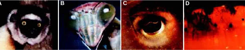
Evolution has generated at least three basically different types of eyes (Fig. 1): the camera-type eye with a single lens projecting onto a retina that is found in vertebrates and cephalopods; the compound eye with multiple ommatidia each consisting of a set of photoreceptor cells and a lens of its own, which is characteristic for insects and other arthropods; and the mirror eye which in the case of the scallop (Pecten) uses both a lens focussing the light onto a distal retina and a reflecting parabolic mirror projecting the light onto a proximal retina. Most of these eyes are positioned on the head of the animal and send their signals to the brain, which processes the information and transmits the appropriate signals to the effector organs, e.g. the muscles. However, in bivalves which do not form a head, the eyes are placed at the edge of the mantle allowing the animal to see when the two shells are slightly opened. In certain annelids which live head down in a tube, the eyes are placed at the hind end, where they are useful. Classical morphological studies have been used to argue for a polyphyletic origin of the various eye types. On the basis of morphological considerations Salvini-Plawen and Mayr (1961) claim “that the earliest invertebrates, or at least those that gave rise to the more advanced phyletic lines, had no photoreceptors” and that “photoreceptors have originated in at least 40 but possibly up to 65
or more phyletic lines”. They strongly adhere to the dogma “that the lens eye of a vertebrate and the compound eye of an insect are independent evolutionary developments”, a notion that you can find in nearly all zoology textbooks. I would like to challenge these ideas on the basis of our recent genetic work and argue for a monophyletic origin of the eyes from precambrian ancestors which already pos-sessed primitive prototypic eyes. From these precambrian ancestors the different phyla with the various eye-types evolved by adaptive radiation. This hypothesis is much more compatible with Darwin’s ideas (1882) than the assumption of a polyphyletic origin of the eyes. Before considering these issues, I would like to present the molecular genetic evidence which argues in favor of this radically different point of view.
Identification of the Master Control Genes for Eye
Devel-opment
homeotic transformations that allowed Lewis to construct the four-winged and the eight-legged fly. For the eye, a number of Droso-phila mutants have been isolated which block eye development by inducing cell death rather than leading to a homeotic transforma-tion. As early as 1915 Hoge isolated the first eyeless (ey) mutation which shows a rather variable phenotype from a total loss of eye facets on one or both sides of the head to more or less reduced eyes. For the mouse, a mutation Small eye was isolated and a similar mutant syndrome Aniridia was described in humans. Ho-mozygous Small eye embryos are eyeless, noseless and suffer serious brain damage, whereas heterozygotes develop to adult mice with reduced eyes, suggesting that the eye is critically affected. Heterozygous Aniridia patients have a similar phenotype with a reduced or no iris at all; and two homozygous mutant human fetuses have been described which were eyeless and noseless, suffered brain damage and died prior to birth. Using the Drosophila paired box as a probe Walther and Gruss (1991) cloned the Pax 6 gene of the mouse which was shown to correspond to the gene affected by the Small eye mutation (Hill et al., 1991). Similarly, the human Aniridia gene was cloned and shown to encode Pax 6 (Ton et al., 1991). Interestingly, the human and the murine Pax 6 proteins are identical in amino acid sequence indicating that there is a strong selective pressure on this protein. The protein contains both a paired and a homeodomain indicating that it is a transcription factor with two different DNA binding domains. It came as a total surprise, when my graduate student Rebecca Quiring accidentally cloned the Pax 6 homolog of Drosophila and it turned out to be the eyeless gene. Again the degree of sequence conservation is very high, 95% in the paired domain and 90% in the homeodomain as compared to the mammalian proteins.
Fig. 1. Different types of eyes. (A) Camera-type eye from the Lemur Propithecus verrauxi. (B) Compound eye of the praying Mantis. (C) Camera-type eye from the Cephalopod Sepia erostrata. (D) Mirror eye from the clam Chlamys nobilis. (Courtesy of Dr. Kazuto Kato. Photographs kindly provided by Masahiro Iijima, Susumu Yamaguchi and Isamu Soyama).
These observations suggested to me that Pax 6 might be the universal master control gene for eye development, implying that the dogma of independent evolution of the vertebrate eye and the insect compound eye might be wrong. Encouraged by the fact that occa-sionally a transdetermination from wing to eye structures can be observed (Gehring et al., 1968) and that the ectopic expression of the Antennapedia gene can induce antenna-to-leg transformations (Schneuwly et al., 1987), I proposed to my collaborators Georg Halder and Patrick Callaerts to try to induce ectopic eyes in other regions of the body of the fly by targeted expression of the Drosophila eyeless gene and its mouse homolog (Pax 6). Using the gal 4 system and suitable genomic enhancers, we succeeded in inducing ectopic eyes on the antennae, wings and legs of the fly (Fig. 2) (Halder et al., 1995). Thus, eyeless alias Pax 6 became the paradigm for a master control gene. It represents a master switch initiating a cascade of at least 2000 genes which are required for eye morphogenesis.
Histologically, the ectopic eyes on the antenna are quite normal and 32 antennal eyes tested electrophysiologically produced an electroretinogram (ERG) suggesting that their photoreceptor cells are functional. From 8 of the antennal eyes a completely normal ERG was recorded indicating that these eyes are functional, whereas the other 24 lacked the on- and off-transients which are characteristic of photoreceptors which are functional but not postsynaptically con-nected to the brain. By following the axons from the antennal eye to the brain, we found that the axons project to the antennal rather than the optic centers of the brain (Callaerts et al., unpubl.), suggesting that these flies might “smell the light”. An interesting thought that we are going to pursue.
In collaboration with Meinrad Busslinger and Thomas Czerny (Czerny et al., 1999) we discovered a second Pax 6 gene in
Fig. 2. (Left) Induction of ectopic eyes by targeted expression of the eyeless gene
on the antennae and wings of Drosophila.
Fig. 3. (Right) Induction of ectopic eyes on the legs of Drosophila by targeted expression of the twin of eyeless gene.
Drosophila called twin of eyeless which arose as a duplication during insect evolution. It is present in holometabolous insects (Drosophila and silk moth), whereas hemimetabolous (Schistocerca) and apterygote insects (springtail) have only a single gene. Twin of eyeless is more similar to vertebrate Pax 6 proteins than eyeless with regard to overall sequence conservation and DNA-binding function, and it is expressed earlier in embryogenesis, but the two genes share a similar expression pattern in the developing visual system and targeted expression of twin of eyeless, like eyeless, induces the formation of ectopic eyes (Fig. 3). Genetic and biochemical evi-dence indicates, however, that twin of eyeless functions upstream of eyeless by directly regulating the eye-specific enhancer of eyeless (Hauck et al., 1999). Twin of eyeless is therefore required for the initiation of eyeless expression in the embryo and acts through eyeless to activate the eye developmental pathway. The isolation of loss-of-function mutants for both eyeless and twin of eyeless mutants indicates that the two genes can partially substi-tute one another and that twin of eyeless is required for the formation of ocelli whereas eyeless is predominantly involved in compound eye morphogenesis.
The Notch signalling pathway defines an evolutionarily con-served cell-cell interaction mechanism that throughout develop-ment controls the ability of precursor cells to respond to developmen-tal signals. By targeting the expression of a dominant negative Notch receptor into the early eye primordia, we have been able to induce an eyeless phenotype, whereas the expression of a constitutively activated Notch receptor leads to the opposite effect and generates considerably enlarged eyes ressembling so-called “hammerhead” flies (Fig. 4), as they occur in certain Drosophila species in nature (Kurata et al., 2000). The dominant activated form of Notch can also induce ectopic eyes at the base of the proboscis which is derived from the antennal disc. The induction of ectopic eyes correlates with the induction of both eyeless and twin of eyeless expression by Notch, but we do not know whether this effect is directly mediated by the Suppressor of hairless transcription factor in the Notch signalling pathway, or whether the effect is indirect. Notch signalling regulates the master control genes eyeless, vestigial and Distal-less which in combination with different homeotic genes can induce the formation of ectopic eyes, wings, antennae and legs respectively (Kurata et al., 2000). Thus, Notch is involved in a common regulatory pathway for the determination of the various Drosophila appendages.
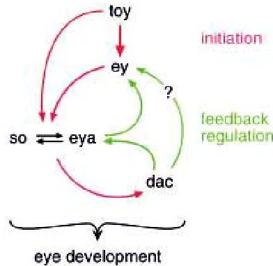
To determine the extent of evolutionary conservation of the eye morphogenetic pathway, we have begun to identify subordinate target genes of eyeless further downstream in the gene regulatory cascade. An important gene in this respect is sine oculis whose expression is induced by eyeless. Sine oculis is a homeobox gene and also functions as a transcription factor. We have obtained evidence from ectopic expression studies in transgenic flies, from transcription activation studies in yeast, and from gel shift assays in vitro, that the Eyeless protein activates transcription of sine oculis by direct interaction with an eye-specific enhancer in the long intron of the sine oculis gene (Niimi et al., 1999). Detailed analysis of this eye-specific enhancer has revealed functional binding sites for both Eyeless and Twin of eyeless proteins, which explains why twin of eyeless can partially substitute for eyeless (Punzo et al., unpubl.). Even though twin of eyeless is acting upstream of eyeless and activates its expression, it also directly controls sine oculis which is downstream of eyeless. Since there is also a positive feedback from sine oculis to eyeless, the eye morphogenetic pathway is by no means linear but rather a complex genetic network (Fig. 5). Further-more, sine oculis contains an ocelli-specific enhancer which is regulated by twin of eyeless and some other transcription factor(s), but not by eyeless (Punzo et al., unpubl.). This explains the observa-tion that eyeless null mutants which possess a funcobserva-tional twin of eyeless gene are still capable of forming normal ocelli.
As indicated in Fig. 5, the genetic cascade specifying eye morpho-genesis involves many players that have to be analyzed one by one. However, modern genomics provides for the first time the means to get a general overview over a morphogenetic pathway. Using DNA microarrays and DNA chips we have begun to analyze the eye morphogenetic pathway. By comparing RNA from leg imaginal discs with leg discs in which an eye morphogenetic field has been induced by ectopic expression of eyeless, RNA from eye versus leg discs, and RNA from leg discs in which eyeless has been induced by a heat shock promoter, we are trying to identify the relevant genes that are activated (directly or indirectly) by eyeless when an eye is induced, and the “leg genes” that are repressed. An example of a microarray resulting from a collaboration with Kevin White is shown in Fig. 6. By hybridizing six candidate genes directly or indirectly induced by Fig. 4.“Hammerhead” flies induced by expression of a constitutively
activated form of the Notch Receptor. Wild-type (left) and Hammerhead
(right) flies with enlarged compound eyes (UAS-Nact ey-GAL4).
eyeless my collaborator Lydia Michaut was able to show that all six genes are expressed early in eye morphogenesis, i.e. in the eye disc anterior to the morphogenetic furrow, where eyeless is expressed, and / or in the morphogenetic furrow when and where differentiation sets in. It is our aim to compare the eye morphogenetic pathways between Drosophila and mouse, being analyzed by Peter Gruss and collaborators. Such a comparison will provide important insights into the parallel evolution of the compound and camera-type eye.
New Perspectives in Eye Evolution
Charles Darwin in “The Origin of Species” had great difficulties with eye evolution and devoted an entire chapter to “Difficulties of the Theory” in which he discusses “Organs of extreme Perfection and Complication”:
“To suppose that the eye with all its inimitable contrivances for adjusting the focus to different distances, for admitting different amounts of light and for the correction of spherical and chromatic aberration, could have been formed by natural selection, seems, I freely confess, absurd in the highest degree”.
But then he continues:
“Reason tells me, that if numerous gradations from a simple and imperfect eye to one complex and perfect can be shown to exist, each grade being useful to its possessor, as is certainly the case; if further, the eye ever varies and the variations be inherited, as is likewise certainly the case; and if such variations should be useful to any animal under changing conditions of life, then the difficulty of believing that a perfect and complex eye could be formed by natural selection, though insuperable by our imagination, should not be considered as subversive of the theory”.
This pushes the question of eye evolution back to the problem of how the first primitive eye, the prototype has evolved. The evolution of an eye prototype would seem to be a highly improbable stochastic event, since selection can only work after the various components are assembled into a prototype that is at least partially functional as a photoreceptor organ.
“The simplest organ which can be called an eye consists of an optic nerve, surrounded by pigment-cells and covered by translucent skin, but without any lens or other refractive body”.
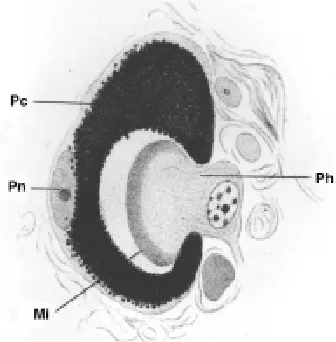
Such primitive eyes are found, for example, in certain flatworms. Hesse (1897) has described the eyes of Planaria torva which consists of three photoreceptor cells and a single pigment cell only (Fig. 7), and there is a planarian species in Japan which has eyes with one photoreceptor cell and one pigment cell only, which corresponds exactly to the Darwinian prototype (Kiyokazu Agata, pers. comm.).
For the present discussion we adopt Darwin’s definition of an eye as an organ consisting of at least two different cell-types, photoreceptor cells and pigment cells. The “eyes” of protists are organelles (and not organs) formed within a single cell and arise by the assembly of mol-ecules within a cell rather than by the assembly of different cells, which is a fundamental difference with respect to the genetic control of morphogenesis.
Since the evolution of a prototypic eye is a highly improbable stochastic event that is not driven by selec-tion, the hypothesis of a polyphyletic origin of the eyes, arising 40 to 65 times independently, is extremely unlikely Fig. 6. DNA microarray probed with RNA from leg discs (green spots) versus leg
discs in which an eye morphogenetic field is induced by ectopic expression of eyeless (red spots). A number of eyeless induced genes (spots) are labelled. Genes expressed in both kinds of discs are labelled yellow. No homology means that the respective gene is unknown. Courtesy of Lydia Michaut and Kevin White.
Fig. 7. Histological section across the eye of Planaria torva (after Hesse, 1897). Mi, microvilli; Pc, pigment cell; Ph, photoreceptor cell; Pn, pigment cell nucleus.
and incompatible with Darwin’s ideas. Furthermore, all three major eye types (the camera-type, the compound eye and the mirror-eye) are found within the same class of molluscs, in the bivalvia, making an independent evolutionary origin even more unlikely.
Xenopus eye structures can be induced by the ectopic expression of the Drosophila homologs (Kurata et al., submitted). In addition, the activated form of the Notch receptor induces a considerable enlargement of the eye as in Drosophila and a duplication of the lens. This indicates that the top of the genetic cascade is highly conserved. At this point it has to be pointed out that there is no functional necessity to use a particular transcription factor like Pax 6 for particular function e.g. eye morphogenesis, since a transcription factor can regulate any gene, if this gene is endowed
environment there is no selective pressure to maintain this gene intact. In contrast, Pax 6 in addition to eye morphogenesis is also involved in morphogenesis of the brain and the nose (or other olfactory organs) and is therefore, indis-pensable even in the absence of light in the environment.
The observation that Pax 6 is expressed during eye development and regeneration in planarians and nemerteans is of course only a correlation and does not provide any evidence for a causal relationship. However, recently the method of RNA interference (RNAi) by injection of double stranded RNA leading to the degrada-tion of the targeted mRNA has been adapted to these worms and we have obtained genetic evidence for the involvement of both Pax 6 and sine oculis homologs in eye morphogenesis of these worms. By injecting double stranded Pax Fig. 8. Scanning electron–micrograph of an ectopic eye on the antennae induced by ectopic
expression of the mouse Small eye gene (= Pax 6). (A) Overview. (B) Higher magnification. Courtesy of A. Hefti (REM Laboratory, University of Basel).
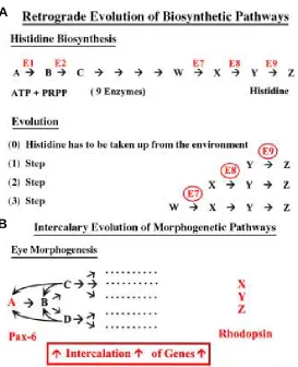
Fig. 9. Models for the evolution of biosynthetic and morphoge-netic pathways.(A) Retrograde Evolution of biosynthetic pathways (after N. Horowitz, 1945), (B) Intercalary Evolution of morphogenetic pathways (after Gehring & Ikeo, 1999).
with the appropriate regulatory elements in its enhancer or promoter. Therefore, if various eye types are controlled by the same Pax 6 gene this is merely for evolutionary (histori-cal) reasons and argues for descent from a common ances-tor.
Until now Pax 6 homologs (true orthologs) have been identified in vertebrates, Amphioxus, ascidians, sea urchins cephalopods, nemerteans, nematodes and platyhelminths, and for Amphioxus, Phallusia, Loligo and Caenorhabditis (Plaza, Dozier & Seimiya unpubl.) we have shown that their Pax 6 genes are capable of inducing ectopic eyes in Droso-phila. The only exception is the Dugesia tigrina gene that has diverged to the extent that it is no longer capable of inducing ectopic eyes in Drosophila (the second Dugesia gene has not been tested yet). In all cases the Pax 6 genes are expressed in the eyes, in the brain and in several cases in chemosen-sory organs.
The case of Caenorhabditis requires some special expla-nation, since this worm has no eyes. There are freeliving marine nematodes which possess eyes, but C.elegans lives underground and like many cave animals has lost its eyes. Why then should it have an intact Pax 6 gene? It has been shown for blind cave animals that their rhodopsin genes rapidly degenerate and become pseudogenes (Iwabe et al., 1996), which is due to the fact that rhodopsin has only one function, i.e. light perception, and in the absence of light in the
6 RNA into the regeneration blastema of regenerating nemerte-ans (Lineus) we have been able to knock out Pax 6 function and to prevent eye regeneration (M. Tarpin, J. Bierne & W.J. Gehring, unpubl.). In the planarian Dugesia tigrina, the situation is more complicated because it possesses two Pax 6 genes. However, we have been able to knock out the sine oculis homolog of Dugesia thereby preventing eye regeneration (Pineda et al., 2000). These findings provide strong evidence that both Pax 6 and sine oculis are structurally and functionally highly conserved in eye evolution.
A
B
How could the Eye Morphogenetic Pathways have
Evolved?
The evolutionary events leading to a complex biological struc-ture like the eye are difficult to reconstruct, since the evolutionary intermediates are frequently missing and often we do not know in which direction evolution has proceeded; from simple to more complex structures, or by reduction of highly complex to rudimen-tary structures. For biosynthetic pathways leading from one or more substrates via numerous enzymatic steps to more complex products, Horowitz (1945) has proposed the mechanism of retro-grade evolution. For example, nine enzymes are involved in histidine biosynthesis in bacteria like Salmonella, starting from ATP and phosphoribosylpyrophosphate, (two substrates that are used in many other biochemical reactions) to synthesize histidine in a linear biochemical pathway. Horowitz hypothesized that the most primitive bacteria had to take up histidine from the environ-ment (as many higher organisms do) and that the first enzyme to evolve was the enzyme (E9) catalyzing the last step in histidine biosynthesis (Fig. 7). The organism possessing E9 had a strong selective advantage when histidine became scarce in the
envi-ronment, since it could use the immediate precursor of histidine as a substrate. The next enzyme to evolve was presumably E8 which could have arisen by gene dupli-cation of E9 and subsequent divergence, and so on until a completely linear and streamlined pathway had been established in a retrograde manner. Recent amino acid sequence and structural compari-sons of these enzymes indicate that the Horowitz model is not entirely correct, but there is evidence that e.g. His A and His F, two enzymes with a β/α barrel structure have evolved by two-fold gene duplication and gene fusion from a common half-barrel ancestor (Lang et al., 2000).
For the evolution of the eye morphoge-netic pathway we have proposed a mecha-nism of intercalary evolution (Fig. 9) (Gehring and Ikeo, 1999). Since both the
A
B
Fig. 10. Eyes of jellyfish medusae. (A) Cladonema eyes at the base of the tentacles. (B) Chironex eye (Courtesy of Volker Schmid).
Fig. 11. Eye organelle of the unicellu-lar dinoflagellate Erythropsis pavillardi.(A) Schematic drawing. (B)
Light micrograph showing the lens and the shielding pigment. (C) Stacked mem-branes resembling the arrangement in photoreceptor cells in the retina of mul-ticellular organisms. After Greuet, 1965. (Courtesy of Marie-Odile Soyer).
A
B
C
highly conserved genes can be recruited for different functions. For example, the homeobox gene Rx plays a major role in vertebrate eye development, but de-spite the fact that Drosophila has a highly conserved Rx homolog, this gene is not expressed in Drosophila eye morphogenesis (Eggert et al., 1998) and seems to serve a function in brain rather than eye develop-ment. Differential expression of conserved genes and recruitment of “new” genes into the eye morphoge-netic pathway provide a likely mechanism for the evolution of different eye-types originating from the same eye prototype.
The Origin of Eyes and Brain
Gregory (1967) has considered the question of whether the eyes or the brain came first in evolution, assuming that the evolution of the mechanism of visual perception must entail the separate elabora-tion of eyes and brain. He regards the informaelabora-tion given by the eyes as of indirect use to living crea-tures, since considerable “computing” is required to make any use of visual information. By contrast touch and chemical senses directly monitor biologically vital features of the environment, and their informa-tion requires but a minimal “computer”. Gregory therefore considers it reasonable to suppose that vision is a lately acquired sense. Furthermore, he supposes that the simple eye took over existing touch neural mechanism. Since probe touch can provide information in three dimensions, the underly-ing “computer” in the brain, was ideally suited to process visual information. Rüdiger Wehner (pers. comm.) has proposed the idea that rhodopsin may be considered as a chemoreceptor with a covalently bound ligand, retinal. In this case the ligand does not have to be bound by the receptor in order to activate it as in chemoreception, but it can be activated by photons. There is in fact some evidence supporting the hypothesis that rhodopsin is derived from chemoreceptors, since Klein et al. (1988) have found significant sequence homology between rhodopsin submitted) suggests that eyeless and twin of eyeless are derived
from a common ancestral gene which is in line with the extensive similarity of their amino acid sequences. These two genes are partially redundant in their function, since they can partially substi-tute each other. However, they also have diverged to the extent that eyeless plays a major role in compound eye development, whereas twin of eyeless is required for the formation of ocelli.
A second case found in Drosophila is Drosocrystallin which like some crystallins in vertebrates has been recruited into the lens developmental pathway from another source (Piatigorsky & Wistow, 1989). In this case, there is strong evidence from sequence data that Drosocrystallin belongs to the family of cuticle proteins, suggesting it has been recruited into the eye developmental pathway by fusion to a lens-specific enhancer (Jansens & Gehring, 1999). Drosocrystallin represents a case for a gene that is not conserved in the course of evolution and contributes to lens development in Drosophila but not in vertebrates. However, even
Fig. 12. Hypothetical evolution of photosensitive cells containing rhodopsin as a light receptor and monophyletic evolution of the various eye-types starting from a Darwin-ian prototype eye consisting of a single photoreceptor cell and a pigment cell assembled under the control of Pax 6 (after Gehring and Ikeo, 1999).
and the cyclic AMP receptor of Dictyostelium, which serves as a chemoattractant receptor. This lends support to the idea that the olfactory and visual systems are closely related and that the visual system has evolved from chemoreception. In this context it is important to point out that Pax 6 is not only involved in eye morphogenesis but also in the development of the nose and other chemosensory organs, and in brain morphogenesis as well.
wasp (Fig. 10), which have highly evolved eyes, but no brain. The eyes are located at the base of the tentacels and transmit their information directly to the muscles without processing by a brain. There are very few interneurons between the eyes and the muscles, and there is only a ring nerve around the bell margin which may coordinate the movements of the tentacles, but there is no real brain. Of course, one may argue that jellyfish originally had a brain, but have reduced it in the course of evolution. However, there is no selective advantage to loose the brain in a freeliving pelagic animal. On the contrary, there is selective pressure to maintain it. Animals living underground or in caves may loose their eyes because of lack of selective pressure, however, they retain their brain. Parasites may even reduce their brains, by adapting to their hosts and become extremely reduced to reproduction “machines”, but freeliving organ-isms can neither dispense with their sensory organs nor their brains. Therefore, I consider it likely that the eyes came before the brain.
This point of view is supported by the evolution of eye organelles in protists. Chlamydomonas, for example, has an eye organelle with a rhodopsin-related photosensitive pigment and a shielding pigment spot which allows it to determine the direction of the incoming light. Analogous to jellyfish, the eye organelle is located directly at the base of the effector organ, the flagellum, and the information is transmitted directly from the eye organelle to the flagellum without an intervening information processing organelle. It is worth pointing out that some dinoflagellates like Erythropsis, have evolved the most elaborate eye organelles consisting of a lens, shielding pigment and retina-like structures with membrane stacks, closeling resembling the eyes of multicellular organisms (Fig. 11). We have no idea how such compli-cated organelles can evolve in single celled organisms. However, it raises the interesting possibility that the metazoan eye may have originated from symbionts, much like mitochondria and chloroplasts.
Concluding Remarks
The discovery of the universal master control genes for eye development has led us to a radically different concept of eye evolution than previously assumed. Our data argue strongly for a monophyletic origin of the various eye types starting from a Darwin-ian eye prototype present before the CambrDarwin-ian explosion and leading to adaptive radiation in most or all phyla by parallel evolution, which can be divergent or convergent (Fig. 12). To base evolutionary studies exclusively on morphological criteria without considering the genetic basis can be misleading. Genetic criteria have also to be applied to the notion of evolutionary homology and this term has to be redefined in a quantitative rather than qualitative way as proposed by Zuckerkandl (1994). Developmental genetics and functional genomics will provide deeper insights into evolution, since DNA not only contains a detailed developmental program, but also vast information about the evolutionary history of organisms.
References
BENDER et al. (1983). Molecular genetics of the bithorax complex in Drosophila melanogaster. Science 221: 23-29.
CARRASCO, A.E., MCGINNIS, W., GEHRING, W.J. and DEROBERTIS E.M. (1984). Cloning of a X. laevis gene expressed during early embryogenesis that codes for a peptide region homologous to Drosophila homeotic genes. Cell 37: 409-414.
CZERNY, T., HALDER, G., CALLAERTS, P., KLOTER, U., SOUABNI, A. GEHRING, W.J. and BUSSLINGER, M. (1999). Twin of eyeless, a second Pax-6 gene of
Drosophila, acts upstream of eyeless in the control of eye development. Mol. Cell 3: 297-307.
DARWIN, CHARLES (1882). The Origin of Species by means of Natural Selection (6th edition, John Murray, London), p. 143-146.
DESPLANS, C. (1999). A view from Switzerland. Science 283: 799.
EGGERT, T., HAUCK, B., HILDEBRANDT, N., GEHRING, W.J. and WALLDORF, U. (1998). Isolation of Drosophila homolog of the vertebrate homeobox gene Rx and its possible role in brain and eye development. Proc. Natl. Acad. Sci. USA 95: 2343-2348.
GARBER, R., KUROIWA, A. and GEHRING, W.J. (1983). Genomic and cDNA clones of the homeotic locus Antennapedia in Drosophila. EMBO J. 2: 2027-2036.
GEHRING, W.J. (1999a). Master control genes in Drosophila and evolution: the homeobox story. Yale University Press, New Haven and London, pp. 236
GEHRING, W.J. (1999b). Lifting the lid on the homeobox discovery. Nature 398: 521.
GEHRING, W.J. and IKEO, K. (1999). Pax 6 mastering eye morphogenesis and eye evolution. Trends Genet. 15: 371-377.
GEHRING, W., MINDEK, G. and HADORN, E. (1968). Auto- und allotypische Differenzierungen aus Blastemen der Halterenscheibe von Drosophila melanogaster nach Kultur in vivo. J. Embryol. Exp. Morph. 20: 307-318.
GREUET, C. (1965). Structure fine de lócelle d'Erythropsis pavillardi Hertwig, pteridinien Warnowiidae Lindemann. C.R. Acad. Sci. (Paris). 261: 1904-1907.
GREGORY, R.L. (1967). Origin of eyes and brains. Nature 28: 369-372.
HALDER, G., CALLAERTS, P. and GEHRING, W.J. (1995). Induction of ectopic eyes by targeted expression of the eyeless gene in Drosophila. Science 267: 1788-1792.
HAUCK, B., GEHRING, W. J. and WALLDORF, U. (1999). Functional analysis of an eye specific enhancer of the eyeless gene in Drosophila. Proc. Natl. Acad. Sci USA 96: 564-569.
HESSE, R. (1897). Untersuchungen über die Organe der Lichtempfindung bei niederen Thieren. II. Die Augen der Plathelminthen. Z. wiss. Zool. Band 62: 527-582.
HILL, R.E. et al. (1991). Mouse Small eye results from mutations in a paired-like homeobox-containing gene. Nature 354: 522-525.
HOGE, M.A. (1915). Another gene in the fourth chromosome of Drosophila. Am.Nat. 49: 47-49.
HOROWITZ, N.H. (1945). On the evolution of biochemical syntheses. Proc. Natl. Acad. Sci. USA 31: 153-157.
IWABE N., KUMA K. and MIYATA T. (1996). Evolution of gene families and relationship with organismal evolution: rapid divergence of tissue-specific genes in the early evolution of chordates. Mol. Biol. Evol. 3: 483-93.
JANSSENS, H. and GEHRING, W.J. (1999). Isolation and characterization of drosocrystallin, a lens crystallin gene of Drosophila melanogaster. Dev. Biol. 207: 204-214.
KLEIN, P.S., SUN, T.J., SAXE III, C.L., KIMMEL, A.R., JOHNSON R.J. and DEVREOTES, P.N. (1988). A chemoattractant receptor control development in Dictyostelium discoideum. Science 241: 1407-1472.
KURATA, S.,GO, M.J., ARTAVANIS-TSAKONAS,S. and GEHRING, W.J. (2000). Notch signalling and the determination of appendage identity. Proc. Natl. Acad. Sci. USA 97: 2117-2122.
LANG, D., THOMA, R., HENN-SAX, M., STERNER, R. and WILMAN, M. (2000). Structural evidence for evolution of the β/α barrel scaffold by gene duplication and fusion. Science 289: 1546-1550.
LEWIS, E.B. (1992). Clusters of master control genes regulate the development of higher organisms. J. Am. Med. Assoc. 267: 1524-1531.
MCGINNES, W., LEVINE, M.S., HAFEN, E., KUROIWA, A. and GEHRING, W.J. (1984). A conserved DNA sequence in homeotic genes of the Drosophila Antennapedia and bithorax complex. Nature 308: 428-433.
MCGINNES, W. and LAWRENCE, P.A. (1999). Historical transformations. Nature 398: 301-302.
PIATIGORSKY, J. and WISTOW, G.J. (1989). Enzyme/crystallins: gene sharing as an evolutionary strategy. Cell 57: 197-199.
PINEDA, D, GONZALEZ, J., CALLAERTS, P., IKEO, K., GEHRING, W.J. and SALO, E. (2000). Searching for the prototypic eye genetic network: sine oculis is essential for eye regeneration in planarians. Proc. Natl. Acad. Sci. USA 97: 4525-4529
SALVINI-PLAWEN, L. and MAYR, E. (1961) in Evolutionary Biology, (Vol. 10) (Hecht, M.K., Steere, W.C. and Wallace, B., eds), pp. 207-263, Plenum Press.
SCHNEUWLY, S., KLEMENZ, R. and GEHRING, W.J. (1987). Redesigning the body plan of Drosophila by ectopic expression of the homoeotic gene Antennapedia. Nature 325: 816-818.
TON, C.C., HIRVONEN, H., MIWA, H., WEIL, M.M., MONAGHAN, P. et al. (1991). Positional cloning and characterization of a paired box- and homeobox-contain-ing gene from the aniridia region. Cell 67: 1059-1074.
WALTHER, C. and GRUSS, P. (1991). Pax 6, a murine paired box gene, is expressed in the developing CNS. Development 113: 1435-1449.
WEINTRAUB, H., TAPSCOTT, S.J., DAVIS R.L., THAYER, M.J., ADAN, M.A., LASSAR, A.B. and MILLER, A.D. (1989). Activation of muscle-specific genes in pigment, nerve, fat, liver and fibroblast cell lines by forced expression of Myo D. Proc. Nat. Acad. Sci. USA 86: 5434-5438.