Journal of Asthma and Allergy
Dove
press
O r i g i n A L r e s e A r c h
open access to scientific and medical research
Open Access Full Text Article
controversial data on simvastatin in asthma:
What about the rat model?
Thomas Tschernig1
Wolfgang Bäumer2
reinhard Pabst1
1institute of Functional and Applied
Anatomy, hannover Medical school, hannover, 2institute of Pharmacology,
Toxicology and Pharmacy, University of Veterinary Medicine, hannover, germany
correspondence: Thomas Tschernig institute of Anatomy and cell Biology, saarland University Medical Faculty, 66421 homburg/saar, homberg, germany Tel +49 684 1162 6143
email [email protected]
Abstract: The effects of simvastatin on lung inflammation in asthma are controversial. Reduction of inflammation and hyperreactivity has been reported in studies using murine models of asthma. In contrast, a clinical study has not found beneficial effects in patients. The rat model of asthma has some distinct advantages and is still widely used in industrial studies. Therefore, the role of simvastatin was investigated in this rat model using intraperitoneal and intratracheal administration. With both simvastatin administration routes, the relative and absolute numbers of neutrophils, eosinophils, and lymphocytes were only partially reduced after increasing dosages (0.1, 1.0, and 10 mg per animal). The most obvious effect was on CD4 T cell numbers, which were reduced in most treatment groups. The results presented here suggest that treatment with simvastatin differs between species, and that it is too early for extrapolation of these data to humans.
Keywords: simvastatin, inflammation, rat, asthma
Introduction
Simvastatin has been shown to be a promising new drug for the treatment of allergic asthma. The first relevant report was published by McKay et al in 2004.1
In that report, 4.0 or 40 mg/kg body weight of the drug was administered to mice either orally or intraperitoneally (IP), leading to a reduction in inflammatory cell infiltrates and eosinophilia in bronchoalveolar lavage (BAL) fluid. Furthermore, ovalbumin (OVA)-induced interleukin (IL)-4 and IL-5 levels in BAL fluid were found to be reduced after treatment. However, the exact mechanism by which statins mediate anti-inflammatory and immunomodulatory actions is not well understood. By inhibiting 3-hydroxy-3-methylglutaryl coenzyme A reductase, the mevalonate synthetic pathway is affected, which results in inhibition of prenylation of proteins. Prenylated proteins play a crucial role in the regulation of cell growth and signal transduction. Additionally, by inhibition of cholesterol synthesis, statins are lipid raft modulators. Lipid rafts act as platforms, bringing together molecules essential for activation of immune cells (eg, the “immunologic synapse”). By these and other described effects, statins exert immunomodulatory actions in vitro and in vivo.2
In 2007 Kim et al analyzed the mechanisms of action of simvastatin in a murine model of asthma.3 This group showed regulation of G proteins, mitogen-activated
protein kinases, and NF-kappaB. A very recent report from Zeki et al found that the mevalonate pathway was partially involved in the action of simvastatin during allergic airway inflammation.4 In contrast, a clinical study has not found therapeutic
anti-inflammatory effects in asthma.
Journal of Asthma and Allergy downloaded from https://www.dovepress.com/ by 118.70.13.36 on 24-Aug-2020
For personal use only.
Number of times this article has been viewed
This article was published in the following Dove Press journal: Journal of Asthma and Allergy
Dovepress Tschernig et al
It is always difficult to compare animal and human studies for many reasons, eg, models, dosage, and duration of treat-ment, as well as read-out parameters. However, the rat as a model for asthma is widely used in industrial studies and offers some specific advantages, as have been summarized previously.6 The main aspect is that the rat, similar to humans,
has a mucosal blood supply from the bronchial arteries. In contrast, mice do not have a bronchial circulation. Data from a Fisher rat model of asthma are presented here after treat-ment with simvastatin 10 mg IP or 0.1 and 1.0 mg simvastatin IP or intratracheally (IT). The IT application was investigated because a local or topical treatment, eg, a TLR2/6 agonist, would be very attractive for clinical application and has been effective in rat model testing.7
Materials and methods
Animals
Female Fisher rats (8–10 weeks of age [Charles River, Sulzfeld] and kept in the central animal laboratory facility [Medical School of Hannover]). Several experiments were performed with animals in numbers from 3–9 per group. Food and water were provided ad libitum. Rats were main-tained in a separated minimal-barrier susmain-tained facility and were microbiologically monitored according to the recom-mendations of the Federation of Laboratory Animal Science Associations. All procedures were approved by the Lower Saxony district government in Hannover, Germany.
Asthma induction
On days 0 and 7, all rats were sensitized with 1 mg OVA (Sigma, Deisenhofen) and 200 mg of Al(OH)3 (Sigma) in 1 mL 0.9% (sterile, pyrogen-free) NaCl (Braun, Melsungen) applied subcutaneously. A second adjuvant, concentrated preparation of 5 × 10⁹ heat-killed Bordetella pertussis bacilli
(donated by the manufacturers, Chiron Behring, Marburg) in 0.4 mL 0.9% NaCl was given IP at the same time. On day 13, the animals were challenged with 250 µL 0.5% OVA IT. Under short ether (Baker, Deventer) anesthesia, they were suspended in a hanging position by a rubber band fixed to the teeth of the upper jaw. The solution was instilled after intubation of the trachea via the oral cavity. The correct position of the tube was checked by blowing air into the lung before application.
Treatment
The animals were treated with 1.0 mg or 0.1 mg simvastatin IT or with 10 mg, 1.0 mg, and 0.1 mg IP one hour before the challenge. The high dose of simvastatin 10 mg (approximately
40 mg/kg) could not be applied via the IT route. Vehicle application served as the treatment control. Simvastatin was supplied by Merck, UK. The animals were sacrificed 25 hours after challenge. Three animals were included in each experi-ment with the following exceptions: statin treatexperi-ment 1 mg IP (two experiments each, n = 6) and statin treatment 1 mg IT (one experiment, n = 1, and one experiment, n = 9).
Animal dissection and preparation
of bronchoalveolar and interstitial cells
The animals were sacrificed on day 15 under deep ether anesthesia, during which the abdominal wall was opened and the animals killed by aortic exsanguination. The trachea was dissected and a cannula was inserted in situ. The lungs were lavaged with 5 × 5 mL cold (4°C) 0.9% NaCl. The fluid was recovered by gentle aspiration. The lavage fluid was pooled, and on average over 90% of the volume was retrieved from all animals. The pooled BAL fluid was centrifuged (400 × g, 10 minutes) washed, centrifuged again, and the resulting cell pellet resuspended in 1 mL phosphate-buffered saline (PBS, containing 1% bovine serum albumin and 0.1% sodium azide). A mechanical method was used for lung cell extraction. To this end, the whole left lung was passed through a metal sieve with two rounded tweezers. The sieve was rinsed with 40 mL PBS and the suspension was centrifuged, washed, and resuspended as described above. The right lung was filled with PBS-diluted embedding medium (1:4) and deep frozen. Cryostat sections were performed and stained with hematoxilyn and eosin.
Differential cell counts
Total cell numbers in BAL fluid and the lung interstitium (complete left lung) were determined in a Neubauer count-ing chamber uscount-ing standard staincount-ing with Tuerk’s solu-tion (Merck, Darmstadt). Eosinophils, neutrophils, and monocytes/macrophages were assessed on slides prepared by centrifuging 1 × 10⁵ cells in anticoagulant (8.8 g NaCl,
Merck, +0.99 g ethylenediaminetetraacetic acid + 50 g bovine serum albumin, in 1 L distilled water) for five minutes at 200 × g. The slides were fixed in acetone for 10 minutes. Cells were morphologically identified after Quik-Diff (DQ, Dade Behring) staining. At least 300 cells were differenti-ated on each slide.
For flow cytometry, cells were transferred onto microti-ter plates (1 × 10⁶ per well). The staining was performed as
described previously.7 In brief, after the cells were washed twice
with PBS (containing 1% bovine serum albumin and 0.1% sodium azide) the first antibody was incubated for 20 minutes at 4°C. The procedure was repeated with the secondary antibody.
Journal of Asthma and Allergy downloaded from https://www.dovepress.com/ by 118.70.13.36 on 24-Aug-2020
Dovepress simvastatin in asthma
The cells were analyzed using a FACScan flow cytometer (Becton Dickinson, Mountain View, CA) focusing on the lymphocyte cluster. CD4+ T cells were identified as T cell
receptor positive/CD8 negative cells. Dendritic cells were identified as CD103/major histocompatibility complex Class II positive cells. All antibodies were monoclonal mouse anti-rat (Serotec, Oxford). Unconjugated antibodies were detected with phycoerythrin (PE)-conjugated secondary antibody (kappa PE, Dianova, Hamburg) and biotinylated primary antibodies with Red 670-streptavidin (Gibco, Gaithersburg, MD). Isotype-matched antibodies served as the control.
statistical analyses
The experiments were performed on different days. Data from different experiments for one group have not been pooled to show the day-to-day variation of the model and treatment. Means and significant differences were calculated using GraphPad Prism 4.00 (Graph Pad Prism Software, San Diego, CA). The Mann-Whitney test was used to compare experimental groups. Values of P , 0.05 were taken as statistically significant.
Results
Tissue inflammation
After the OVA challenge in the sensitized Fisher rats, strong peribronchial and perivascular leukocyte cuffs were observed in controls as well as in treated animals. Eosinophils were abundant in lung tissue in the control groups, and also after IP and IT simvastatin.
Leukocyte accumulation
in bronchioalveolar lavage
fluid and lung tissue
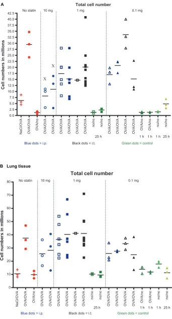
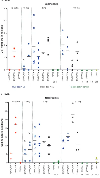
Cellular analysis of BAL fluid and lung tissue revealed asthma-like inflammation, as indicated by a strong leukocyte accumulation in BAL fluid, as well as in lung tissue of OVA-challenged rats (Figure 1). The number of total cells in BAL fluid was reduced after treatment with simvastatin 10 mg IP, but no significant differences were detected in the numbers of total cells between the control group and the treatment group after treatment with simvastatin 0.1 or 1 mg IP or IT (Figure 1A). No differences were seen in lung tissue (Figure 1B). Eosinophil numbers were reduced after one experiment using simvastatin 10 mg IP and after another using simvastatin 0.1 mg IT. In other experiments, simvastatin treatment had no effect on numbers of eosinophils in BAL fluid (Figure 2A). Neutrophils in BAL fluid were reduced again only after treatment with simvastatin
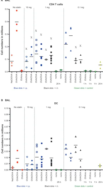
10 mg IP (Figure 2B). The number of CD4+ T cells was reduced
in most of the experiments or at least showed a tendency towards lower numbers after the different routes and dosages of simvastatin (Figure 3A). In contrast, no effect was seen on the numbers of dendritic cells (Figure 3B).
Discussion
In this study, the effect of different dosages and application routes of simvastatin was investigated in allergic airway inflammation in Fisher rats. There are several reports demonstrating anti-inflammatory and immunomodulatory actions of statins in different in vitro and in vivo models.2,8,9
One potential mechanism by which statins mediate anti-inflammatory and immunomodulatory actions is the inhibi-tion of 3-hydroxy-3-methylglutaryl coenzyme A reductase in the mevalonate synthetic pathway which results in impaired prenylation of proteins. Prenylated proteins have pleiotropic effects in the regulation of cell growth and signal transduc-tion. Statins also act as modulators of lipid rafts which are membrane microdomains playing a substantial role in immune cell interaction.2 It has to be noted that statins may
differ in their anti-inflammatory efficacy. It was demon-strated that lovastatin and fluvastatin inhibit granulocyte-macrophage colony-stimulating factor-stimulated human eosinophil adhesion to intercellular adhesion molecule-1. Interestingly, simvastatin, mevastatin, and pravastatin failed to show inhibitory actions in this in vitro assay.9
We wanted to test the efficacy of simvastatin in a rat model of asthma because, in contrast with mice, rats have a bronchial circulation, and in this respect rat physiology is closer to that of humans than that of the mouse.6 The data on
leukocyte subsets in BAL fluid or left lung tissue are presented as absolute numbers to avoid misleading conclusions when relative numbers (percentages) are given. A robust effect of simvastatin has been shown in murine asthma models.1,3,4
Simvastatin was associated with a reduction in inflamma-tory cells and cytokine numbers in those reports. Similar results were obtained for lovastatin, another statin. However, although inhibition of eosinophil influx could be confirmed in a more recent study, no effects on typical chemokines (eg, eotaxin, TARC, LTB4, and the Th2 cytokines, IL-4 and Il-13, were found.8 Simvastatin also failed as a therapeutic drug in a
clinical study. The same lack of efficacy was also shown for atorvastatin. Even the effect of atorvastatin as add-on therapy to inhaled glucocorticoids was only moderate.10 In the present
study, a limited therapeutic effect was shown in the Fisher rat asthma model. A high dose of simvastatin 10 mg per rat, which is approximately 40 mg/kg body weight, was injected IP and
Journal of Asthma and Allergy downloaded from https://www.dovepress.com/ by 118.70.13.36 on 24-Aug-2020
Dovepress Tschernig et al
Total cell number
NaCl/OVA OVA/OV
A
OVA/n
o
OVA/OV
A
OVA/OV
A
OVA/OV
A
OVA/OV
A
OVA/OV
A
OVA/OV
A
no/n
o
no/n
o
OVA/OV
A
OVA/OV
A
OVA/OV
A
OVA/OV
A
OVA/n
o
OVA/n
o
no/n
o
no/n
o
0.0 2.5 5.0 7.5 10.0 12.5 15.0 17.5 20.0 22.5 25.0 27.5 30.0 32.5 35.0 37.5 40.0
42.5 0.1 mg
25 h 1 h 1 h 1 h 25 h
Blue dots = i.p. Black dots = i.t. Green dots = control
Cell numbers in million
s
No statin 10 mg 1 mg
A
Total cell number
NaCl/OVA OVA/OVA OVA/no OVA/OVA OVA/OVA OVA/OVA OVA/OVA OVA/OVA OVA/OVA
no/n
o
no/no
OVA/OVA OVA/OVA OVA/OVA OVA/OVA OVA/no OVA/no
no/n
o
no/n
o
0 10 20 30 40 50 60 70 80
Blue dots = i.p. Black dots = i.t. Green dots = control
C
el
l n
um
be
rs
in
m
ill
io
ns
0.1 mg
25 h 1 h 1 h 1 h 25 h
No statin 10 mg 1 mg
B Lung tissue
Figure 1 single data and mean value of total cell numbers in (A) BAL fluid and (B) in separated lung tissue. There were three animals in each experiment with the following exceptions: statin treatment 1 mg iP (two experiments each, n = 6) and statin treatment 1 mg iT (one experiment, n = 1 and one experiment, n = 9). The reduction in total cell numbers in BAL fluid after simvastatin 10 mg treatment was significant compared with the control group (P, 0.05). no differences were found between cell numbers isolated from lung tissue.
Abbreviations: BAL, bronchioalveolar lavage; iP, intraperitoneal; iT, intratracheal.
Journal of Asthma and Allergy downloaded from https://www.dovepress.com/ by 118.70.13.36 on 24-Aug-2020
Dovepress simvastatin in asthma
A BAL
Eosinophils
NaCl/OVA OVA/OVA OVA/no OVA/OVA OVA/OVA OVA/OVA OVA/OVA OVA/OVA OVA/OVA
no/n
o
no/n
o
OVA/OVA OVA/OVA OVA/OVA OVA/OVA OVA/no OVA/no
no/n o no/n o 0 1 2 3 4 5
Blue dots = i.p. Black dots = i.t. Green dots = control
Cell numbers in million
s
0.1 mg
25 h 1 h 1 h 1 h 25h
No statin 10 mg 1 mg
B BAL
Neutrophils N aC l/O V A O V A /O V A O V A /n o O V A /O V A O V A /O V A O V A /O V A O V A /O V A O V A /O V A O V A /O V A no /n o no /n o O V A /O V A O V A /O V A O V A /O V A O V A /O V A O V A /n o O V A /n o no /n o no /n o 0 5 10 15 20 25 30 35
Blue dots = i.p. Black dots = i.t. Green dots = control
Cell numbers in million
s
0.1 mg
25 h 1 h 1 h 1 h 25 h
No statin 10 mg 1 mg
Figure 2 single data and mean values of (A) eosinophils and (B) neutrophils in BAL fluid. A significant reduction in eosinophil numbers in BAL fluid was found compared with the control groups in the first experiment after simvastatin 10 mg IP and in the first experiment after simvastatin 0.1 mg IT (P, 0.05). Neutrophils were reduced significantly after simvastatin 10 mg.
Abbreviations: BAL, bronchioalveolar lavage; iP, intraperitoneal; iT, intratracheal.
Journal of Asthma and Allergy downloaded from https://www.dovepress.com/ by 118.70.13.36 on 24-Aug-2020
Dovepress Tschernig et al
A BAL
CD4 T cells
NaCl/OVA OVA/OVA OVA/no OVA/OVA OVA/OVA OVA/OVA OVA/OVA OVA/OVA OVA/OVA
no/n
o
no/n
o
OVA/OVA OVA/OVA OVA/OVA OVA/OVA OVA/no OVA/no
no/n
o
no/n
o
0.0 0.1 0.2 0.3 0.4 0.5
Blue dots = i.p. Black dots = i.t. Green dots = control
Cell numbers in million
s
0.1 mg
25 h 1 h 1 h 1 h 25 h
No statin 10 mg 1 mg
DC
NaCl/OVA OVA/OVA OVA/no OVA/OVA OVA/OVA OVA/OVA OVA/OVA OVA/OVA OVA/OVA no/no no/no
OVA/OVA OVA/OVA OVA/OVA OVA/OVA OVA/no OVA/no
no/n
o
no/n
o
0.00 0.01 0.02 0.03 0.04 0.05 0.06 0.07 0.08 0.09 0.10
Blue dots = i.p. Black dots = i.t. Green dots = control
Cell numbers in million
s
B BAL
0.1 mg
25 h 1 h 1 h 1 h 25 h
No statin 10 mg 1 mg
Figure 3 single data and mean values for (A) cD4 T cells and (B) dendritic cells in BAL fluid. CD4 T cells were significantly reduced in all simvastatin treatment groups except for the two simvastatin 0.1 mg iP groups (P, 0.05). no differences were found between dendritic cell numbers.
Abbreviations: BAL,bronchioalveolar lavage; iP, intraperitoneal; Dc, dendritic cells.
Journal of Asthma and Allergy downloaded from https://www.dovepress.com/ by 118.70.13.36 on 24-Aug-2020
Journal of Asthma and Allergy
Publish your work in this journal
Submit your manuscript here: http://www.dovepress.com/journal-of-asthma-and-allergy-journal
The Journal of Asthma and Allergy is an international, peer-reviewed open-access journal publishing original research, reports, editorials and commentaries on the following topics: Asthma; Pulmonary physi-ology; Asthma related clinical health; Clinical immunology and the immunological basis of disease; Pharmacological interventions and
new therapies. Issues of patient safety and quality of care will also be considered. The manuscript management system is completely online and includes a very quick and fair peer-review system, which is all easy to use. Visit http://www.dovepress.com/testimonials.php to read real quotes from published authors.
Dovepress
Dove
press
simvastatin in asthma
led to a decreased total number of cells in BAL fluid. This was also seen in neutrophils, indicating that neutrophils were the main cell type 24 hours after challenge. Eosinophils are a very important read-out parameter in allergic airway inflammation. A clear-cut effect of simvastatin on the number of eosinophils in BAL fluid was not observed. A reduction in eosinophil numbers was seen in one experiment using IP injection and in another using the IT route. However, in other experiments, no effects on eosinophil numbers were detected. However, T cells, and especially CD4+ T cells, are also very important
key players in the pathophysiology of asthma.11 CD4+ T cells
were found to be reduced in the present study, indicating that simvastatin might have a modulatory effect. This might be of interest in the context of T cells producing IL-17.
The discussed mechanisms of simvastatin actions are different. In addition to the aforementioned mechanisms of action for statins, some special actions for simvastatin have been reported. There is a blockade of certain adhesion mol-ecule interactions (lymphocyte function-associated antigen-1, intercellular adhesion molecule-1) and cytokine secretion (interferon-gamma) and also modulation of the so-called mevalonate pathway.3 Additionally, distinct inhibitory effects
on mitogen-activated protein kinases, small G proteins (eg, Ras, Rho, Rac1, Rac2), and the proinflammatory transcrip-tion factor NFκB (but not activator protein-1) by simvastatin were observed in a murine model of asthma. However, the present study focused on endpoints of inflammation and has not analyzed cytokine secretion or transcription factors which might limit the conclusions that could be drawn.
Conclusion
The complete spectrum of simvastatin effects and their related mechanisms has not been fully elucidated yet. One general problem in experimental asthma models is the time point of intervention. Should the treatment be started prior to sensitization or be started during the sensitization phase? The options are to treat prior to the challenge, prior to each challenge if repeated challenges are performed, or even after the challenge. What might best reflect the normal patient situ-ation? Should the treatment be performed before both, ie, the sensitization and challenge phases? Another difficult issue is
the pharmacokinetics of the drug. How long is its duration of action and what is its bioavailability? These questions have to be addressed in all studies dealing with the treatment of experimental asthma. However, the present strategy was to treat once and locally before challenge. Our results support the anti-inflammatory effects of simvastatin, but its role in the treatment of asthma has to be elucidated further.
Acknowledgements
The authors thank S Weber and K Westermann for their excellent technical assistance, S Fryk for proofreading, and D Stelte and M Peter for assistance with the figures.
Disclosure
The authors report no conflict of interest in this work.
References
1. McKay A, Leung BP, McInnes IB, Thomson NC, Liew FY. A novel anti-inflammatory role of simvastatin in a murine model of allergic asthma. J Immunol. 2004;172(5):2903–2908.
2. Hothersall E, McSharry C, Thomson NC. Potential therapeutic role for statins in respiratory disease. Thorax. 2006;61(8):729–734.
3. Kim DY, Ryu SY, Lim JE, Lee YS, Ro JY. Anti-inflammatory mecha-nism of simvastatin in mouse allergic asthma model. Eur J Pharmacol. 2007;557(1):76–86.
4. Zeki AA, Franzi L, Last J, Kenyon NJ. Simvastatin inhibits airway hyperreactivity: Implications for the mevalonate pathway and beyond. Am J Respir Crit Care Med. 2009;180(8):731–740.
5. Menzies D, Nair A, Meldrum KT, Fleming D, Barnes M, Lipworth BJ. Simvastatin does not exhibit therapeutic anti-inflammatory effects in asthma. J Allergy Clin Immunol. 2007;119(2):328–335.
6. Tschernig T, Neumann D, Pich A, Dorsch M, Pabst R. Experimental bronchial asthma – the strength of the species rat. Curr Drug Targets. 2008;9(6):466–469.
7. Luehrmann A, Deiters U, Skokowa J, et al. In vivo effects of a synthetic 2-kilodalton macrophage-activating lipopeptide of Myco-plasma fermentans after pulmonary application. Infect Immun. 2002;70(7):3785–3792.
8. Chiba Y, Sato S, Misawa M. Lovastatin inhibits antigen-induced airway eosinophilia without affecting the production of inflammatory mediators in mice. Inflamm Res. 2009;58:363–369.
9. Robinson AJ, Kashanin D, O’Dowd F, Fitzgerald K, Williams V, Walsh GM. Fluvastatin and lovastatin inhibit granulocyte macrophage-colony stimulating factor-stimulated human eosinophil adhesion to inter-cellular adhesion molecule-1 under flow conditions. Clin Exp Allergy. 2009;39(12):1866–1874.
10. Hothersall EJ, Chaudhuri R, McSharry C, et al. Effects of atorvastatin added to inhaled corticosteroids on lung function and sputum cell counts in atopic asthma. Thorax. 2008;63(12):1070–1075.
11. Finotto S. T-cell regulation in asthmatic diseases. Chem Immunol Allergy. 2008;94:83–92.
Journal of Asthma and Allergy downloaded from https://www.dovepress.com/ by 118.70.13.36 on 24-Aug-2020