Article
Silver Nanoparticle-Based Paper Packaging to combat
Black Anther Disease in Orchid Flowers
Bang-on Nokkrut 1, Sawitree Pisuttipiched 1, Somwang Khantayanuwong 1 and Buapan Puangsin 1,*
1 Department of Forest Products, Faculty of Forestry, Kasetsart University, Bangkok 10900, Thailand; [email protected] (B.N.); [email protected] (S.P.); [email protected] (S.K.)
* Correspondence: [email protected] (B.P.); Tel.: +66 (0) 2942 8109 ext. 1918
Abstract: Metal nanoparticle has been reported to have a high antimicrobial activity against fungi, bacteria, and yeasts. In this study, silver nanoparticles (AgNPs) were synthesized using a chemical reduction method, at 90 oC, and used as an antifungal coating in paper packaging, to control the growth of C. gloeosporioides in cut orchid flowers during shipping. AgNPs were characterized by UV-Vis spectroscopy and atomic force microscope (AFM). The results indicated that the shape of AgNPs was spherical and homogenous with an average size of 47 nm. Twenty and 50 particles per million (ppm) concentration of AgNPs, mixed with starch, were prepared as the coating solution. The paper coated with 50 ppm AgNPs exhibited a significant antifungal activity against C. gloeosporioides compared to 20 ppm AgNPs coating. The AgNPs coated paper had a better water resistance and mechanical properties compared to paper without coating. We observed a significant reduction in the number of anthers, of orchid inflorescences, infected by C. gloeosporioides, when stored in the coated boxes. The current study demonstrates that paper boxes coated with AgNPs are a potential solution to control the infection of C. gloeosporioides in the storage of cut orchid flowers.
Keywords: Black anther disease; Orchid cut flower; Silver nanoparticles
1. Introduction
Orchid is one of the most important commercial ornamental plants in Thailand, especially the cut flowers and potted plants [1, 2]. Around 1300 species and 180-190 genera of orchid have been reported that are grown widely across the country [3]. Thailand is one of the world’s largest exporters
of cut orchid flowers and has had a long history of trading in orchid around the world [2]. The total cut orchid flower export business is valued at around 59-70 million US$ (approximately 1,949-2,307 million Baht) and has been not stable during the last 5 years [2]. In addition, the exports of cut orchid flowers experienced a decrease by about 24.2 million tons valued at approximately 67.5 million US$ (approximately 2,228 million Baht), in 2017. Black anther, caused by the phytopathogen Colletotrichum gloeosporioides, is one of the major problems plaguing the postharvest quality of cut orchid flowers, especially during the rainy season [3, 4]. The symptoms of this disease include a black colored spot on the anther of an orchid cut flower [5], leading to a low quality and short vase life, and a reduction in the export value.
Synthesized fungicides, such as thiabendazole, prochloraz, azoxystrobin, and chlorothalonil, have been commonly used to control the growth of C. gloeosporioides in orchid, during the postharvest shelf life, but their excessive use has caused the fungi to become resistant to fungicides. At the same time, there have been increasing concerns about the related consumer safety [5, 6]. Studies related to the development of an antimicrobial packaging paper, with properties that can prolong the shelf life during storage or transportation, while maintaining an acceptable quality, have been gaining the attention of researchers [7, 8]. Different types of antimicrobial agents, such as silver nanoparticles (AgNPs) [6, 9, 10] zinc pyrithione [11], benzimidazole [12], organic acids [13], borate [14], and plant extraction [15], have been reported to be suitable for use with paper boxes in order to control the growth of C. gloeosporioides. However, the organic and natural biological antimicrobials were showed to be less stable at higher temperatures and high volatility, compared to inorganic ones [16, 17], which may result in only a limited application of the coated paper.
Therefore, the main objective of this study was to synthesize AgNPs by chemical reduction method and using it as an antifungal agent. The packaging paper was coated with an appropriate amount of AgNPs mixed with a starch solution, to increase its antifungal properties. The morphology, basis weight, thickness, mechanical properties, and water resistance of the paper were evaluated. The antifungal activity of coated paper, to combat C. gloeosporioides by disc diffusion, were evaluated. The efficacy, of the antifungal packaging, in inhibiting the proliferation of black anther disease in stored cut orchid flowers, was also tested.
2. Materials and Methods
2.1. Materials
Uncoated paper (134 g/m2), commonly used for orchid storage, was obtained from Mahachai Kraft
Paper Co. Ltd. (Samutsakorn, Thailand). Hydrophobic starch (FILMKOTE 370TM) was supplied by National Starch and Chemical (Thailand) Co. Ltd. (Samutprakan, Thailand). Chemicals to prepare AgNPs, Silver nitrate (AgNO3) and Sodium hydroxide (NaOH), were purchased from Merck Co.
(Darmstadt, Germany). Trisodium citrate (Na3C6H5O7) and sodium borohydride (NaBH4) were
purchased from Ajax Finechem Co. (Victoria, Australia). Potato dextrose agar (PDA) was purchased from HiMedia Laboratories Pvt. Ltd. (Mumbai, India). C. gloeosporioides was obtained from the Department of Agriculture, Ministry of Agriculture and Cooperatives (Bangkok, Thailand). The cut orchid flowers (Dendrobium sonia) were collected from the Siamtaiyoo farm Co. Ltd. (Samut Sakhon, Thailand).
2.2. Synthesis of silver nanoparticles
AgNPs were prepared by chemical synthesis method of Agnihotri et al. [18]. Briefly, a mixture aqueous solution containing 24 ml of sodium borohydride (2×10⁻³ mol dm-3) and 24 ml of trisodium
citrate (4.28×10⁻³ mol dm-3) was heated to 60 oC for 30 min, in the dark. Two ml of silver nitrate (1×10⁻³
mol dm-3) solution was then added to the mixture, and heated from 60 to 90 oC. The reaction was
allowed to continue for an additional 20 min and then cooled to room temperature. UV–visible properties of the synthesized AgNPs solution were measured, using a spectrophotometer (UV-1800, Shimadzu Corp., Kyoto, Japan), at wavelengths ranging between 300–700 nm. The dimensions of AgNPs were examined by an atomic force microscope (MFP-3D (Bio), Asylum Research Corp., Santa Barbara, CA, USA).
2.3. Preparation of antifungal coating solution
The antifungal coating solution was prepared using 8 g of hydrophobic starch, added to 100 ml of deionized water. The mixture was then heated and stirred at 90± 3 oC for 30 min. The obtained starch
solution was cooled down to 65± 3 oC and the AgNP solution added in the desired quantity (0, 20, and
50 ppm) before use.
2.4. Preparations of antifungal coating papers
The blended solution was coated on multiple papers (180x180 mm) using the bar coating method. The coated papers were then dried in an oven at 105±2 oC for 15 min. All the coated papers were
prepared at a constant coated weight of 4±0.5 g/m2. The morphology of both the surface and
cross-section of the paper samples were examined by a field emission scanning electron microscope (FE-SEM) (Su8020, Hitachi, Tokyo, Japan), at an accelerating voltage of 5 kV. The paper samples were sputtered with a 10 nm platinum coating.
2.5. Antifungal activity of the coated paper
The coated paper discs, of diameter 6 mm, were placed at the center of the PDA medium. This plate was incubated at 25 oC for 7 days and the growth diameter measured 3, 5, and 7 days after incubation
with the experiment being repeated 5 times. The percentage inhibition was calculated from the equation
Inhibition (%) = (A-B /A) ×100, (1)
where A is the fungal colony radius of the control plate containing PDA without the paper samples and B is the colony radius in the test plate containing PDA and the paper samples.
2.6. Basis weight and thickness
Basis weight and thickness of the uncoated and coated papers was measured according to ISO 536: 1995 and ISO 534: 2005 standards, respectively. The weight of 10 individual papers was measured and the mean values calculated. The thickness of the papers was measured using a micrometer (Lorentzen & Wettres, Stockholm, Sweden). Each paper was randomly measured at different 5 positions and the mean thickness value, of a single paper, was calculated.
2.7. Tensile and bursting test
A universal testing machine (Vantage NX, Thwing-Albert Instrument Co. Ltd., Philadelphia, USA) was used to test the tensile strength according to ISO 1924-2: 2008 standard. The gauge length was 10 cm and the crosshead speed set at 50 mm/min. The papers were cut to a width of 15±0.1 mm and length 180±1 mm. For the bursting strength of the papers was examined using a burst test machine (MTA-2000, Regmed Indústria Técnica de Precisão Ltda., Osasco, Brazil), according to ISO 2758: 2001 standard. The measurement was done on 10 replicates of samples paper.
2.8. Water absorptiveness
The water absorptiveness of the uncoated and coated papers was determined using the Cobb method according to ISO 535: 1991 standard. The papers were cut into squares of size 14×14 cm and clamped inside the ring of a Cobb tester, having an area of 100 m2. Hundred ml of distilled water was
poured into the ring and allowed the water to be absorbed for 120 s. The excess distilled water was then poured out and the wet paper was placed between blotting papers in order to remove the excess surface water on the paper. The Cobb value was measured as the amount of distilled water in g/m2 and the
experiment was repeated 10 times.
2.9. Antifungal activity of the coated paper on the development of C. gloeosporioides in cut orchid flowers The cut orchid flowers (Dendrobium sonia) were collected from Siamtaiyoo farm Co. Ltd. (Samut Sakhon, Thailand) to be used in the experiments. The healthy cut orchid flowers were selected with a long stem (45 cm), flower with approximately 7±1 blooms, and 5±1 buds per stem (export quality grade). The boxes used for shipping the flowers, of size of 52 cm high × 40 cm wide × 60 cm long, obtained from Siamtaiyoo farm Co. Ltd. (Samut Sakhon, Thailand), were used during the packaging test. The entire inside surface of the boxes was attached with the AgNPs coated sample papers, using a double sided tape. The 40 stems of freshly cut orchid flowers were prepared for the box packaging test, with and without AgNPs coating. From each box, one bloom of the flowers was selected and wounded on the anther by puncturing it using a sterilized pin. Twenty µL of spore suspension (106 spore/ml) was then dropped into the wound. The box samples were stored at a room temperature of 25±2 oC and 50 % RH
for 7 days. Table 1 shows the treatments used during the packaging test with the experiment repeated 5 times. The percentage of infection was calculated from the equation
Infection (%) = (B/A) ×100, (2)
Table 1 The treatments used during the packaging test.
Treatment Packaging Pulsing solution
T0 Uncoated Distilled water
T1 Uncoated 8 - HQS 225 ppm + AgNO330 ppm + Sucrose 4%
T2 Uncoated 8 - HQS 225 ppm + AgNPs20 ppm + Sucrose 4%
T3 AgNPs coated 8 - HQS 225 ppm + AgNO330 ppm + Sucrose 4%
T4 AgNPs coated 8 - HQS 225 ppm + AgNPs20 ppm + Sucrose 4%
2.10. Statistical analysis
All the data were statistically analyzed using a completely randomized design (CRD). A one way analysis of variance (ANOVA) was performed and means were compared for significant difference in each treatment, using the Duncan’s new multiple ranges test (DMRT) (p<0.05).
3. Results
3.1. Synthesis of AgNPs
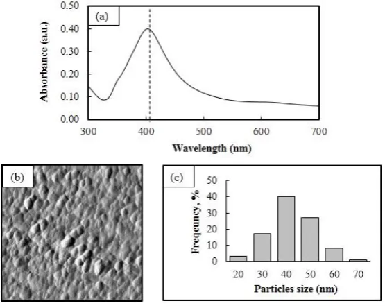
The synthesis of AgNPs was carried out using the chemical reduction of sodium borohydride and trisodium citrate. The formation of light yellow colored AgNPs in an aqueous solution was evaluated by UV-Vis spectroscopy. It was observed that the UV-visible spectrum of AgNPs was obtained at a wavelength of 403 nm (Figure 1a). The free electrons, as a result of after AgNPs preparation, give rise to a surface plasmon resonance (SPR) absorption band because of the combined vibration of free electrons from AgNPs in resonance with a light wave [19]. Generally, the absorption of AgNPs depending on the particles size [20]. Previous reports revealed that the absorption peaks between wavelengths of 400-430 nm and can be attributed to AgNPs of size ranging between 20 and 60 nm [18, 21, 22]. The particle size of AgNPs was measured and further confirmed by atomic force microscope (AFM). The size of synthesized AgNPs varied from 20 to 70 nm, with an average value of 47±9.06 nm and the synthesized AgNPs were topologically spherical in shape (Figure 1b, 1c).
3.2. Antifungal activity of paper coated by disc diffusion
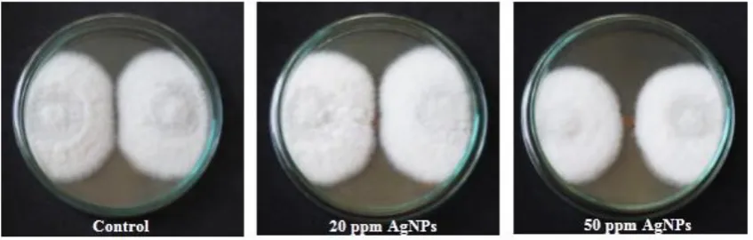
In this study, the antifungal activity of paper coated with 20 and 50 ppm of AgNPs, was investigated, against C. gloeosporioides, using the growth of colony diameter method and results are shown in Figure 2 and Table 2. As seen in the figure, the growth of the fungi in a paper coated with 50 ppm AgNPs was significantly inhibited (p<0.05) compared with paper coated with 20 ppm, after incubation for 2 days. According to these results, a 50 ppm coating of AgNPs resulted in maximum inhibition of fungi on the coated paper.
Figure 2. Antifungal activity in a control plate (no AgNP coating) and test plate with different
concentrations of AgNPs coated paper,for 7 days and after 2 days of incubation.
3.3. Morphology of uncoated and coated paper
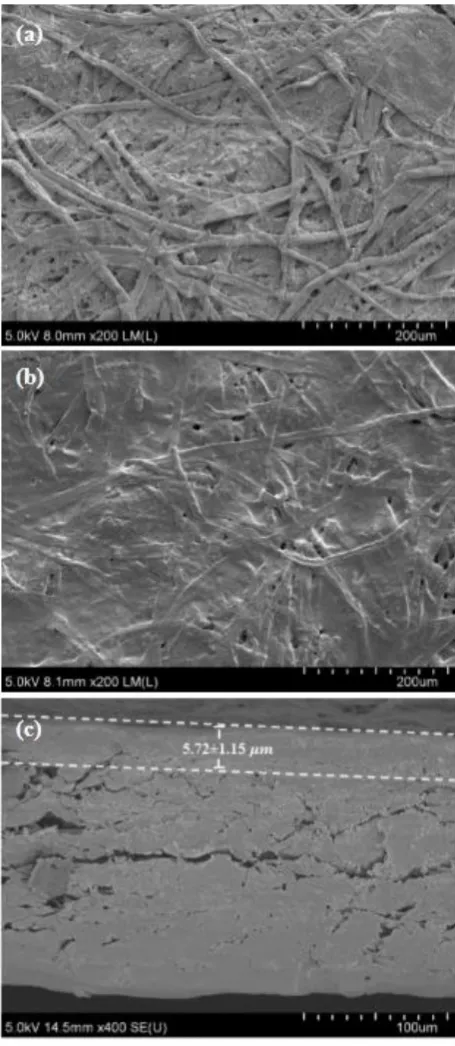
FE-SEM micrographs of the surface of an uncoated paper indicated that the surface was rough and had a porous fibrous structure, as shown in Figure 3a. After coating the paper with a solution containing AgNPs at about 4.0 g
/
m2 the coating weight, the paper showed a relatively smoothersurface compared to the uncoated paper (Figure 3b). This smoothness is probably a result of the coating solution filling the pores on the entire surface of the paper making it homogeneous (Figure 3c) [23, 24].
Table 2 Inhibition efficacy (% relative to control) ofAgNPs coated paper (20 and 50 ppm) on C
.
gloeosporioides.Days
Inhibition (%relative to control)1 AgNPs coated paper (ppm)
20 50
1 ND ND
2 12.00±2.70a 36.00±2.18b
3 3.92±1.79a 18.95±2.73b
4 4.88±2.44a 11.22±2.99b
5 4.26±1.73a 10.08±2.21b
6 2.72±1.42a 6.80±3.27b
7 0.00±0.00a 3.33±1.83b
Remark: Data are presented as mean ± standard deviation, followed by the same letters in the row, indicating that the numbers are not significantly different (p>0.05, based on DMRT), ND –
Not determined.
3.4. Basis weight and thickness
were used as a baseline value. When the paper was coated with coating bar (No.3), the basis weight and thickness of was 137 g
/
m2 and 180 µm, respectively. This indicated that the weight of coatedpaper increased by 4 g
/
m2.Table 3 The basis weight, thickness, coated weight, and coating thickness of an uncoated and coated paper.
Properties Paper
Uncoated Coated
Basis weight
(
g/
m2)
133.
88±1.
25a 137.
61±1.
34bThickness
(
µm)
174.28±0.95a 180.00±0.72bCoating weight
(
g/
m2)
ND 3.73±0.32Coating thickness
(
µm)
ND 5.72±1.15Figure 3. FE- SEM micrographs of (a) uncoated paper, (b) paper coated with AgNPs, and (c) cross-section of the surface coated with a layer of AgNPs.
3.5. Tensile and burst strength
3.08±0.08 kPa m2/g (p<0.05), when the paper was coated. This was due to the solution creating an
excellent film on surface of the paper, which increased the bursting strength [25].
Table 4 The bursting and tensile index of the uncoated and coated paper.
Properties Paper
Uncoated Coated
Tensile index (Nm/g)
-MD 56.33±2.33a 56.02±3.56a
-CD 23.74±1.02a 25.94±0.98b
Bursting index (kPa m2/g) 2.79±0.13a 3.08±0.08b
Remark: Data are presented as mean ± standard deviation followed by the same letters in the row, indicating that the numbers are not significantly different (p>0.05, based on the DMRT).
3.6. Water absorptiveness
The results of water resistancetest, of an uncoated and coated paper, using the Cobb test, are shown in Figure 4. The top side of the coated paper indicated to a significant difference (p<0.05) when compared with the uncoatedpaper, and the values decreased from 34.97±1.35 g/m2 for the uncoated
paper to 32.81±0.86 g/m2 for coated paper. For the bottom side, the uncoated and coated paper, the
water absorptiveness was not significantly different (p>0.05), with the values decreasing from 39.82±1.58 g/m2 for the uncoatedpaper to 38.65±0.78 g/m2 for the coated paper.The results indicate
that the hydrophobic starch can reduce water absorptiveness, and can effectively improve the hydrophobicity and moisture barrier properties of hydrophilic films [26, 27].
Figure 4. Water absorptiveness values for an uncoated and coated paper,as obtained from the
Cobb’s test.
3.7. Effect of AgNPs coating on C. gloeosporioides
Table 5 shows the percentage of infected orchid flowers during the packaging test conducted for 7 days. The highest level of infection percentage was calculated at 40% for the T0 (control) treatment.
While T3 and T4 treatments were found to have a lower infection percentage at 12.5% and significant
difference (p<0.05) compared to T0,T1,and T2 treatments. Therefore, packaging that was coated with
50 ppm of AgNPs combined with a pulsing solution (containing AgNO3 and AgNPs) could inhibit
gloeosporioides hyphae formation was observed on anther for treatments T3 and T4. This validated the
above speculation that the paper coated with AgNPs, at a concentration of 50 ppm, has a good antifungal activity with.
Table 5The percentage of infected cut orchid flowers during the packaging test conducted for 7 days.
Treatments Packaging Pulsing solution Infection1 (%)
T0 (control) Uncoated Distilled 40.00±0.00a
T1 Uncoated AgNO3 29.17±1.44a
T2 Uncoated AgNPs 31.67±3.82a
T3 AgNPs coated AgNO3 12.50±0.00b
T4 AgNPs coated AgNPs 12.50±0.00b
Remark: 1Average±standard deviation followed by the same letters in column indicating that the
numbers are not significantly different (p>0.05, based on DMRT).
Figure 5. Photographs of cut orchid flowers (left) and anther (right) after stored for 7 days during the packaging test. Hyphae formations can be observed for treatments T0,T1,and T2. On
the other hand, no C. gloeosporioides hyphae formation was observed on anther for T3 and T4
4.Conclusion
Synthesized AgNPs, spherical in shape and of size 47 nm, were prepared using chemical reduction and a stabilizing solution at 90 oC. Results from UV- Visible spectrography and AFM
analysis confirmed the presence of AgNPs and their topology. Paper coated with 50 ppm of AgNPs had an excellent antifungal activity against C. gloeosporioides, under culture conditions. The AgNPs-strach coating increased the water resistance, tensile strength in CD, and bursting strength of the paper. After the packaging test, the resultant paper, coated with AgNPs, was successful in decreasing the infection caused by C. gloeosporioides on cut orchid flowers. Therefore, AgNP coated paper has a great potential to be applied widely in the production of paper products suitable for preventing C. gloeosporioides infection that plagues cut orchid flowers and other crops.
Author Contributions: Conceptualization, B.N.and B.P.; Methodology, B.N.; Software, B.N. and B.P.; Validation, B.N., S.P., S.K. and B.P.; Formal Analysis, B.N. and B.P.; Resources, B.N. and B.P.; Data Curation, S.P. and S.K.; Writing-Original Draft Preparation, B.N. and B.P.;Writing-Review &
Editing, B.N. and B.P.; Visualization, B.N. and B.P.; Supervision, B.P.; Project Administration, B.N.; Funding Acquisition, B.N. and B.P.
Funding: Funding: This work was supported by the Graduate Scholarship from the National Research Council of Thailand (NRCT2561001).
Acknowledgments: We are grateful to Dr. Jantana Praiboon for her helpful technical assistance. Conflicts of Interest: The authors declare that they do not have any conflicts of interests.
References
1. Chowdappa, P.; Chethana, C.S.; Bharghavi, R.; Sandhya, H.; Pant, R.P. Morphological and molecular characterization of Colletotrichum gloeosporioides (Penz) Sac. isolates causing anthracnose of orchids in India. Biotechnol. Bioinf. Bioeng. 2012, 2, 567-572.
2. Office of Agricultural Economic, Exporting value of orchid cut flower in Thailand on 2015-2017. Available online: http://oldweb.oae.go.th/production.html/ (accessed on 11 March 2018).
3. Thammasiri, K. Thai orchid genetic resources and their improvement. Horticulturae. 2016, 2, 1-13.
4. Wittayawannakul,W.; Tassakorn, T.; Bhasabutra, T. Postharvest control of black anther disease in dendrobium. Agricultural Sci. J. 2012, 43, 584-587.
5. Tassakorn, T.; Bhasabutra, T.; Wittayawannakul, W. Chemical control of black anther disease on dendrobium orchids. Available online: http://www.doa.go.th/research/ showthread.php?tid=1781/ (accessed on 11 March 2018)
6. Chowdappa, P.; Gowda, S.; Chethana, C.S.; Madhura, S. Antifungal activity of chitosan-silver nanoparticle composite against Colletotrichum gloeosporioides associated with mango anthracnose. Afr. J. Microbiol. Res. 2014, 8, 1803-1812.
7. Junqueira-Gonçalves, M. P.; Alarcón, E.; Niranjan, K. Development of antifungal packaging for berries extruded from recycled PET. Food Control. 2013, 33, 455-460.
8. Rudra, S.G.; Singh, V.; Jyoti, S.D.; Shivhare, U.S. Mechanical properties and antimicrobial efficacy of active wrapping paper for primary packaging of fruits. Food Biosci. 2013, 3, 49–58.
9. Lamsal, K.; Kim, S.W.; Jung, J.H.; Kim, Y.S.; Kim, K. S.; Lee, Y.S. Application of silver nanoparticles for the control of Colletotrichum species in vitro and pepper anthracnose disease in field. Mycobiology. 2011, 39, 194-199.
10. Aguilar-Méndez, M.A.; Martín-Martínez, E.S.; Ortega-Arroyo, L.; Cobián-Portillo, G.; Sánchez-Espíndola, E. Synthesis and characterization of silver nanoparticles: effect on phytopathogen Colletotrichum gloesporioides. J Nanopart Res. 2013, 13, 2525- 2532.
11. Nokkrut, B.; Pisutpiched, S.; Puangsin, B. Effect of quaternary ammonium compounds and zinc pyrithione on antimicrobial properties in packaging containers for orchid cut flowers. Thai J. For. 2017, 36, 136-144. 12. Chaichompoo,W.; Phichai, K. Effect of plant extract on growth inhibition of Colletotrichum sp.. Research
13. Kang, H.C.; Park, Y.H.; Go, S.J. Growth inhibition of a phytopathogenic fungus, Colletotrichum species by acetic acid. Microbiol. Res. 2003, 158, 321–326.
14. Shi, X.; Li, B.; Qin, G.; Tian, S. Mechanism of antifungal action of borate against Colletotrichum gloeosporioides related to mitochondrial degradation in spores. Postharvest Biol Tec. 2012, 67, 138–143.
15. Bussaman, P.; Namsena, P.; Rattanasena, P.; Chandrapatya, A. Effect of crude leaf extracts on Colletotrichum gloeosporioides (Penz.) Sacc. Psyche A Journal of Entomology. 2012, 1, 1-6.
16. Echegoyen, Y.; Nerín, C. Nanoparticle release from nano-silver antimicrobial food containers. Food Chem Toxicol. 2013, 62, 16–22.
17. Carbone, M.; Donia, D.T.; Sabbatella, G.; Antiochia, R. Silver nanoparticles in polymeric matrices for fresh food packaging. J. King Saud Univ., Sci. 2016, 28, 273–279.
18. Agnihotri, S.; Mukherji, S.; Mukherji, S. Size-controlled silver nanoparticles synthesized over the range 5– 100 nm using the same protocol and their antibacterial efficacy. RSC Adv. 2014, 4, 3974–3983.
19. Xia, Y.; Xiong, Y.; Lim, B.; Skrabalak, S.E. Shape-controlled synthesis of metal nanocrystals: simple chemistry meets complex physics?. Angew. Chem. Int. Ed. 2009, 48, 60-103.
20. Pacioni, N.L.; Borsarelli, C.D.; Rey, V.; Veglia, A.V. Synthetic routes for the preparation of silver nanoparticles. In Silver nanoparticle applications; Alarcon, E.I., Griffith, M., Udekwu, K.I., Eds.; Springer International Publishing: Basel, Switzerland, 2015; Volume 1, pp. 13-45, ISBN 978-3-319-11261-9.
21. Paramelle, D.; Sadovoy, A.; Gorelik, S.; Free, P.; Hobley, J.; Fernig, D.G. Rapid method to estimate the concentration of citrate capped silver nanoparticles from UV-visible light spectra. Analyst. 2014, 139, 4855-4861.
22. Tomaszewska, E.; Soliwoda, K.; Kadziola, K.; Tkacz-Szczesna, B.; Celichowski, G.; Cichomski, M.; Szmaja, W.; Grobelny, J. Detection limits of DLS and UV-Vis spectroscopy in characterization of polydisperse nanoparticles colloids. J Nanomater. 2013, 1-10.
23. Li, Y.; Zhang, J.; Li, H.; Gu, W.j.; He, B. Analysis of the Coating surface Properties of Coated Paper. In Proceedings of the 2015 International Conference on Materials, Environmental and Biological Engineering (MEBE 2015), Guilin, China, 28-30 March 2015; pp. 250-253.
24. Amini, E.; Azadfallah, M.; Layeghi, M.; Talaei-Hassanloui, R. Silver-nanoparticle-impregnated cellulose nanofiber coating for packaging paper. Cellulose. 2016, 23, 557–570.
25. Bruun, S.E. Starch. In Pigment coating and surface sizing of surface; Lehtnen, E., Eds.; Fapet Oy: Jyväskylä, Finland, 2000; Volume 1, pp. 241-249, ISBN 952-5216-11-X.
26. Butinaree, S.; Jinkarn, T.; Yoksan, R. Effects of biodegradable coating on barrier properties of paperboard food packaging. JMMM. 2008, 18, 219-222.
27. Biricik, Y.; Sonmez, S.; Ozden, O. Effects of surface sizing with starch on physical strength properties of paper. Asian J. Chem. 2011, 23, 3151-3154.