Multicellular Group Formation in Saccharomyces cerevisiae
Fisher, R.M. & Regenberg, B.
Section for Ecology and Evolution, Department of Biology, University of Copenhagen
Key words: yeast, multicellularity, adhesion, major evolutionary transition
Abstract
Understanding how and why cells cooperate to form multicellular organisms is a central aim of evolutionary biology. Multicellular groups can form through clonal development (where
daughter cells stick to mother cells after division) or by aggregation (where cells aggregate to form groups). These different ways of forming groups directly affect relatedness between individual cells, which in turn influences the degree of cooperation and conflict within the multicellular group. It is hard to study the factors that favoured multicellularity by focusing only on obligately multicellular organisms, like complex animals and plants, because the factors that favour multicellular cooperation cannot be disentangled, as cells cannot survive and reproduce independently. We propose bakers yeast, Saccharomyces cerevisiae,as an ideal model for studying the very first stages of the evolution of multicellularity. This is because it can form multicellular groups both clonally and through aggregation and uses a family of proteins called ‘flocculins’ that determine the way in which groups form, making it particularly amenable to lab experiments. We briefly review current knowledge about multicellularity in S. cerevisiae and then propose a framework for making predictions about the evolution of multicellular
phenotypes in yeast based on social evolution theory. We finish by suggesting outstanding questions and potentially fruitful avenues for future research.
Introduction
Multicellular organisms dominate the world we see around us, and yet they are formed from millions of individual cells that specialize on different tasks and cooperate to form a cohesive body. Understanding how and why cells cooperate to form multicellular structures is a central aim of evolutionary biology, because multicellularity has arisen many times across the tree of life, and has led to some of the most important species radiations for both biological complexity and diversity.
The evolution of obligate multicellularity, like we see in animals and plants, has been called a ‘major evolutionary transition in individuality’ because cells are entirely mutually dependent on each other and conflict between them is so minimal that they can be considered a new individual (Figure 1) [1-3]. However, this transition has only ever occurred in species that have clonal multicellular development – when daughter cells remain attached to mother cells after
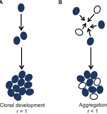
division, meaning cells will be genetically identical, leading to clonal relatedness (Figure 2A). Multicellularity can also arise as a consequence of cell aggregation, however this has never led to obligate multicellularity (Figure 2B). For example, the slime mold Dictyostelium discoideum and other species that form groups through aggregation remain able to switch between unicellularity and multicellularity, making them facultatively multicellular (Figure 1). As a consequence, they have a lower number of cell types and are generally smaller, compared to species that form multicellular groups through clonal development [4].
Figure 1:Stages of major evolutionary transitions. Major evolutionary transitions involve independent units (genes, cells or organisms) joining together to form a social group, which then becomes a new individual through the evolution of mutual dependence [12,55]. S. cerevisiae is able to form multicellular groups through cooperation, but remains facultatively multicellular – i.e. it has not made the major evolutionary transition to obligate multicellularity.
Figure 2: Modes of multicellular group formation.A: Cells can form multicellular groups through clonal development, where daughter cells remaining attached to mother cells after cell division. This guarantees that cells will be clonally related to each other (relatedness, r = 1). B: Cells can aggregate to form a multicellular group of cells. These can be genetically similar or dissimiliar cells (relatedness, r < 1).
Facultative multicellularity
Obligate multicellularity
Saccharomyces cerevisiae
Aggregation r < 1 Clonal development
r = 1
There is a growing and convincing pool of evidence suggesting that the way in which multicellular groups form is key for understanding when and how major evolutionary transitions occur, through its effect on relatedness between the interacting cells [3-6] (Figure 2). However, it is hard to study major evolutionary transitions by focusing only on obligately multicellular organisms, because the factors that favour multicellular cooperation cannot be disentangled, as cells cannot survive and reproduce independently (Figure 1). Obligately multicellular species may have also undergone secondary changes that make the origins of multicellularity unclear. Hence, factors that favour multicellularity are best studied in facultative multicellular species. Many examples of this are found across the tree of life, but very few concrete examples exist where species are able to form multicellular groups through both aggregation and clonal development. This makes it difficult to investigate the mechanisms and consequences of the two types of group formation experimentally in one species.
Here, we propose bakers yeast, Saccharomyces cerevisiae,as an ideal model for studying the very first stages of the evolution of multicellularity as a major evolutionary transition in individuality (Figure 1). This is because: (1) it is able to switch between unicellularity and multicellularity, (2) it can do this through both modes of group formation (clonal development and aggregation) and, (3) it is a well-studied, tractable model organism. In this paper, we briefly review current knowledge about group formation and multicellularity in S. cerevisiae and then propose a framework for making predictions about the evolution of multicellular phenotypes in yeast based on social evolution theory. We suggest terminology that is general and useful, and we finish by suggesting outstanding questions and potentially fruitful avenues for future
research.
Multicellularity in yeast: a major evolutionary transition?
Throughout this article, we use a very broad definition of multicellularity (see Glossary). This is necessary in order to capture the variety of multicellular behaviours both in S. cerevisiae and other yeast species, but also across the tree of life. We define a phenotype as ‘multicellular’ in the simplest possible way – if multiple cells are in physical contact. This allows us to
encompass both facultative and obligate multicellularity and is an umbrella term capturing every possible type of multicellular phenotype.
The term ‘multicellular organism’ should only be used for a restricted set of species that are obligately multicellular and have undergone a major evolutionary transition – that are unable to switch between reproducing as unicellular and multicellular forms (e.g. metazoans). We consider obligately multicellular species, e.g. animals and plants, as having undergone a major evolutionary transition in individuality because multicellularity is a developmentally determined part of the life cycle and cells cannot survive independently (Figure 1) (see Glossary).
How do yeasts fit into this framework? Yeast are a polyphyletic group of species within the Kingdom Fungi. They are predominantly unicellular, although many yeasts are known to switch between unicellular and multicellular lifestyles depending on environmental factors, so we classify them as facultatively multicellular. Yeasts have evolved at least 5 times independently within Kingdom Fungi [7] and many of the most important fungal pathogens and
biotechnologically useful species are yeasts. S. cerevisiae is perhaps the most famous, displaying a startling variety of multicellular phenotypes, including pseudohyphae, biofilms and flocs (Figure 3) [8-11] that are also common in other yeasts [12].
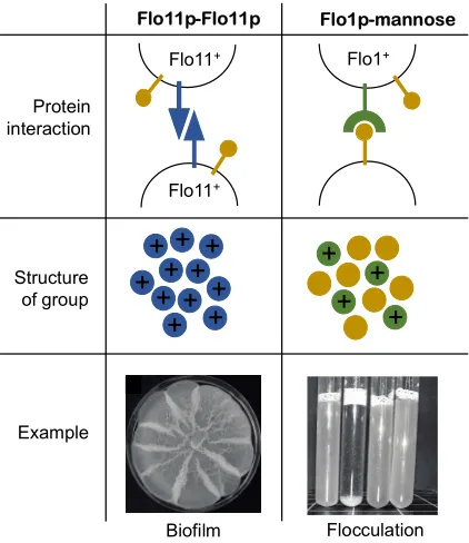
Figure 3:Flocculins determine the structure of multicellular groups. Homophilic (self-self) and heterophilic (self-non-self) interactions of Flo11p and other flocculins (Flo1p, Flo5p, Flo9p and Flo10p). The left panel shows the way Flo11p (coloured in blue) adheres to other Flo11p on neighbouring cell walls. Flo11p does not interact directly with mannose residues (coloured in yellow). Flo11p will only adhere to Flo11p, creating self-self adherance and therefore clonal groups of cells. An example of this can be found in clonal biofilms. The right panel shows the way Flo1p (coloured in green) can adhere to mannose residues which are expressed by all cells, meaning multicellular groups can contain cells of different genotypes. Images: Surface spreading biofilm on semisolid complex growth medium after two weeks growth (as described in [20], strain CLIB326_1) and flocculation of diploid yeast in liquid complex medium (second tube from left, strain from L to R: SFL1/sfl1Q320STOP; sfl1Q320STOP/sfl1Q320STOP;
sfl1Q320STOP/SFL1; SFL1/SFL1 in the CEN.PK strain background described in [53]).
Biofilm Flocculation
+
+
+
+
+
+
+ +
+ + +
+
+
+
+
Flo11p-Flo11p Flo1p-mannose
Protein interaction
Flo11+
Flo11+
Flo1+
Structure of group
How does Saccharomyces cerevisiae become multicellular?
In order to be multicellular, cells need to be able to adhere to one another. In S. cerevisiae adhesion is conferred by a family of proteins called flocculins. They comprise seven different functional FLO genes, coding for five proteins involved in multicellularity [13,14] and two proteins specific for conjunction of haploid cells in mating [15]. Flocculins are cell wall proteins that are anchored to the cell membrane and protrude from the cell wall to confer cell-cell and cell-surface adhesion [16].
In S. cerevisiae, flocculins can be broadly split into two types, based on the structure of their amino terminal A-domain (the part of the protein responsible for adhesion) [9]. Flo1p, Flo5p, Flo9p, and Flo10p confer general adhesion, by sticking to mannose residues that protrude from the surfaces of other cells [17,18] and these flocculins are generally expressed when cells are not growing. In contrast, Flo11p confers very specific adhesion, through a homophilic Flo11p-Flo11p interaction and is expressed during growth (Figure 3) [19]. So, whilst the other flocculins make cells generally ‘sticky’, FLO11 produces a protein that will only adhere to other cells expressing FLO11 [20].
Therefore, flocculins in S. cerevisiae produce two distinct ways of sticking together and forming multicellular groups (Figure 2). FLO1, 5, 9, and 10 result in aggregative multicellular group formation – cells expressing them will stick to other cells in a general ‘sticky’ response regardless of their genotype. On the other hand, expression of FLO11 will lead to clonal group formation between related cells, usually a mother and daughter cell after division. This is because the daughter receives FLO11 mRNA from her mother during development [21] and because FLO11 is only expressed during growth. This special quality of the flocculins found in S. cerevisiae means that flocculin expression corresponds almost exactly to two distinct ways of forming multicellular groups (Figure 2); aggregative and clonal group formation.
Flocs
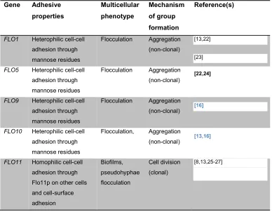
Table 1:The genetic basis of multicellularity in yeast. Flocculin genes involved in multicellular group formation in S. cerevisiae, adhesive properties of the flocculins, the multicellular phenotypes produced and the way in which multicellular groups are formed.
Gene Adhesive properties Multicellular phenotype Mechanism of group formation Reference(s)
FLO1 Heterophilic cell-cell adhesion through mannose residues
Flocculation Aggregation (non-clonal)
[13,22]
[23]
FLO5 Heterophilic cell-cell adhesion through mannose residues
Flocculation Aggregation (non-clonal)
[22,24]
FLO9 Heterophilic cell-cell adhesion through mannose residues
Flocculation Aggregation (non-clonal) [16]
FLO10 Heterophilic cell-cell adhesion through mannose residues
Flocculation, Aggregation
(non-clonal) [13,16]
FLO11 Homophilic cell-cell adhesion through Flo11p on other cells and cell-surface adhesion Biofilms, pseudohyphae flocculation Cell division (clonal) [8,13,25-27] Pseudohyphae
Pseudohyphal growth is a filamentous growth form that allows diploid cells of S. cerevisiae to grow in a nitrogen-limited environment through long thin filaments with little increase in their biomass [10]. Pseudohyphae are comprised of a chain of elongated cells that remain loosely attached after unipolar budding . As with biofilm formation, the Flo11p protein is essential for pseudohyphal growth [25] (Table 1) but the exact role of Flo11p is unclear, since cells in pseudohyphal colonies are not attached to each other as in biofilm colonies [10].
Biofilms
Surface spreading biofilms on semi-solid 0.3% agar are particularly interesting because of the large variety of growth forms found in natural isolates [20,34]. Recently, Regenberg et al. (2016) showed that when grown on semi-solid 0.3% agar, certain strains of S. cerevisiae form differentiated biofilms [20]. Such biofilms are created through a FLO11 epigenetic switch where both Flo11+ and Flo11- cells are produced simultaneously in one population of cells [9,20,35]. These biofilms outgrew others without the epigenetic Flo11 switching mechanism, which were either solely Flo11+ or solely Flo11-. This research shows that conditional differentiation between adhesive and non-adhesive cells can allow cells to outgrow competitors through cooperation in a multicellular biofilm and that differentiation between cells might be selected for in very early stages of multicellularity, but without necessarily leading to obligate multicellularity.
Why is group formation important?
The way in which multicellular groups form has fundamental consequences for behaviour, complexity and social evolution, because it has direct implications on the genetic relatedness between interacting cells [3,4]. When groups form through aggregation, cells are likely to be genetically different and so the resulting multicellular group will contain cells that are genetically unrelated (or at least non-clonal) (Figure 2B). In contrast, when groups form through cell division, by the daughter cell remaining attached to the mother, cells will be clonally related to each other (Figure 2A). Relatedness is known to be an important force shaping social
behaviour, as cells that are genetically related will be more likely to engage in cooperative behaviours, compared to cells that are unrelated [36]. For example, Pseudomonasaeruginosa show higher levels of cooperative siderophore production when they are interacting with relatives, compared to when they are interacting with non-relatives [37]. There is in fact compelling comparative evidence that clonal relatedness between cells has always been a necessary condition for the evolution of complex, obligate multicellularity like we see in animals and plants, and some lineages of fungi and algae [4].
Unlike many other species, S. cerevisiae is able to form multicellular groups both by aggregation and through cell division, resulting in different multicellular phenotypes (Figure 3). When groups are formed through budding, as is the case for biofilms and pseudohyphae, the cells in the multicellular group will be clonal (all else being equal). However, flocculation can occur between genetically dissimilar cells, meaning that relatedness will be less than clonal and variable. Therefore, the way in which these various multicellular groups form has
consequences for cell-cell relatedness, and this means we can make several predictions about the social interactions we may expect.
must overcome in order to maintain cooperation and ensure the benefits of cooperation are returned to other cooperative cells.
The way in which the multicellular groups form will have a profound influence on whether or not cheats even have the potential to invade. Biofilms and pseudohyphae, where cells are clonally related to one another, should intrinsically be able to withstand the effects of cheating, simply because the way in which the groups form will exclude cheating cells. Furthermore, Flo11+ cells can only adhere to other cells expressing Flo11+ (Figure 3). This means clonal biofilms expressing FLO11 have the inherent capacity to protect against invasion by other genotypes (that do not express FLO11). This is not the case for flocculation. This is because flocs are formed through aggregation of potentially unrelated cells (Figure 2B) [39]. Cells in the floc adhere to each other through expression of FLO1, but the flocculating Flo1+ cells can be of different origin, leading to flocs comprised of genetically different cells. Non-producers can still adhere because Flo1p is able to stick to mannose residues produced by all cells, not just by other producers (Figure 3) [39]. Cheats could therefore reap the benefits of flocculating without paying the cost of expressing FLO1 [39]. In fact, there is evidence that loss-of-function mutants can occur in and spread through natural populations of yeast expressing the FLO1 homologue, FLO5 [40]. This provides support for the prediction that cheating mutants can only spread in multicellular groups that have formed through aggregation, and not through clonal development.
Saccharomyces in the laboratory
S. cerevisiae has been used historically as model for eukaryotic genetics due to the ease by which it can be cultured in the lab and by which it lent itself to genetic studies of gene linkage [41,42]. A major advantage of using S. cerevisiae as a model is the valuable strain collections and genetic tools developed by a large community of yeast geneticists over the past 30 years, which makes S. cerevisiae one of the most well understood eukaryotic organisms at the molecular level [43-47].
There are several aspects in particular that make S. cerevisiae a desirable and tractable model organism for studying the evolution of multicellularity. Firstly, there are robust methods for transformation of S. cerevisiae with exogenous DNA, and making all types of chromosomal mutations including insertions, deletions, and substitutions[46]. Furthermore, there are now mutant strain collections where any one of the approximately 6000 genes have been deleted in otherwise functional strains [43,44]. One strain collection is made in the 1278b genetic background that expresses FLO11 naturally, which has allowed for the identification of genes and proteins involved in biofilm and pseudohyphal growth [27,44].
allows researchers to see the cellular level structure of multicellular phenotypes such as biofilms, and to investigate how cells expressing different adhesive properties interact. Finally, the sequenced genomes of S. cerevisiae strains allow for comparative genomics studies [47,48], that have already revealed that, for example, FLO1 and FLO11 are among the fastest evolving genes in the yeast genome [49].
These methods, among others, mean that we can ask important questions about the evolution of multicellularity using S. cerevisiae that may not be possible with other organisms. For example, phenotypes and behaviors found in nature can be manipulated and studied in genetic tractable strains [44]. Recently, S. cerevisiae has been used as a model for studying the very beginnings of multicellularity, including the molecular factors underlying and
environmental factors selecting for multicellularity [20].Another example is the identification of the sucrose-degrading enzyme invertase as a public good [39,50], allowing us to ask questions about public good production in cooperative multicellular groups and the potential for cheats to invade. Finally, several studies have used S. cerevisiae in experimental evolution of
multicellularity in the lab, exploiting the tractability, genetic tools and fast-generation times of S. cerevisiae [51-53].
Concluding remarks
There has been a wealth of research on multicellularity in yeast on mechanisms [17,19], genetics [18] and social evolution [20,39,50,51,54]. However, we suggest that there is an opportunity to synthesize this research within the major evolutionary transitions framework and to capitalise on an incredibly useful experimental system for studying the first stages of
multicellularity.
We believe that multicellular group formation in S. cerevisiae, through the expression of flocculins, provides an ideal system for studying multicellularity. Firstly, the facultative nature of multicellularity in S. cerevisiae means it is possible to study and manipulate the benefits and costs of group formation in controlled experiments. Secondly, flocculin proteins allow us to study the effect of different modes of group formation on multicellular cooperation. For example, we can use the flocculin proteins that confer aggregative (e.g. Flo1p) and clonal (Flo11p) adhesion as an opportunity to study the effect of different modes of group formation on cooperative behaviours in the same species. This could provide a complementary lab system to the comparative research showing how crucial group formation is in determining subsequent multicellular evolution.
Acknowledgements
Glossary of terms
Multicellularity
When multiple individual cells are in contact. This includes cells sticking together transiently through production of a sticky substance, coordinated groups of cells that show cooperative behaviours such as production of public goods, and obligate groups of cells forming
multicellular organisms like we see in animals and plants.
Obligate multicellularity
When individual cells are obligately part of a multicellular body, and cannot survive and reproduce outside of the multicellular body. Obligate multicellularity is developmentally determined, and not a response to environmental conditions.
Facultative multicellularity
When individual cells can become part of a multicellular body in response to environmental conditions, and then can revert to being unicellular again. They do not rely on being multicellular in order to survive and reproduce.
Major evolutionary transition in individuality
A major evolutionary transition in individuality occurs when individual units (e.g. genes, cells or individuals) cooperate and form a new, more complex individual, that can subsequently only reproduce as a whole.
Multicellular organism
References
1. Grosberg, R. K. & Strathmann, R. R. 2007 The Evolution of Multicellularity: A Minor Major Transition? Annu Rev Ecol Evol S38, 621–654.
(doi:10.1146/annurev.ecolsys.36.102403.114735)
2. Maynard Smith, J. & Szathmáry, E. 1995 The Major Transitions in Evolution. Oxford University Press, USA.
3. West, S., Fisher, R. M., Gardner, A. & Kiers, E. T. 2015 Major evolutionary transitions in individuality. Proceedings of the National Academy of Sciences of the United States of America (doi:10.1073/pnas.1421402112)
4. Fisher, R. M., Cornwallis, C. K. & West, S. 2013 Group Formation, Relatedness, and the Evolution of Multicellularity. Current Biology23, 1120–1125. (doi:10.1016/j.cub.2013.05.004)
5. Boomsma, J. J. 2009 Lifetime monogamy and the evolution of eusociality. pp. 3191– 3207.(doi:10.1098/rstb.2009.0101)
6. Queller, D. C. 2000 Relatedness and the fraternal major transitions. Philos. Trans. R. Soc. Lond., B, Biol. Sci.355, 1647–1655. (doi:10.1098/rstb.2000.0727)
7. Nagy, L. G. et al. 2014 Latent homology and convergent regulatory evolution underlies the repeated emergence of yeasts. Nat Commun5. (doi:10.1038/ncomms5471)
8. Reynolds, T. B. & Fink, G. R. 2001 Bakers' yeast, a model for fungal biofilm formation. Science
291, 878–881.
9. Brückner, S. & Mösch, H.-U. 2011 Choosing the right lifestyle: adhesion and development in Saccharomyces cerevisiae. FEMS Microbiology Reviews36, no–no. (doi:10.1111/j.1574-6976.2011.00275.x)
10. Gimeno, C. J., Ljungdahl, P. O., Styles, C. A. & Fink, G. R. 1992 Unipolar cell divisions in the yeast S. cerevisiae lead to filamentous growth: regulation by starvation and RAS. Cell68, 1077– 1090.
11. LINDQUIST, W. 1952 Cell Surface Constituents and Yeast Flocculation. Nature170, 544–545.
12. Vallejo, J. A., Sánchez-Pérez, A., Martínez, J. P. & Villa, T. G. 2013 Cell aggregations in yeasts and their applications. Appl. Microbiol. Biotechnol.97, 2305–2318. (doi:10.1007/s00253-013-4735-y)
13. Guo, B., Styles, C. A., Feng, Q. H. & Fink, G. R. 2000 A Saccharomyces gene family involved in invasive growth, cell-cell adhesion, and mating. Proceedings of the National Academy of Sciences of the United States of America97, 12158–12163. (doi:10.1073/pnas.220420397)
14. Caro, L., Tettelin, H., Vossen, J. H., Ram, A., VandenEnde, H. & Klis, F. M. 1997 In silicio identification of glycosyl-phosphatidylinositol-anchored plasma-membrane and cell wall proteins of Saccharomyces cerevisiae. Yeast13, 1477–1489.
(doi:10.1002/(SICI)1097-0061(199712)13:15<1477::AID-YEA184>3.0.CO;2-L)
15. Erdman, S., Lin, L., Malczynski, M. & Snyder, M. 1998 Pheromone-Regulated Genes Required for Yeast Mating Differentiation. The Journal of Cell Biology140, 461–483.
(doi:10.2307/1618450?refreqid=search-gateway:7ae53039fab33d362cd114f682725875)
16. Teunissen, A. W. & STEENSMA, H. Y. 1995 Review: the dominant flocculation genes of Saccharomyces cerevisiae constitute a new subtelomeric gene family. Yeast11, 1001–1013. (doi:10.1002/yea.320111102)
17. Goossens, K. V. Y. et al. 2015 Molecular Mechanism of Flocculation Self-Recognition in Yeast and Its Role in Mating and Survival. MBio6, e00427–15. (doi:10.1128/mBio.00427-15)
basis of flocculin-mediated social behavior in yeast. Proceedings of the National Academy of Sciences of the United States of America107, 22511–22516. (doi:10.1073/pnas.1013210108)
19. Kraushaar, T., Brückner, S., Veelders, M., Rhinow, D., Schreiner, F., Birke, R., Pagenstecher, A., Mösch, H.-U. & Essen, L.-O. 2015 Interactions by the Fungal Flo11 Adhesin Depend on a Fibronectin Type III-like Adhesin Domain Girdled by Aromatic Bands. Structure/Folding and Design23, 1005–1017. (doi:10.1016/j.str.2015.03.021)
20. Regenberg, B., Hanghøj, K. E., Andersen, K. & Boomsma, J. J. 2016 Clonal yeast biofilms can reap competitive advantages through cell differentiation without being obligatorily multicellular. Proceedings of the Royal Society B: Biological Sciences283, 20161303.
(doi:10.1098/rspb.2016.1303)
21. Wolf, J. J., Dowell, R. D., Mahony, S., Rabani, M., Gifford, D. K. & Fink, G. R. 2010 Feed-Forward Regulation of a Cell Fate Determinant by an RNA-Binding Protein Generates Asymmetry in Yeast. Genetics185, 513–U175. (doi:10.1534/genetics.110.113944)
22. Hodgson, J. A., Berry, D. R. & Johnston, J. R. 1985 Discrimination by heat and proteinase treatments between flocculent phenotypes conferred on Saccharomyces cerevisiae by the genes FLO1 and FLO5. J. Gen. Microbiol.131, 3219–3227. (doi:10.1099/00221287-131-12-3219)
23. TEUNISSEN, A., HOLUB, E., VANDERHUCHT, J., VANDENBERG, J. A. & STEENSMA, H. Y. 1993 Sequence of the Open Reading Frame of the Fl01 Gene From Saccharomyces-Cerevisiae. Yeast9, 423–427. (doi:10.1002/yea.320090413)
24. BIDARD, F., Blondin, B., DEQUIN, S., VEZINHET, F. & Barre, P. 1994 Cloning and Analysis of a Flo5 Flocculation Gene From S-Cerevisiae. Curr. Genet.25, 196–201.
25. Lo, W. S. & Dranginis, A. M. 1998 The cell surface flocculin Flo11 is required for pseudohyphae formation and invasion by Saccharomyces cerevisiae. Mol. Biol. Cell9, 161–171.
26. Lo, W. S. & Dranginis, A. M. 1996 FLO11, a yeast gene related to the STA genes, encodes a novel cell surface flocculin. The Journal of Bacteriology178, 7144–7151.
27. Andersen, K., Bojsen, R., Sorensen, L. G. R., Nielsen, M. W., Lisby, M., Folkesson, A. & Regenberg, B. 2014 Genetic Basis for Saccharomyces cerevisiae Biofilm in Liquid Medium. G3 (Bethesda)4, 1671–1680. (doi:10.1534/g3.114.010892)
28. Bony, M., Barre, P. & Blondin, B. 1998 Distribution of the flocculation protein, Flop, at the cell surface during yeast growth: The availability of Flop determines the flocculation level. Yeast14, 25–35. (doi:10.1002/(SICI)1097-0061(19980115)14:1<25::AID-YEA197>3.0.CO;2-C)
29. O'Toole, G., Kaplan, H. B. & Kolter, R. 2000 Biofilm formation as microbial development. Annu. Rev. Microbiol.54, 49–79. (doi:10.1146/annurev.micro.54.1.49)
30. Hall-Stoodley, L., Costerton, J. W. & Stoodley, P. 2004 Bacterial biofilms: from the natural environment to infectious diseases. Nat Rev Microbiol2, 95–108. (doi:10.1038/nrmicro821)
31. Blankenship, J. R. & Mitchell, A. P. 2006 How to build a biofilm: a fungal perspective. Current Opinion in Microbiology9, 588–594. (doi:10.1016/j.mib.2006.10.003)
32. Bojsen, R., Regenberg, B. & Folkesson, A. 2014 Saccharomyces cerevisiae biofilm tolerance towards systemic antifungals depends on growth phase. BMC Microbiol14, 305.
(doi:10.1186/s12866-014-0305-4)
33. Andersen, K., Bojsen, R. K., Sorensen, L. G., Nielsen, M. W., Lisby, M., Folkesson, A. & Regenberg, B. 2014 Genetic Basis for Saccharomyces cerevisiae Biofilm in Liquid Medium. Genes, Genomes, Genetics, 1–10. (doi:10.1534/g3.114.010892/-/DC1)
34. Hope, E. A. & Dunham, M. J. 2014 Ploidy-Regulated Variation in Biofilm-Related Phenotypes in Natural Isolates of Saccharomyces cerevisiae. G3 (Bethesda)4, 1773–1786.
35. Bumgarner, S. L., Neuert, G., Voight, B. F., Symbor-Nagrabska, A., Grisafi, P., van Oudenaarden, A. & Fink, G. R. 2012 Single-Cell Analysis Reveals that Noncoding RNAs Contribute to Clonal Heterogeneity by Modulating Transcription Factor Recruitment. Mol. Cell45, 470–482. (doi:10.1016/j.molcel.2011.11.029)
36. Hamilton, W. D. 1964 The genetical evolution of social behaviour. I. J. Theor. Biol.7, 1–16. (doi:10.1016/0022-5193(64)90038-4)
37. Griffin, A. S., West, S. & Buckling, A. 2004 Cooperation and competition in pathogenic bacteria. Nature430, 1024–1027. (doi:10.1038/nature02744)
38. Ghoul, M., Griffin, A. S. & West, S. 2014 Toward an evolutionary definition of cheating. Evolution
68, 318–331. (doi:10.1111/evo.12266)
39. Smukalla, S. et al. 2008 FLO1 Is a Variable Green Beard Gene that Drives Biofilm-like Cooperation in Budding Yeast. Cell135, 726–737. (doi:10.1016/j.cell.2008.09.037)
40. Di Gianvito, P., Tesniere, C., Suzzi, G., Blondin, B. & Tofalo, R. 2017 FLO5 gene controls flocculation phenotype and adhesive properties in a Saccharomyces cerevisiae sparkling wine strain. Sci Rep7. (doi:10.1038/s41598-017-09990-9)
41. Cherry, J. M. et al. 1997 Genetic and physical maps of Saccharomyces cerevisiae. Nature387, 67–73.
42. Guthrie, C. & Fink, G. R. 2004 Guide to Yeast Genetics and Molecular and Cell Biology. Gulf Professional Publishing.
43. Giaever, G. et al. 2002 Functional profiling of the Saccharomyces cerevisiae genome. Nature
418, 387–391. (doi:10.1038/nature00935)
44. Ryan, O. et al. 2012 Global Gene Deletion Analysis Exploring Yeast Filamentous Growth. Science337, 1353–1356. (doi:10.1126/science.1224339)
45. Huh, W. K., Falvo, J. V., Gerke, L. C., Carroll, A. S., Howson, R. W., Weissman, J. S. & O'Shea, E. K. 2003 Global analysis of protein localization in budding yeast. Nature425, 686–691. (doi:10.1038/nature02026)
46. Goffeau, A. et al. 1996 Life with 6000 genes. Science274, 546–563–7.
47. Liti, G. et al. 2009 Population genomics of domestic and wild yeasts. Nature458, 337–341. (doi:10.1038/nature07743)
48. Peter, J. et al. 2018 Genome evolution across 1,011 Saccharomyces cerevisiae isolates. Nature
556, 339–. (doi:10.1038/s41586-018-0030-5)
49. Verstrepen, K. J., Jansen, A., Lewitter, F. & Fink, G. R. 2005 Intragenic tandem repeats generate functional variability. Nat. Genet.37, 986–990. (doi:10.1038/ng1618)
50. Koschwanez, J. H., Foster, K. R. & Murray, A. W. 2011 Sucrose utilization in budding yeast as a model for the origin of undifferentiated multicellularity. Plos Biol9, 1–10.
(doi:10.1371/journal.pbio.1001122)
51. Ratcliff, W. C., Denison, R. F., Borrello, M. & Travisano, M. 2012 Experimental evolution of multicellularity. Proceedings of the National Academy of Sciences of the United States of America
109, 1595–1600. (doi:10.1073/pnas.1115323109)
52. Ratcliff, W. C., Fankhauser, J. D., Rogers, D. W., Greig, D. & Travisano, M. 2015 Origins of multicellular evolvability in snowflake yeast. Nat Commun6. (doi:10.1038/ncomms7102)
53. Torbensen, R., Moller, H. D., Gresham, D., Alizadeh, S., Ochmann, D., Boles, E. & Regenberg, B. 2012 Amino Acid Transporter Genes Are Essential for Dependent and
54. Wloch-Salamon, D. M., Fisher, R. M. & Regenberg, B. 2017 Division of labour in the yeast: Saccharomyces cerevisiae. Yeast34, 399–406. (doi:10.1002/yea.3241)