Article
A Deep Transfer Learning Model with Classical Data
Augmentation and CGAN to Detect COVID-19 from
Chest CT Radiography Digital Images
Mohamed Loey 1*, Florentin Smarandache 2, Nour Eldeen M. Khalifa 3
1 Department of Computer Science, Faculty of Computers Artificial Intelligence, Benha University, Benha
13511, Egypt; [email protected] (M.L.)
2 Department of Mathematics, University of New Mexico, Gallup Campus, NM 87301, USA;
[email protected] (F.L.)
3 Department of Information Technology, Faculty of Computers & Artificial Intelligence, Cairo University,
Cairo 12613, Egypt; [email protected] (N.E.M.K.)
*Correspondence Author: [email protected].
Received: - April 2020; Accepted: - April 2020; Published: date
Abstract: The coronavirus disease 2019 (COVID-19) is the fastest transmittable virus caused by severe acute respiratory syndrome coronavirus 2 (SARS-CoV-2).The detection of COVID-19 using artificial intelligence techniques and especially deep learning will help to detect this virus in early stages which will reflect in increasing the opportunities of fast recovery of patients worldwide. This will lead to release the pressure off the healthcare system around the world. In this research, classical data augmentation techniques along with CGAN based on a deep transfer learning model for COVID-19 detection in chest CT scan images will be presented. The limited benchmark datasets for covid-19 especially in chest CT images is the main motivation of this research. The main idea is to collect all the possible images for covid-19 that exists until the very writing of this research and use the classical data augmentations along with CGAN to generate more images to help in the detection of the COVID-19. In this study, five different deep convolutional neural network-based models (AlexNet, VGGNet16, VGGNet19, GoogleNet, and ResNet50) have been selected for the investigation to detect the coronavirus infected patient using chest CT radiographs digital images. The classical data augmentations along with CGAN improve the performance of classification in all selected deep transfer models. The Outcomes show that ResNet50 is the most appropriate classifier to detect the COVID-19 from chest CT dataset using the classical data augmentation and CGAN with testing accuracy of 82.91%.
Keywords: COVID-19, 2019 novel coronavirus, SARS-CoV-2, Deep Transfer Learning, Convolutional Neural Network, Machine Learning, CGAN.
1. Introduction
At the end of February 2003, the Chinese population was infected with a Severe Acute Respiratory Syndrome (SARS) virus causing in Guangdong province in China. SARS was named SARS-CoV and confirmed as a member of the beta coronavirus subgroup [1]. In 2019, Wuhan in China infected by a 2019 novel coronavirus that killed more than hundreds and infected over thousands of humans within few days of the 2019 novel coronavirus epidemic. The World Health Organization (WHO) named The 2019 novel virus as Wuhan coronavirus (2019-nCov) which can cause respiratory disease and severe pneumonia [2]. In 2020, the International Committee on Taxonomy of Viruses (ICTV) announced the Wuhan coronavirus as Severe Acute Respiratory
Syndrome CoronaVirus-2 (SARS-CoV-2) and the disease as Coronavirus disease 2019 (COVID-19) [3–5]. The family of coronaviruses is alpha (α), beta (β), gamma (γ), and delta (δ) coronavirus. 2019-nCov was reported to be a member of the β group of coronaviruses. An epidemic of SARS coronavirus affected 26 countries and outcomes in more than 8000 cases in 2003. An epidemic of SARS-CoV-2 infected more than 1.5 million individuals with death-rate of 4%, across 150 countries, till the date of this writing. The transmission rate of SARS-CoV-2 is higher than SARS coronavirus because of S protein in the RBD region of SARS-CoV-2 may have enhanced its transmission [6].
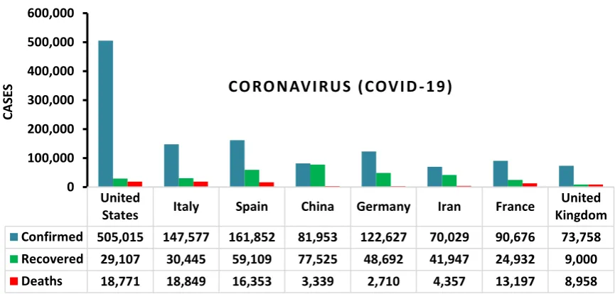
In 2012, Middle East Respiratory Syndrome (MERS) was reported in Saudi Arabiaas an illness caused by a coronavirus. SARS and MERS are Betacoronaviruses (β-CoVs or Beta-CoVs) that transmitted to people from some cats and Arabian camels respectively [7,8]. The sale of wild animals may be the source of coronavirus infection. The discovery of multiple offspring of pangolin coronavirus and their similarity to SARS-CoV-2 suggests that pangolins should be considered as possible hosts of novel coronaviruses. WHO recommendations to reduce the risk of transmission of Coronavirus from animals to humans in wild animal markets [9]. Human-to-human transmission of coronavirus Coronavirus transmission from different cases outside China, namely in Italy [10], US [11], Germany [12], and Vietnam [13], Nepal [14]. On 11 April 2020, SARS-CoV-2 Confirmed more than 1.7 million cases, 400000 recovered cases, and 100000 death cases. Figure 1 shows some statistics about recovered and death cases of COVID-19 [15].
Fig. 1. Statistics of COVID-19 in some countries
Deep Transfer Learning (DTL) is a deep learning technique that reused a trained deep learning model that inspired by neurons of the brain [16]. DTL is quickly becoming a critical technique in image/video classification and detection. DTL improves such a medical system to realize higher outcomes, widen illness scope, and implementing applicable real-time medical image [17,18] disease detection systems. In 2012, Krizhevsky and et al. and Ciregan et al. [19,20] showed how Convolutional Neural Networks (CNN/ ConvNet) based on Graphics Processing Unit (GPU) can enhance many image benchmark classification such as MNIST [21], Chinese characters [22], NORB
United
States Italy Spain China Germany Iran France
United Kingdom Confirmed 505,015 147,577 161,852 81,953 122,627 70,029 90,676 73,758 Recovered 29,107 30,445 59,109 77,525 48,692 41,947 24,932 9,000 Deaths 18,771 18,849 16,353 3,339 2,710 4,357 13,197 8,958
0 100,000 200,000 300,000 400,000 500,000 600,000
CAS
(jittered, cluttered) [23], traffic signs [24], large-scale ImageNet [25], Arabic digits recognition [26], and Arabic handwritten characters recognition [27]. In the following years, various advances in CNN further decreased the error rate on the image/video classification competition. Many DTL models were introduced as AlexNet [20], VGGNet [28], GoogleNet [29], ResNet [30], Xception [31], DenseNet
[32]
,
Inception-V3 [33].
This section conducts the recent scientific papers for applying deep learning in the field of medical chest computerized tomography (CT) classification. Christe et al. [34] introduced a computer-aided detection method based on deep learning was able to detect idiopathic pulmonary fibrosis with similar accuracy to a human reader. The CAD system used for the automatic classification of CT images into 4 radiological diagnostic categories. The model was achieved an F-score (harmonic mean for precision and recall) of 80%. In [35], the authors introduced a novel system for automated classify of Interstitial Lung Abnormality patterns in computed tomography (CT) images. The proposed system was an ensemble of deep convolutional neural networks (DCNNs) that detect more features by incorporating dimensional architectures. The outcome of the ensemble is the sensitivity of 91,41% and specificity of 98,18%.
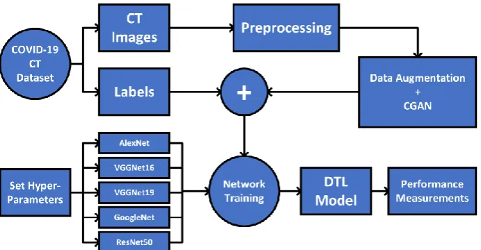
In this paper, we introduced a deep transfer learning (DTL) models to classify COVID-19 chest CT scan digital images. To input adopting CT images of the chest to the deep convolutional neural network (DCNN), we enriched the medical chest CT images using classical data augmentation and CGAN to generate more CT images. After that, a classifier is used to ensemble the class (COVID/NonCOVID) outputs of the classification outcomes. The proposed DTL model was evaluated on the COVID-19 CT scan images dataset. The novelty of this research is conducted as follows: i) The introduced DTL models have end-to-end structure without classical feature extraction and selection methods. ii) We show that data augmentation and Conditional Generative Adversarial Network (CGAN) is an effective technique to generate CT images. iii) Chest CT images are one of the best tools for the classification of COVID-19. iv) The DTL models have been shown to yield very high accuracy in the limited dataset COVID-19. The rest of the paper is organized as follows. In section 2, discusses the dataset used in our research. In section 3, introduces the proposed models, while section 4 illustrates the achieved outcomes and its discussion. Finally, section 5 provides conclusions and directions for further research.
2.
Dataset
Table 1. Number of images for each class in Covid-19 CT dataset
Dataset
Train set Validation set Test setCOVID NonCOVID COVID NonCOVID COVID NonCOVID
COVID19 191 234 60 58 94 105
COVID19 +Aug 2292 2808 720 696 94 105
COVID19 +CGAN 2191 2234 210 208 94 105
COVID19 +Aug+CGAN 4292 4808 870 846 94 105
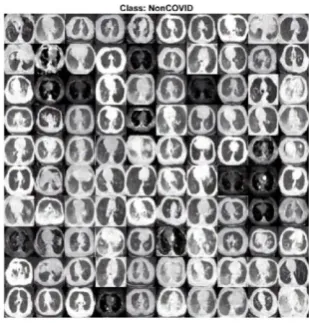
Fig. 2. Samples of the used COVID-19/NonCOVID CT images used in this research
3. Proposed Model
Fig. 3. The proposed architecture of the classical data augmentation along with CGAN and DTL models
Algorithm 1 Proposed Algorithm
Require: COVID-19 CT scan Images (𝑋, 𝑍); where 𝑍 = {𝑧/𝑧 ∈ { COVID; NonCOVID }}
Output: The trained DTL model that classifies the COVID-19 CT image 𝑥 ∈ 𝑋
Preprocessing:
• Resize the image to dimension 256 × 256
• Perform data augmentation: Generate COVID-19 CT images
• Perform CGAN: Generate COVID-19 CT images
• Mean normalize each COVID-19 CT scan images
Import a set of DTL models D= { AlexNet, VGGNet16, VGGNet19, GoogleNet, ResNet50}
Replace the last fully connected layer of each model by a layer of (2 × 1) dimension.
foreach ∀𝑚 ∈ 𝑀 do
𝜇 = 0.001
for epochs = 1 to 50 do
foreach mini-batch (𝑋𝑖; 𝑍𝑖) ∈ (Xtrain; Ztrain) do
Update the parameters of the model 𝑚(·)
if the validation error is not improving for five epochs then
𝜇 = 𝜇 × 0.01
end
end
end
end
foreach ∀𝑥 ∈ 𝑍𝑡𝑒𝑠𝑡 do
the output of all models, 𝑚 ∈ 𝑀
end
Algorithm 1 introduces the proposed model in detail below. Let 𝑀 = {AlexNet, VGGNet16, VGGNet19, GoogleNet, ResNet50} be the set of DTL models. Each DTL is fine-tuned with the COVID-19 CT Images dataset (𝑋, 𝑍); where 𝑋 the set of 𝑁 images, each of size, 256 × 256, and 𝑍 contain the corresponding labels, 𝑍 = {𝑧/𝑧 ∈ {COVID; NonCOVID}}. The dataset divided to train, validate, and test, training set (Xtrain; Ztrain) , validate set (Xval; Zval) , test set (Xtest; Ztest). The training data
then divided into mini-batches, each of size 𝑛 = 32, such that (𝑋𝑖; 𝑍𝑖) ∈ (Xtrain; Ztrain); 𝑖 = 1,2, … , 𝑁 𝑛
and iteratively optimizes (fine-tuning) the DCNN model 𝑑 ∈ 𝐷 to reduce the empirical loss as illustrated in equation (1).
𝐿(𝑤, 𝑋𝑖) =
1
𝑛 ∑ 𝑙(𝑚(𝑥, 𝑤), 𝑧)
𝑥∈𝑋𝑖,𝑧∈𝑍𝑖
(1)
3.1 Deep Transfer Learning
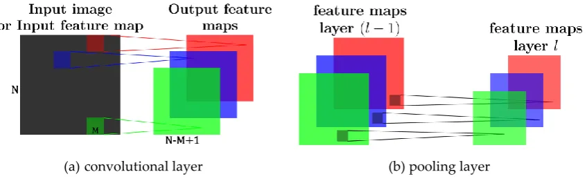
Deep Transfer Learning (DTL) is the most successful reuse type of deep convolutional neural network model for image/video classification. A single DTL model contains many different layers of convolution and pooling layer that work on feature extraction from image/video and more complex deep features in deeper layers.
(a)convolutional layer (b) pooling layer
Fig. 4. Illustration of the convolutional and pooling layer which produce feature maps
Let layer 𝑙 be a convolutional layer. Suppose that we have some 𝑁 × 𝑁 square neuron nodes which are followed by a convolutional layer. If we use an 𝑀 × 𝑀 filter (mask) 𝑊 then convolutional layer output will be of size (𝑁 − 𝑀 + 1) × (𝑁 − 𝑀 + 1) which produces 𝑘-feature maps that are illustrated in Fig. 4. The convolutional layer acts as a feature extractor that grabs features of the inputs. The convolution layer extract features from the image like edges, lines, and corners. To compute the pre-nonlinearity input to some unit. Then, the input of layer 𝑙 − 1
comprises is computed in equation (2):
𝑍𝑖𝑙= 𝐵𝑖𝑙+ ∑ ∑ 𝑊𝑖 𝑋(𝑖+𝑎)(𝑗+𝑏)𝑙−1 𝑁
𝑏=1 𝑁
𝑎=1
(2)
where 𝐵𝑖𝑙 is a bias matrix and 𝑊𝑖 is the mask of size
𝑀 × 𝑀
. Then, the convolutional layer applies its activation function in equation (3):𝑁𝑒𝑡 = 𝜎(𝑍𝑖𝑙) (3)
where 𝜎(. ) is called non-linearity, function applied to achieve non-linearity in DTL, which contains many types such as tanh, sigmoid, Rectified Linear Units (ReLU). In our method, we utilize ReLU in equation (4) as the activation function for faster training process:
𝜎(𝑢) = max(0, 𝑢) (4)
The loss function is the criterion for the training process. Our loss function in equation (5) is defined as the sum of the cross-entropy loss and the box regression loss:
𝐿𝑜𝑠𝑠(𝑠, 𝑡) = 𝐿𝑜𝑠𝑠𝑐𝑙𝑠(𝑠𝑐∗) +λ[𝑝∗> 0]𝐿𝑟𝑒𝑔(𝑣, 𝑣∗) (5)
where 𝑠𝑐∗ denotes the predicted score class 𝑐∗ while 𝑣 and 𝑣∗ denote [𝑣𝑥, 𝑣𝑦, 𝑣𝑤, 𝑣ℎ] of bounding boxes. λ[𝑝∗> 0] indicates that we only consider the boxes of non-background (the box is background if 𝑝∗= 0). This loss function contains two parts for bounding box regression loss
𝐿𝑜𝑠𝑠𝑟𝑒𝑔 𝑎𝑛𝑑 classification loss 𝐿𝑜𝑠𝑠𝑐𝑙𝑠 and, in equation (6-8):
𝐿𝑜𝑠𝑠𝑐𝑙𝑠(𝑠𝑐∗) = − log(𝑠𝑐∗) (6)
𝐿𝑜𝑠𝑠𝑟𝑒𝑔(𝑣, 𝑣∗) = ∑ 𝑅𝐿1(𝑣𝑖− 𝑣𝑖∗)
𝑖∈(𝑥,𝑦,𝑤,ℎ) (7)
where:
𝑅𝐿1(𝑢) = {
0.5𝑢2, 𝑖𝑓 |𝑢| < 0
|𝑢| − 0.5, 𝑜𝑡ℎ𝑒𝑟𝑤𝑖𝑠𝑒 (8)
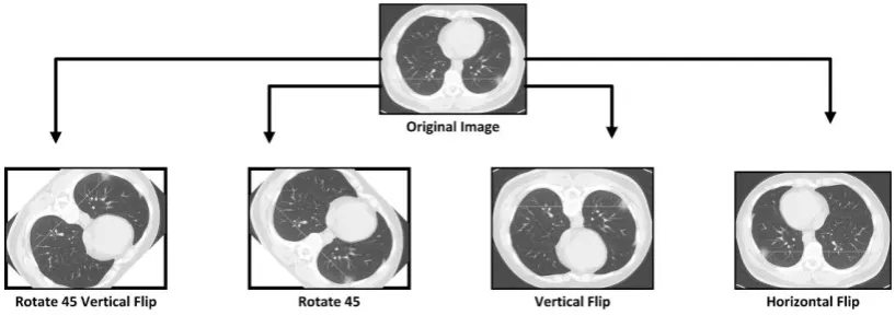
3.2 Data Augmentation
The main idea behind this research is to perform a transfer learning with augmented COVID-19 CT images. ,To increase the performance of the proposed transfer learning models, training data amount and validate data amounts is a very important factor. The most popular classical data augmentation method is to perform a combination of the affine image transformations [37]. Different methods for classical data augmentation such as rotation, shifting, flipping, zooming, transformation, add noise were selected to be applied in the original dataset. Figure 5 shows examples of COVID-19 CT augmented images. The achieved performance measurement will be discussed in the experimental results section.
Fig. 5. Perform augmentation methods to increase limited COVID-19 CT scan images
3.3 Conditional Generative Adversarial Network
Fig. 6. Conditional Generative Adversarial Network model
Fig. 7. The structure of the proposed CGAN network
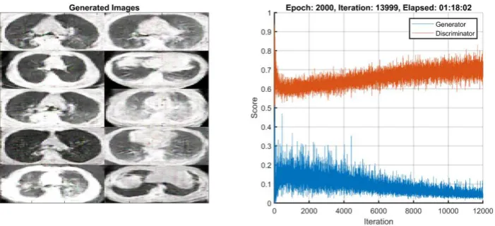
The CGAN network helped in overcoming the overfitting problem caused by the limited number of CT images in the COVID-19 dataset. Figure 7 presents samples of the output of the GAN network for the covid-19 class. Moreover, it increased the dataset images to be 10 times larger than the original one. The dataset number of images reached 4425 images in the train set and 418 in the validation set after using the CGAN network for 2 classes. This will help in achieving better testing accuracy and performance matrices. The achieved performance measurement will be discussed in the experimental results section.
Fig. 9. Samples of COVID-19 CT images generated by the CGAN model.
4. Experimental Results
The proposed model is trained on a high-end Graphics Processing Unit (GPU). The GPU used (NVIDIA RTX 2070) contains 2304 CUDA core and comes with the CUDA Deep Neural Network library (CuDNN) for GPU learning. The deep learning package TensorFlow machine learning and Matlab as back end library. The proposed model has been tested under four different scenarios, the first scenario is to test the DTL models with original COVID-19 CT dataset, the second scenario with data augmentation, the third one with CGAN, and the last one combines all three scenarios. All the test experiment scenarios included the two classes (COVID/NonCOVID). Every scenario consists of the validation phase and the testing phase as shown in Table 2.
Table 2. Configuration of DTL models
Model Layers Batch size Momentum Epoch Learning Rate Optimizer
AlexNet VGGNet16 VGGNet19 GoogleNet ResNet50
8 16 19 22 50
32 0.9 50 0.001 Adam
technique, which updates weights parameters. This optimizer technique is a combination of Root Mean Square Propagation (RMSprop) and Stochastic Gradient Descent (SGD) with momentum. To avoid deep learning network overfitting problems, we utilize this problem by using the dropout method [39] as well as the early-stopping technique [40] to select the most appropriate training iteration.
4.1 Verification and Testing Accuracy Measurement
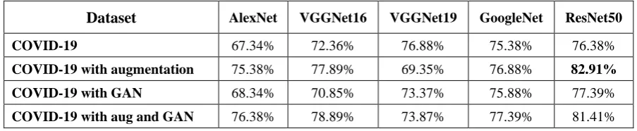
Testing accuracy is one of the estimations which demonstrates the performance measurement of any DTL models. The confusion matrix also is one of the performance measurements which give more insights into the achieved testing accuracy. The first DTL model will be investigated is AlexNet along with four scenarios as shown in Figure 10. Figure 10 shows that the highest testing accuracy is 76.4% when the COVID-19 CT dataset is augmented with data augmentation along with CGAN. The Second DTL model will be investigated with VGGNet16. Figure 10 shows that the highest testing accuracy is 78.9% when the COVID-19 CT dataset is augmented with classical data augmentation along with CGAN. The Third DTL model will be investigated with VGGNet19. Figure 10 shows that the highest testing accuracy is 76.9% when the COVID-19 CT dataset is not augmented. The Fourth DTL model will be investigated with GoogleNet. Figure 10 shows that the highest testing accuracy is 77.4% when the COVID-19 CT dataset is augmented with the classical data augmentation along with CGAN.
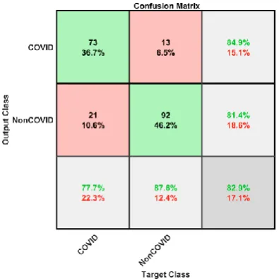
The Final DTL model will be investigated with ResNet50. Figure 10 shows that the highest testing accuracy is 82.9% when the COVID-19 CT dataset is augmented with classical data augmentation as shown in Figure 11. Table 3 summarizes the testing accuracy for the different deep transfer learning models for 2 classes with the four scenarios. Table 3 illustrates according to testing accuracy, the Resnet50 achieved the highest accuracy with 82.9%, this is due to the large number of parameters in the Resnet50 architecture which contains millions of parameters which are not larger than VGGNet and GoogleNet but the VGGNet and GoogleNet only include 16, and 22 layers while the Resnet50 includes 50 layers.
Table 3. DTL testing accuracy for the different four scenarios
Dataset
AlexNet VGGNet16 VGGNet19 GoogleNet ResNet50COVID-19 67.34% 72.36% 76.88% 75.38% 76.38%
COVID-19 with augmentation 75.38% 77.89% 69.35% 76.88% 82.91%
COVID-19 with GAN 68.34% 70.85% 73.37% 75.88% 77.39%
COVID-19 with aug and GAN 76.38% 78.89% 73.87% 77.39% 81.41%
Confusion Matrix of AlexNet
COVID
83
54
69
24
74
43
60
13
NonCOVID
11
51
25
81
20
62
34
92
COVID
58
19
71
21
58
22
59
7
NonCOVID
36
86
23
84
36
83
35
98
Confusion Matrix of VGGNet19
COVID
66
18
83
50
50
9
67
25
NonCOVID
28
87
11
55
44
96
27
80
Confusion Matrix of GoogleNet
COVID
65
20
70
22
71
25
67
18
NonCOVID
29
85
24
83
23
80
27
87
Confusion Matrix of ResNet50
COVID
62
15
73
13
58
9
76
19
NonCOVID
32
90
21
92
36
96
18
86
COVID-19
COVID-19 with
Augmentation
COVID-19 with
CGAN
COVID-19 with
Augmentation
and CGAN
Fig. 10. DTL Confusion matrices for two classes with different scenarios
Fig. 11. Confusion matrix of highest accuracy for ResNet50 in COVID-19 with classical data augmentation
4.2 Performance Evaluation and Discussion
Accuracy =
(TP+FP)+( 𝑇𝑁+𝐹𝑁) TP+TN (9)where TP (True Positives) is the count of correctly labeled instances of the class under observation, FP (False Positives) is the count of miss-classified labeled of rest of the classes, TN (True Negatives) is the count of correctly labeled instances of rest of the classes, and FN (False Negatives) is the count of miss-classified labeled of the class under observation.
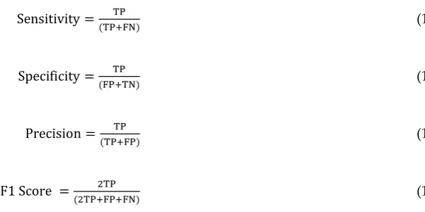
Sensitivity = TP
(TP+FN) (10)
Specificity = TP
(FP+TN) (11)
Precision = TP
(TP+FP) (12)
F1 Score = 2TP
(2TP+FP+FN) (13)
Figure 12 presents the performance metrics for different scenarios with DTL models for the COVID-19 CT dataset. The highest sensitivity of 88.3% (Table 4) is achieved by scenario-1 and scenario-2 (COVID-19 only and with augmentation) based on AlexNet and VGGNet19 that refers to the test's ability to correctly classify COVID-19 CT patients who do have the condition. In the example of a CT scan medical test used to classify and detect a COVID-19 disease, the detection rate (sensitivity) of the test is the proportion of people who test positive for the COVID-19 malady among those who have the COVID-19 malady. A negative result in a test with a high detection rate is useful for getting rid of the COVID-19 CT malady.
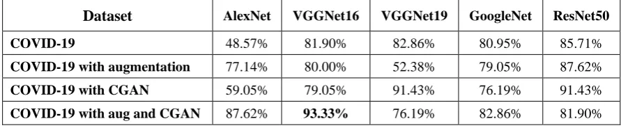
A test with high specificity would be able to determine the human that does not have the COVID-19 as shown in Table 5. Sensitivity and specificity can be summarized by a single quantity called the balanced accuracy as shown in Table 6, which is defined as the mean of both measures in equation (14):
Balanced accuracy =Sensitivity+Specificity
2 (14)
(a) COVID-19 (b) COVID-19 with Augmentation
(c) COVID-19 with CGAN (d) COVID-19 with Augmentation and CGAN
Fig. 12. Performance measurements for COVID-19 CT in four scenarios
Table 4. Testing Sensitivity for the different 4 scenarios
Dataset
AlexNet VGGNet16 VGGNet19 GoogleNet ResNet50COVID-19 88.30% 61.70% 70.21% 69.15% 65.96%
COVID-19 with augmentation 73.40% 75.53% 88.30% 74.47% 77.66%
COVID-19 with CGAN 78.72% 61.70% 53.19% 75.53% 61.70%
COVID-19 with aug and CGAN 63.83% 62.77% 71.28% 71.28% 80.85%
Table 5. Testing Specificity for the different 4 scenarios
Dataset
AlexNet VGGNet16 VGGNet19 GoogleNet ResNet50COVID-19 48.57% 81.90% 82.86% 80.95% 85.71%
COVID-19 with augmentation 77.14% 80.00% 52.38% 79.05% 87.62%
COVID-19 with CGAN 59.05% 79.05% 91.43% 76.19% 91.43%
COVID-19 with aug and CGAN 87.62% 93.33% 76.19% 82.86% 81.90% 0 0.1 0.2 0.3 0.4 0.5 0.6 0.7 0.8 0.91 SC O R E PERFORMANCE MEASUREMENT C O V I D - 1 9 D A T A S E T
AlexNet VGGNet16 VGGNet19 GoogleNet ResNet50 0 0.2 0.4 0.6 0.8 1 SC O R E PERFORMANCE MEASUREMENT C O V I D - 1 9 D A T A S E T W I T H
A U G M E N T A T I O N
AlexNet VGGNet16 VGGNet19 GoogleNet ResNet50 0 0.1 0.2 0.3 0.4 0.5 0.6 0.7 0.8 0.91 SC O R E PERFORMANCE MEASUREMENT C O V I D - 1 9 D A T A S E T W I T H G A N
AlexNet VGGNet16 VGGNet19 GoogleNet ResNet50 0 0.2 0.4 0.6 0.8 1 SC O R E PERFORMANCE MEASUREMENT C O V I D - 1 9 D A T A S E T W I T H A U G M E N T A T I O N A N D G A N
AlexNet
VGGNet16
VGGNet19
GoogleNet
Table 6. Testing Balanced accuracy for the different 4 scenarios
Dataset
AlexNet VGGNet16 VGGNet19 GoogleNet ResNet50COVID-19 68.44% 71.80% 76.54% 75.05% 75.84%
COVID-19 with augmentation 75.27% 77.77% 70.34% 76.76% 82.64%
COVID-19 with CGAN 68.89% 70.38% 72.31% 75.86% 76.57%
COVID-19 with aug and CGAN 75.73% 78.05% 73.74% 77.07% 81.38%
As shown in Table 6, the balanced accuracy for different scenarios. The table also indicates that ResNet50 is the best classifier to detect the COVID-19 in CT dataset with classical data augmentation along with CGAN. The classical data augmentation along with CGAN improves the performance of classification in all deep transfer models (AlexNet, VGGNet16, VGGNet19, GoogleNet, ResNet50). The other bottleneck is the limited size of the COVID-19 CT database. Predictably the performance of deep transfer models can be further improved if more data are collected in the future. Although, we have achieved promising accuracy rates, however, the proposed model in this study needs to be tested on larger scale datasets that include different COVID-19 CT images to increase the testing accuracy and extend it in other medical applications. As future work, we plan to classify COVID-19 using a neutrosophic approach [42] and deep learning.
5. Conclusion and future works
In 2019, World infected by a 2019 novel coronavirus that killed more than thousands and infected over millions of humans within few months of the 2019 novel coronavirus epidemic. In this paper, classical data augmentations along with CGAN with deep transfer learning for COVID-19 detection in limited chest CT scan images is presented. The number of COVID-19 CT images of the collected dataset was 742 images for two types of labels. The classical data augmentation and CGAN help to increase the CT dataset and overcoming the overfitting problem. Moreover, five deep transfer learning models (AlexNet, VGGNet16, VGGNet19, GoogleNet, ResNet50) were selected in this paper for investigation. Using a combination of classical data augmentation and CGAN with deep transfer learning improve testing accuracy, and performance measurements such as sensitivity, specificity, precision, accuracy, and F1 score. The results show that ResNet50 with classical data augmentation along with CGAN is the best classifier to detect the COVID-19 from chest CT dataset. As future work, we plan to
approach the COVID-19 study from a neutrosophic environment with deep learning.