International Journal of Advanced Research in Computer Science
RESEARCH PAPER
Available Online at www.ijarcs.info
Implementing Wireless Capsule Endoscopy WCE in Digestive System Diagnostics
Hussam Ghunaim
Computer Science and Engineering Department University of Bridgeport,
Bridgeport, USA
Prof. Christian Bach
Technology Management Department University of Bridgeport,
Bridgeport, USA
Abstract: The purpose of this research is to discuss the revolutionary endoscopy method WCE that would enhance the diagnostic accuracy and reliability level. Additionally, a comparison has been made with other currently in practice endoscopy methods to single out the strengths and advantages of such endoscopy method. The limitation of this research caused by limited up to date data due to the restrict privacy policy normally adopted by hospitals regarding releasing patients information. This limitation will impose a partially outdated comparison results and conclusions. However, the past trends showed a steady increase in the number of medical facilities that decided to approve the usage of the WCE. These trends are derived from direct interactions with various medical communities. This paper originality and value comes from the fact that increasing number of patients showed a serious reluctant toward continuing all their prescribed medical testing or procedures. Consequently, serious implication can be expected affecting those patients’ health. WCE if understood correctly by both patients and doctors will have a positive impact on the success of diagnostic and treatment statistics.
Keywords: WCE; Endoscopy; wireless endoscopy; 3D Trajectory; image processing
I. INTRODUCTION
All The human digestive system has many organs; such as: the esophagus, stomach, small intestine, and large intestine, figure1. Therefore, many different types of diseases have been discovered and diagnosed through the years. Endoscopes have been the most important diagnostic tool used to examine the upper and the lower parts of the digestive system [4]. Nevertheless, these traditional endoscopes suffer from severe limitation due to their inability to visualize the entire digestive system [5].
Figure 1: Human Digestive System. [1]
Other surgical approaches are in use today and can provide a high level of diagnostic data [6]. However, due to their cost and after surgery complications, they are not so common to both doctors and patients. These concerns are magnified when dealing with either old or young (less than 3 years old) patients.
Wireless Capsule Endoscopy (WCE) was proposed as a promising alternative. WCE was approved by FDA in 2001 in United States. Thousands of patients have benefited from this revolutionary technology worldwide [7]. However, the costs associated with the scarcity of professionals experienced enough to use this technology considered as one of the major hindering factors till today.
WCE is easy to be swallowed by an average human being as WCE has relatively very small dimensions: 11 X 24 mm, figure 2 [8]. Therefore, there is no need to procedure preparations protocols for all ages range.
Figure 2: WCE Wireless Capsule Endoscopy Dimensions [2]
The detailed engineering of the WCE is beyond the scope of this paper. Nevertheless, it is beneficial to give brief introduction on how it works. The front end of the WCE has micro camera that capture stream of images while it is on the move normally called as the “Optical Dome”. Of course, cameras need light to function properly. That is why there is a light unit built mainly from powerful LEDs (Light Emitting Diodes) [9]. The camera has to be able to transmit all captured images wirelessly to a receiver. Because of the small size of the WCE, the transmitter has a short range, thus, the patient must carry it with him/her all the time.
WCE camera can be configured by several parameters. However, the most interesting factor is number of captured images or frames per second (FPS) which will count to the total time needed to view these images. Usually, this time is about 6 – 8 hours of continuous video stream. As a result, some medical professionals find it unpractical to analyze the information derived from WCE devices. When WCE finishes its work, doctors will be able to download all captured images (typically from 50,000 to 100,000) and analyze them.
One of the main disadvantages of using WCE is that the resulted video is too long for any doctor to watch and analyze. The problem is now under substantial research activities on how the captured images can be summarized or compressed. On other words, the possibility of using artificial intelligence in designing an automated computer system that is able to detect suspicious structures within the digestive system [5]. There are already available many algorithms for objects detection and tracking that might be implemented in such systems.
II. SCIENTIFIC METHOD
Scientific methods can be seen as a collection of techniques that are used to examine and study a specific phenomenon to acquire new knowledge or simply proving or disapproving exist theory or knowledge. The term scientific comes from the fact that these methods must include empirical and measurable evidences. Such evidences are always subject to specific principles of reasoning. The major difference that differentiates scientific methods from other methods of study is its reliance on scientific facts rather personal judgments and assumptions.
In scientific approach, researchers normally start with proposing a hypothesis. Then, this hypothesis will undergo through a series of experiments to collect data describing how this particular hypothesis relates all dependent and independents variables. At the end, a researcher can use these results and outcomes to either prove the hypothesis or to deny it and call for additional research study.
III. RESEARCH METHOD
The research method adopted in this work was literature review. Because now days, thousands of publications can be found that addressed WCE technology from all known facets. As a consequence, it is worthwhile to spend some time and goes through these works and come up with a new insight that might be useful to people.
IV. THE PAPER MODEL
Figure 3 represents the paper model where it shows the dependent variable along with the corresponding five independent variables. The model also shows the hypotheses made in this paper.
Figure 3: The Model of Implementing WCE in Digestive System Diagnostic.
A. Goal
WCE is the only comprehensive digestive system diagnostic tool available today: “Wireless capsule endoscopy (WCE) has been gradually applied in hospitals due to its great advantage that it can directly view the entire small bowel in human body compared with traditional endoscopies and other imaging techniques for gastrointestinal diseases [10] p17.”
This new revolutionary diagnostic method has attracted tremendous attention worldwide from academic as well as from medical institutions [11]. Several medical institutions in many countries have reported a positive feedback from both patients and medical staff regarding the increasing tendency towards adapting WCE in their diagnostic plans [12].
However, some reports have demonstrated that side effects do exist in case of WCE usage, such as: abdominal pain, fever, weight loss, and anemia. These problems trigger the need for more in depth research to figure out the significance of such problems on the overall performance of using WCE and the possible solutions for them.[13]
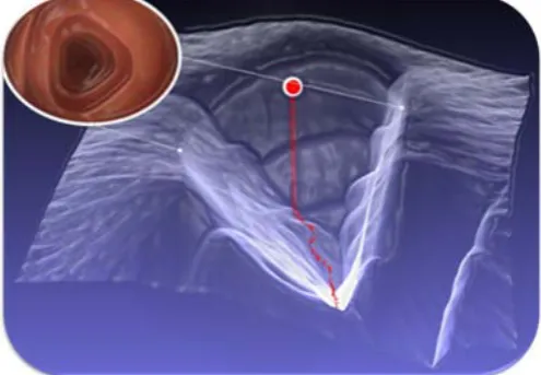
Figure 4: WCE 3D Trajectory Sample [3]
defibrillators (ICD) and other electro medical devices [14] p5.”
What follows is the detailed discussion of the dependent and independent variables in the proposed model.
B. Factor 1: WCE is a Painless Procedure
WCE is the only painless endoscopy procedure: “The development of wireless capsule endoscopy allows painless imaging of the small intestine [15].” In a study for Hull and Church, has been found that other methods of endoscopy could be as painful as one can think of. In many times this a reason why patients refuse to accept this procedure, putting their health at risks. “Colonoscopy is sometimes painful for the patient and often difficult for the endoscopist, but it is hard to predict how difficult or painful the examination will be [16] p12. ”
In a recent study for [17], a questionnaire was distributed to 180 patients were each one of them returned at least one response (94 %). Additionally, 79 % have done all required parts. 14 % out of the total participating patients have expressed their experience with endoscopy as a painful process. In on other hand, 59% find it as an oppressive. While doctors argue this type of endoscopy did not cause any bodily reactions, some patients (47 %) reported a throat ache. Other group of patients has expressed and increase level of anxiety, depression before administering endoscopy regardless of which type.
C. Factor 2: WCE is an Advancement in Endoscopy
WCE is a revolutionary advancement in endoscopy filed: “Wireless capsule endoscopy (WCE) is a new technology for small bowel imaging [18] p9.” Baopu and Meng have summarized in their recent paper: “Interpretation of the produced video data for the digestive tract on each patient is left to naked eyes of medical staff. Such a process is very tedious and time-consuming with the average inspection time about two hours for a whole WCE video [19] p2.” Such argument leads to the conclusion that WCE with its capabilities has a significant influence on the future of endoscopy for its surpassing characteristics.
WCE innovation opened the door wide for advanced algorithms techniques. “The wireless capsule endoscopy (WCE) invented by Given Imaging has been gradually used in hospitals due to its great breakthrough that it can view the entire small bowel for gastrointestinal diseases [20] p21.” One example of such advanced algorithms is objects detection and tracking algorithms.
Additionally, an interesting field of study resulted from such needs is called Video Summarization. In this technique, an algorithm is applied to filter out all faulty, out of scope images form the captured images stream to increase the reliance level on the remaining images only, “Video summarization, aimed at reducing the amount of data that must be examined in order to retrieve the information desired from information in a video, is an essential task in video analysis and indexing applications [21].”
D. Factor 3: WCE has a 3D Capability
WCE is the only available endoscopy technology with 3D capability: “Wireless Capsule Endoscopy (WCE) is an endoscopy technology that allows medical personnel to view the digestive tract non-invasively. Physicians can detect diseases such as blood-based abnormalities, polyps, ulcers and Crohn’s disease [22] p7.” Due to many difficulties facing WCE, such as limited illumination and irregular motion of the capsule endoscope [23], 3D become a potential solution. A recent paper for [24] has demonstrated the possibility for utilizing image segmentation to obtain a 3D reconstruction of the mucosal tissues. This kind of reconstruction requires sophisticated mathematical models that normally consumes large amount of computing processing power. Currently such advancements are still under intense research to increase efficiency and decrease resources consumptions and hence the overall costs.
E. Factor 4: WCE Images are Numerical Representation
Computer aided images are just numerical representations: “A color WCE image is a snapshot of the digestive tract at a given time. However, in a computer-aided diagnosis system, the image content semantics needs to be translated in numerical ways for interpretation. There are several ways to represent the numerical form of an image known as image abstraction [8] p3.” Following this methodology makes it possible to automatically analyze images and detect interesting textures, that is, any abnormal structure that can be defines as a medial issue [25]. Doctors usually spend long time do such analysis manually and generally they report that this process became unreliable after a short while. [26] pointed that designing a computer aided detection system is feasible, though has it is limitation as well.
F. Factor 5: Most of WCE Images are Wasteful
Most of the WCE resultant images are wasteful. A study for [27] has found that only less than 5% of the total captured images of a WCE video, which is normally in the range of 55,000 – 100,000 frames, typically have abnormalities. Consequently, it is very important to design an algorithm that can automatically distinguish abnormal findings from normal ones. Interestingly, while WCE in move inside the patient digestive system, it faces a lot of asserting factors, such as, guts movements. Such movements make the WEC turn and flip the thing that cause it capture unusual images and hence waste [28]. Limited illumination yet is another problem. Although a separate illumination source has been provided to the WCE, it still not enough to capture high clear images compare with other traditional endoscopy methods [29].
V. DISCUSSION OF THE MODEL
awaiting further insights and research. While this paper was limited to five of the most important and influential factors on the success of implanting WCE, many other still unaddressed here for practical reason. As providing a comprehensive literature describing WCE technology will require hundreds of pages and of course this is beyond the scope of this paper.
However, this technology still relatively recent in practice and consequently suffers several drawbacks. One problem that is highly recommended for future research is `movement control`. The actual motion of the WCE is derived mainly by peristaltic propulsion of the gastro-intestinal GI movements where they are definitely unpredictable. This leads to the necessity of solving this problem without affecting the applicability of WCE usage and being in the patient body for many hours.
Other problems come from the fact that we cannot assume that the patient intestines will be completely empty without expecting any remaining that will be likely to obscure the captured images clearness. Many physicians have reported that they were confused about the rambling frames captured by small intestine junctions. WCE because of its compact size, it suffers from an engineering limitation known as narrow field view. This can limit the analytical ability of physicians related to how much information can be actually revealed by captured images. WCE uses an optical dome to capture images where this dome can be covered with any kind of remaining.
Yet another limitation imposed by the use of WEC caused by the lengthy stream of images that can be over 50,000 images per patient. Physicians need to spend in average several hours to analyze the full range of captured images which is a great challenge. This can be another interesting area for future research which suggests the design of an automated system that is able to recognize suspicious features within the entire digestive system. These suspicious features can be anything as tumors, bleedings, ulcers etc. This stream of research has the opportunity to benefit from already proven feasibility of objects detection and traction algorithms. Many of these algorithms found their use in industrial quality assurance applications, security monitoring systems and even in deep space observation activities.
3D reconstruction of the captured images can be promising in terms of overcoming some of the previous drawbacks. There are a decent number of serious practical implementations of such algorithms. Kolar has tried to design an active vision system using traditional hardware [31]. The design included the integration of an image sensor along with a pattern projector.
VI. IMPORTANCE OF MODEL
This work is important because it provides an explanation of relatively new technology that may people still not aware of it. Even for people who had a chance to look at it, they still have not exposed entirely to the facts that lie behind WCE technology, as [32] have claimed this can be seen as one the reason why WCE still did not receive attention that it deserves from both research and medical practice communities. Doctors generally tend to use what they already know and skilled in using. Therefore, they are somehow reluctant in delving inside
new technologies that they do not yet have a complete control on its effects or interactions with other environmental factors.
From this point of view, we believe in the importance of spreading positive understanding among all interested communities including the general public. This kind of understanding will help greatly the implementation of WCE as we except that more people will become convinced in its volubility and effective improvement of people health.
VII. CONCLUSION
After considering different facets of the newly developed WCE technology, we came to the conclusion that WCE is to dominant the field of endoscopy in near future [33]. [34] has claimed that while many other endoscopy technologies are still in practice widely and on every day basis, continuous evaluation reports and surveys addressing the endless side effects on patients’ health physically and psychologically.
On the other hand, WCE as is still relatively recent technology, it is therefore the most expensive compared to other endoscopy technologies. Such burden has a significant impact on adopting WCE especially in countries with low economy level [35]. However, as new advancements and developments are published in this interesting and exciting field on daily basis, we strongly believe that the costs of implementing WCE should decrease dramatically in the coming few years. Nevertheless, other resources such as charity organizations and helping centers can be investigated to support the application of such new technology to needed individuals around the world.
VIII. ACKNOWLEDGMENT
I would like to take this opportunity to show my appreciation to Prof. Christian Bach for his consistent help and support. Without such help, this work would not be completed or published in one of the prestigious journals around the word.
IX. REFERENCES:
[1] International, Q., The Visual Dictionary Online Source: Www.Visualdictonary.Com.
[2] Chen, Y., A Computer-Aided Diagnostic System For Wireless Capsule Endoscopy. 2012.
[3] Ciuti, G., Et Al. Intra-Operative Monocular 3d Reconstruction For Image-Guided Navigation In Active Locomotion Capsule Endoscopy. In Biomedical Robotics And Biomechatronics (Biorob), 2012 4th Ieee Ras & Embs International Conference On. 2012: Ieee.
[4] Saif, J. And H. Krawczyk. Parallel Procedures For Roi Identification In Endoscopic Images. In Parallel Computing In Electrical Engineering, 2002. Parelec '02. Proceedings. International Conference On. 2002.
[5] Westerhof, J., J.J. Koornstra, And R.K. Weersma, Can We Reduce Capsule Endoscopy Reading Times? Gastrointestinal Endoscopy, 2009. 69(3, Part 1): P. 497-502.
In Life Science Systems And Applications Workshop, 2009. Lissa 2009. Ieee/Nih. 2009.
[7] Penna, B., Et Al. A Technique For Blood Detection In Wireless Capsule Endoscopy Images. In Proc Of The 17th European Signal Processing Conference (Eusipco’09). Glasgow, Scotland. 2009: Citeseer.
[8] Chen, Y., A.K. Lakshmanan, And J. Lee, Bleeding Detection In Wireless Capsule Endoscopy Videos Using Temporal Features.
[9] Lau, P.Y. And P.L. Correia. Detection Of Bleeding Patterns In Wce Video Using Multiple Features. In Engineering In Medicine And Biology Society, 2007. Embs 2007. 29th Annual International Conference Of The Ieee. 2007: Ieee. [10] Li, B. And M.Q.H. Meng, Bowel Polyp Detection In Capsule
Endoscopy Images With Color And Shape Features. Biologically Inspired Robotics, 2012: P. 205.
[11] Ciuti, G., A. Menciassi, And P. Dario, Capsule Endoscopy: From Current Achievements To Open Challenges. Biomedical Engineering, Ieee Reviews In, 2011. 4: P. 59-72.
[12] Costamagna, G., Et Al., A Prospective Trial Comparing Small Bowel Radiographs And Video Capsule Endoscopy For Suspected Small Bowel Disease. Gastroenterology, 2002. 123(4): P. 999-1005.
[13] Fireman, Z., Et Al., Diagnosing Small Bowel Crohn’s Disease With Wireless Capsule Endoscopy. Gut, 2003. 52(3): P. 390-392.
[14] Leighton, J.A., Et Al., Safety Of Wireless Capsule Endoscopy In Patients With Implantable Cardiac Defibrillators. American Journal Of Gastroenterology, 2005. 100(8): P. 1728-1735. [15] Mylonaki, M., A. Fritscher-Ravens, And P. Swain, Wireless
Capsule Endoscopy: A Comparison With Push Enteroscopy In Patients With Gastroscopy And Colonoscopy Negative Gastrointestinal Bleeding. Gut, 2003. 52(8): P. 1122-1126. [16] Hull, T. And J. Church, Colonoscopy—How Difficult, How
Painful? Surgical Endoscopy, 1994. 8(7): P. 784-787. [17] Kruijshaar, M., Et Al., The Burden Of Upper Gastrointestinal
Endoscopy In Patients With Barrett's Esophagus. Endoscopy, 2006. 38(09): P. 873-878.
[18] Scapa, E., Et Al., Initial Experience Of Wireless-Capsule Endoscopy For Evaluating Occult Gastrointestinal Bleeding And Suspected Small Bowel Pathology. The American Journal Of Gastroenterology, 2002. 97(11): P. 2776-2779. [19] Baopu, L. And M.Q.H. Meng. Comparison Of Several Image
Features For Wce Video Abstract. In Intelligent Robots And Systems (Iros), 2011 Ieee/Rsj International Conference On. 2011.
[20] Li, B. And M.Q.-H. Meng, Computer-Based Detection Of Bleeding And Ulcer In Wireless Capsule Endoscopy Images By Chromaticity Moments. Computers In Biology And Medicine, 2009. 39(2): P. 141-147.
[21] Gianluigi, C. And S. Raimondo, An Innovative Algorithm For Key Frame Extraction In Video Summarization. Journal Of Real-Time Image Processing, 2006. 1(1): P. 69-88.
[22] Karargyris, A., O. Karargyris, And N. Bourbakis. 3d Representation Of The Digestive Tract Surface In Wireless Capsule Endoscopy Videos. In Bioinformatics And Bioengineering (Bibe), 2010 Ieee International Conference On. 2010: Ieee.
[23] Yichen, F., M.Q.H. Meng, And L. Baopu. 3d Reconstruction Of Wireless Capsule Endoscopy Images. In Engineering In Medicine And Biology Society (Embc), 2010 Annual International Conference Of The Ieee. 2010.
[24] Prasath, V.B.S., Et Al. Mucosal Region Detection And 3d Reconstruction In Wireless Capsule Endoscopy Videos Using Active Contours. In Engineering In Medicine And Biology Society (Embc), 2012 Annual International Conference Of The Ieee. 2012.
[25] Li, B. And M.Q.-H. Meng, Texture Analysis For Ulcer Detection In Capsule Endoscopy Images. Image And Vision Computing, 2009. 27(9): P. 1336-1342.
[26] Stefanini, C., A. Menciassi, And P. Dario, Modeling And Experiments On A Legged Microrobot Locomoting In A Tubular, Compliant And Slippery Environment. The International Journal Of Robotics Research, 2006. 25(5-6): P. 551-560.
[27] Nawarathna, R., Et Al., Abnormal Image Detection Using Texton Method In Wireless Capsule Endoscopy Videos, In Medical Biometrics, D. Zhang And M. Sonka, Editors. 2010, Springer Berlin Heidelberg. P. 153-162.
[28] Ben-Soussan, E., Et Al., Factors That Affect Gastric Passage Of Video Capsule. Gastrointestinal Endoscopy, 2005. 62(5): P. 785-790.
[29] Yang, L., Et Al. Design Of A Video Image Recorder System For Wireless Capsule Endoscopes Based On Dsp. In World Congress On Medical Physics And Biomedical Engineering 2006. 2007: Springer.
[30] Colm, O. And J.S. Barkin, Wireless Capsule Endoscopy In The Evaluation Of The Esophagus. Current Gastroenterology Reports, 2004. 6(3): P. 210-212.
[31] Kolar, A., Et Al., A Systemfor An Accurate 3d Reconstruction In Video Endoscopy Capsule. Eurasip Journal On Embedded Systems, 2009. 2009: P. 12.
[32] Mow, W.S., Et Al., Initial Experience With Wireless Capsule Enteroscopy In The Diagnosis And Management Of Inflammatory Bowel Disease. Clinical Gastroenterology And Hepatology, 2004. 2(1): P. 31-40.
[33] Karargyris, A., A Novel Synergistic Diagnosis Methodology For Identifying Abnormalities In Wireless Capsule Endoscopy Videos. 2010, Wright State University.
[34] Eilertsen, M.-E.B., Children And Adolescents Surviving Cancer: Psychosocial Health, Quality Of Life And Social Support. 2011, Norwegian University Of Science And Technology.
![figure 2 [8]. Therefore, there is no need to procedure preparations protocols for all ages range](https://thumb-us.123doks.com/thumbv2/123dok_us/696288.1077304/1.595.46.275.501.676/figure-need-procedure-preparations-protocols-ages-range.webp)